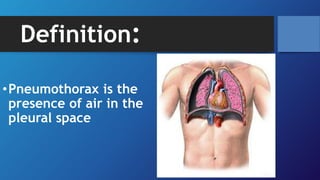
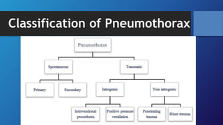
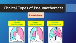
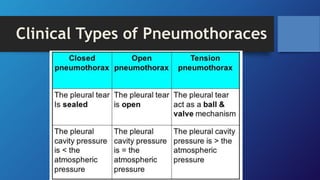
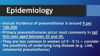
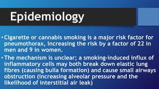
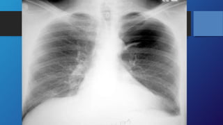
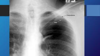
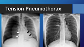
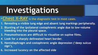
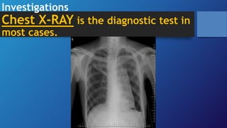
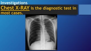
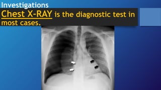
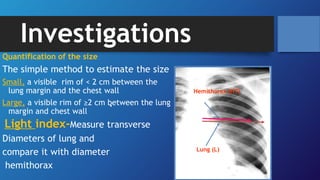
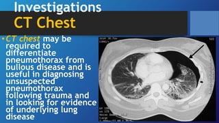
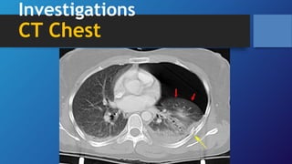
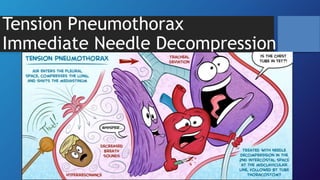
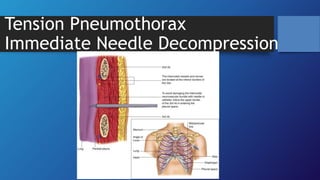
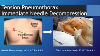
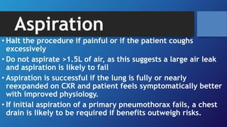
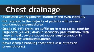
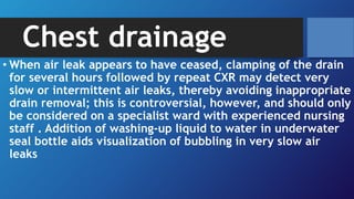
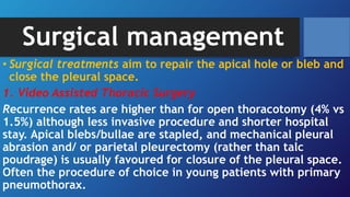
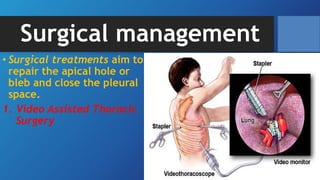
Pleural effusion, sometimes referred to as “water on the lungs,” is the build-up of excess fluid between the layers of the pleura outside the lungs. The pleura are thin membranes that line the lungs and the inside of the chest cavity and act to lubricate and facilitate breathing.
Normally, the pleural space contains a small amount of fluid (5 to 15 mL), which acts as a lubricant that allows the pleural surfaces to move without friction.
But if fluid builds up from either increased production or inadequate removal pleural effusion results.
Pleural effusion B/L or unilateral (parapneumonic process)
Refers to any significant collection of fluid within pleural space.
Any imbalance in formation, absorption lead accumulation of pleural fluid. Common condition:
CHF
Bacterial pneumonia
Malignancy(chest tumor)
Pulmonary embolism
Pleura effusion is a condition refers to a collection of fluid in the pleural space. It is almost secondary to other conditions.
This PowerPoint presentation provides an in-depth overview of pneumothorax, a medical condition that occurs when air leaks into the pleural cavity, causing the lung to collapse. The presentation covers the causes, symptoms, and diagnostic procedures for pneumothorax, including chest x-rays and CT scans.
The presentation also discusses the various treatment options available for pneumothorax, such as thoracentesis, chest tube insertion, and surgery. The benefits and risks of each treatment are also explained in detail, providing the audience with a comprehensive understanding of the condition and its management.
In addition, the presentation includes several case studies and real-life examples to help illustrate the impact of pneumothorax on patients and the importance of early diagnosis and treatment. It is an ideal resource for medical professionals, students, and anyone interested in learning more about this common medical condition.
Overall, this PowerPoint presentation provides a valuable resource for understanding pneumothorax, its causes, symptoms, and treatment options, helping to improve patient outcomes and quality of care.
MANAGEMENT OF ATRIOVENTRICULAR CONDUCTION BLOCK.pdfJim Jacob Roy
Cardiac conduction defects can occur due to various causes.
Atrioventricular conduction blocks ( AV blocks ) are classified into 3 types.
This document describes the acute management of AV block.
Report Back from SGO 2024: What’s the Latest in Cervical Cancer?bkling
Are you curious about what’s new in cervical cancer research or unsure what the findings mean? Join Dr. Emily Ko, a gynecologic oncologist at Penn Medicine, to learn about the latest updates from the Society of Gynecologic Oncology (SGO) 2024 Annual Meeting on Women’s Cancer. Dr. Ko will discuss what the research presented at the conference means for you and answer your questions about the new developments.
HOT NEW PRODUCT! BIG SALES FAST SHIPPING NOW FROM CHINA!! EU KU DB BK substit...GL Anaacs
Contact us if you are interested:
Email / Skype : kefaya1771@gmail.com
Threema: PXHY5PDH
New BATCH Ku !!! MUCH IN DEMAND FAST SALE EVERY BATCH HAPPY GOOD EFFECT BIG BATCH !
Contact me on Threema or skype to start big business!!
Hot-sale products:
NEW HOT EUTYLONE WHITE CRYSTAL!!
5cl-adba precursor (semi finished )
5cl-adba raw materials
ADBB precursor (semi finished )
ADBB raw materials
APVP powder
5fadb/4f-adb
Jwh018 / Jwh210
Eutylone crystal
Protonitazene (hydrochloride) CAS: 119276-01-6
Flubrotizolam CAS: 57801-95-3
Metonitazene CAS: 14680-51-4
Payment terms: Western Union,MoneyGram,Bitcoin or USDT.
Deliver Time: Usually 7-15days
Shipping method: FedEx, TNT, DHL,UPS etc.Our deliveries are 100% safe, fast, reliable and discreet.
Samples will be sent for your evaluation!If you are interested in, please contact me, let's talk details.
We specializes in exporting high quality Research chemical, medical intermediate, Pharmaceutical chemicals and so on. Products are exported to USA, Canada, France, Korea, Japan,Russia, Southeast Asia and other countries.
Tom Selleck Health: A Comprehensive Look at the Iconic Actor’s Wellness Journeygreendigital
Tom Selleck, an enduring figure in Hollywood. has captivated audiences for decades with his rugged charm, iconic moustache. and memorable roles in television and film. From his breakout role as Thomas Magnum in Magnum P.I. to his current portrayal of Frank Reagan in Blue Bloods. Selleck's career has spanned over 50 years. But beyond his professional achievements. fans have often been curious about Tom Selleck Health. especially as he has aged in the public eye.
Follow us on: Pinterest
Introduction
Many have been interested in Tom Selleck health. not only because of his enduring presence on screen but also because of the challenges. and lifestyle choices he has faced and made over the years. This article delves into the various aspects of Tom Selleck health. exploring his fitness regimen, diet, mental health. and the challenges he has encountered as he ages. We'll look at how he maintains his well-being. the health issues he has faced, and his approach to ageing .
Early Life and Career
Childhood and Athletic Beginnings
Tom Selleck was born on January 29, 1945, in Detroit, Michigan, and grew up in Sherman Oaks, California. From an early age, he was involved in sports, particularly basketball. which played a significant role in his physical development. His athletic pursuits continued into college. where he attended the University of Southern California (USC) on a basketball scholarship. This early involvement in sports laid a strong foundation for his physical health and disciplined lifestyle.
Transition to Acting
Selleck's transition from an athlete to an actor came with its physical demands. His first significant role in "Magnum P.I." required him to perform various stunts and maintain a fit appearance. This role, which he played from 1980 to 1988. necessitated a rigorous fitness routine to meet the show's demands. setting the stage for his long-term commitment to health and wellness.
Fitness Regimen
Workout Routine
Tom Selleck health and fitness regimen has evolved. adapting to his changing roles and age. During his "Magnum, P.I." days. Selleck's workouts were intense and focused on building and maintaining muscle mass. His routine included weightlifting, cardiovascular exercises. and specific training for the stunts he performed on the show.
Selleck adjusted his fitness routine as he aged to suit his body's needs. Today, his workouts focus on maintaining flexibility, strength, and cardiovascular health. He incorporates low-impact exercises such as swimming, walking, and light weightlifting. This balanced approach helps him stay fit without putting undue strain on his joints and muscles.
Importance of Flexibility and Mobility
In recent years, Selleck has emphasized the importance of flexibility and mobility in his fitness regimen. Understanding the natural decline in muscle mass and joint flexibility with age. he includes stretching and yoga in his routine. These practices help prevent injuries, improve posture, and maintain mobilit
micro teaching on communication m.sc nursing.pdfAnurag Sharma
Microteaching is a unique model of practice teaching. It is a viable instrument for the. desired change in the teaching behavior or the behavior potential which, in specified types of real. classroom situations, tends to facilitate the achievement of specified types of objectives.
Flu Vaccine Alert in Bangalore Karnatakaaddon Scans
As flu season approaches, health officials in Bangalore, Karnataka, are urging residents to get their flu vaccinations. The seasonal flu, while common, can lead to severe health complications, particularly for vulnerable populations such as young children, the elderly, and those with underlying health conditions.
Dr. Vidisha Kumari, a leading epidemiologist in Bangalore, emphasizes the importance of getting vaccinated. "The flu vaccine is our best defense against the influenza virus. It not only protects individuals but also helps prevent the spread of the virus in our communities," he says.
This year, the flu season is expected to coincide with a potential increase in other respiratory illnesses. The Karnataka Health Department has launched an awareness campaign highlighting the significance of flu vaccinations. They have set up multiple vaccination centers across Bangalore, making it convenient for residents to receive their shots.
To encourage widespread vaccination, the government is also collaborating with local schools, workplaces, and community centers to facilitate vaccination drives. Special attention is being given to ensuring that the vaccine is accessible to all, including marginalized communities who may have limited access to healthcare.
Residents are reminded that the flu vaccine is safe and effective. Common side effects are mild and may include soreness at the injection site, mild fever, or muscle aches. These side effects are generally short-lived and far less severe than the flu itself.
Healthcare providers are also stressing the importance of continuing COVID-19 precautions. Wearing masks, practicing good hand hygiene, and maintaining social distancing are still crucial, especially in crowded places.
Protect yourself and your loved ones by getting vaccinated. Together, we can help keep Bangalore healthy and safe this flu season. For more information on vaccination centers and schedules, residents can visit the Karnataka Health Department’s official website or follow their social media pages.
Stay informed, stay safe, and get your flu shot today!
Ethanol (CH3CH2OH), or beverage alcohol, is a two-carbon alcohol
that is rapidly distributed in the body and brain. Ethanol alters many
neurochemical systems and has rewarding and addictive properties. It
is the oldest recreational drug and likely contributes to more morbidity,
mortality, and public health costs than all illicit drugs combined. The
5th edition of the Diagnostic and Statistical Manual of Mental Disorders
(DSM-5) integrates alcohol abuse and alcohol dependence into a single
disorder called alcohol use disorder (AUD), with mild, moderate,
and severe subclassifications (American Psychiatric Association, 2013).
In the DSM-5, all types of substance abuse and dependence have been
combined into a single substance use disorder (SUD) on a continuum
from mild to severe. A diagnosis of AUD requires that at least two of
the 11 DSM-5 behaviors be present within a 12-month period (mild
AUD: 2–3 criteria; moderate AUD: 4–5 criteria; severe AUD: 6–11 criteria).
The four main behavioral effects of AUD are impaired control over
drinking, negative social consequences, risky use, and altered physiological
effects (tolerance, withdrawal). This chapter presents an overview
of the prevalence and harmful consequences of AUD in the U.S.,
the systemic nature of the disease, neurocircuitry and stages of AUD,
comorbidities, fetal alcohol spectrum disorders, genetic risk factors, and
pharmacotherapies for AUD.
Title: Sense of Smell
Presenter: Dr. Faiza, Assistant Professor of Physiology
Qualifications:
MBBS (Best Graduate, AIMC Lahore)
FCPS Physiology
ICMT, CHPE, DHPE (STMU)
MPH (GC University, Faisalabad)
MBA (Virtual University of Pakistan)
Learning Objectives:
Describe the primary categories of smells and the concept of odor blindness.
Explain the structure and location of the olfactory membrane and mucosa, including the types and roles of cells involved in olfaction.
Describe the pathway and mechanisms of olfactory signal transmission from the olfactory receptors to the brain.
Illustrate the biochemical cascade triggered by odorant binding to olfactory receptors, including the role of G-proteins and second messengers in generating an action potential.
Identify different types of olfactory disorders such as anosmia, hyposmia, hyperosmia, and dysosmia, including their potential causes.
Key Topics:
Olfactory Genes:
3% of the human genome accounts for olfactory genes.
400 genes for odorant receptors.
Olfactory Membrane:
Located in the superior part of the nasal cavity.
Medially: Folds downward along the superior septum.
Laterally: Folds over the superior turbinate and upper surface of the middle turbinate.
Total surface area: 5-10 square centimeters.
Olfactory Mucosa:
Olfactory Cells: Bipolar nerve cells derived from the CNS (100 million), with 4-25 olfactory cilia per cell.
Sustentacular Cells: Produce mucus and maintain ionic and molecular environment.
Basal Cells: Replace worn-out olfactory cells with an average lifespan of 1-2 months.
Bowman’s Gland: Secretes mucus.
Stimulation of Olfactory Cells:
Odorant dissolves in mucus and attaches to receptors on olfactory cilia.
Involves a cascade effect through G-proteins and second messengers, leading to depolarization and action potential generation in the olfactory nerve.
Quality of a Good Odorant:
Small (3-20 Carbon atoms), volatile, water-soluble, and lipid-soluble.
Facilitated by odorant-binding proteins in mucus.
Membrane Potential and Action Potential:
Resting membrane potential: -55mV.
Action potential frequency in the olfactory nerve increases with odorant strength.
Adaptation Towards the Sense of Smell:
Rapid adaptation within the first second, with further slow adaptation.
Psychological adaptation greater than receptor adaptation, involving feedback inhibition from the central nervous system.
Primary Sensations of Smell:
Camphoraceous, Musky, Floral, Pepperminty, Ethereal, Pungent, Putrid.
Odor Detection Threshold:
Examples: Hydrogen sulfide (0.0005 ppm), Methyl-mercaptan (0.002 ppm).
Some toxic substances are odorless at lethal concentrations.
Characteristics of Smell:
Odor blindness for single substances due to lack of appropriate receptor protein.
Behavioral and emotional influences of smell.
Transmission of Olfactory Signals:
From olfactory cells to glomeruli in the olfactory bulb, involving lateral inhibition.
Primitive, less old, and new olfactory systems with different path
Basavarajeeyam is an important text for ayurvedic physician belonging to andhra pradehs. It is a popular compendium in various parts of our country as well as in andhra pradesh. The content of the text was presented in sanskrit and telugu language (Bilingual). One of the most famous book in ayurvedic pharmaceutics and therapeutics. This book contains 25 chapters called as prakaranas. Many rasaoushadis were explained, pioneer of dhatu druti, nadi pareeksha, mutra pareeksha etc. Belongs to the period of 15-16 century. New diseases like upadamsha, phiranga rogas are explained.
NVBDCP.pptx Nation vector borne disease control programSapna Thakur
NVBDCP was launched in 2003-2004 . Vector-Borne Disease: Disease that results from an infection transmitted to humans and other animals by blood-feeding arthropods, such as mosquitoes, ticks, and fleas. Examples of vector-borne diseases include Dengue fever, West Nile Virus, Lyme disease, and malaria.
2. Learning Objectives:
• By the end of this lecture, you should be able to :
1. Define Pneumothorax
2. Classify Pneumothorax
3. Discuss the epidemiology of pneumothorax
4. List the causes of pneumothorax
5. List the clinical features and signs of pneumothorax
6. Mention the modalities of investigating pneumothorax and their
interpretations
7. Emergency treatment and definitive treatment of
pneumothorax.
7. Epidemiology
•Annual incidence of pneumothorax is around 9 per
100,000
•Primary pneumothoraces occur most commonly in tall
thin men aged between 20 and 40.
•They are less common in women (♂:♀ ~5:1) — consider
the possibility of underlying lung disease (e.g. LAM,
catamenial pneumothorax)
8. Epidemiology
• Cigarette or cannabis smoking is a major risk factor for
pneumothorax, increasing the risk by a factor of 22 in
men and 9 in women.
• The mechanism is unclear; a smoking-induced influx of
inflammatory cells may both break down elastic lung
fibres (causing bulla formation) and cause small airways
obstruction (increasing alveolar pressure and the
likelihood of interstitial air leak)
9. Epidemiology
• More common in patients with Marfan’s syndrome and
homocystinuria
• May rarely be familial (Birt–hogg–Dubé syndrome;
autosomal dominant mutation in folliculin gene; causes
renal and skin tumours and pulmonary cysts).
10. Causes and pathophysiology
•Primary Spontaneous Pneumothorax
•Pathogenesis is poorly understood; pneumothoraces
are presumed to occur following an air leak from
apical subpleural blebs and bullae, although small
airway inflammation is often also present and may
contribute by increasing airways resistance, causing
‘emphysema-like changes’ (ELC).
15. Tension Pneumothorax
•Tension pneumothoraces occur when intrapleural air
accumulates progressively in such a way as to exert
positive pressure on mediastinal and intrathoracic
structures.
•It is a life-threatening occurrence requiring rapid
recognition and treatment is required if a
cardiorespiratory arrest is to be avoided.
17. Causes and pathophysiology
•Secondary Spontaneous Pneumothorax
•Underlying diseases include: COPD (60% of cases),
asthma, ILD, necrotizing pneumonia, TB, PCP, CF, LCH,
LAM, Marfan’s syndrome, oesophageal rupture, lung
cancer, catamenial pneumothorax, and pulmonary
infarction
•Pneumothorax may be the first presentation of the
underlying disease.
18. Clinical features
•Classically presents with:
1. Acute onset of pleuritic chest pain and/or
breathlessness.
2.Breathlessness is often minimal in young patients and
is more severe in secondary spontaneous
pneumothorax
20. Clinical features
•Signs of pneumothorax include:
1. Tachycardia
2. Hyperinflation
3. Reduced expansion,
4. Hyperresonant percussion note
21. Clinical features
•Signs of pneumothorax include:
5. Quiet breath sounds on the pneumothorax side.
these are frequently absent in small pneumothoraces.
6. Hamman’s sign refers to a ‘click’ on auscultation in
time with the heart sounds, due to movement of
pleural surfaces with a left-sided pneumothorax
22. Clinical features
•Signs of pneumothorax include:
7. May feel ‘bubbles’ and ‘crackles’ under
the skin of the torso and neck if there is
subcutaneous emphysema.
Presents in ventilated patients with acute
clinical deterioration and hypoxia or
increasing inflation pressures.
23. Investigations
•Chest X-RAY is the diagnostic test in most cases.
1. Revealing a visible lung edge and absent lung markings peripherally.
2. Blunting of the ipsilateral costophrenic angle due to low-volume
bleeding into the pleural space.
3. Pneumothoraces are difficult to visualize on supine films.
4. Look for a sharply delineated heart border.
5. Hemidiaphragm and costophrenic angle depression (‘deep sulcus
sign’),
6. Increased lucency on the affected side
27. Investigations
• Width of the rim of air surrounding the lung on CXR may be
used to classify pneumothoraces into small (rim of air
measured at level of hilum ≤2cm) and large (>2cm).
• A 2cm rim of air approximately equates to a 50%
pneumothorax in volume
• Tiny pneumothoraces that are not apparent on PA CXR may
be visible on lateral chest or lateral decubitus radiographs
• CXR appearance may also show features of underlying lung
disease, although this can be difficult to assess in the
presence of a large pneumothorax
28. Investigations
Quantification of the size
The simple method to estimate the size
Small, a visible rim of < 2 cm between the
lung margin and the chest wall
Large, a visible rim of ≥2 cm between the lung
margin and chest wall
Light index-Measure transverse
Diameters of lung and
compare it with diameter
hemithorax
28
Hemithorax (HT)
Lung (L)
29. Investigations
CT Chest
•CT chest may be
required to
differentiate
pneumothorax from
bullous disease and is
useful in diagnosing
unsuspected
pneumothorax
following trauma and
in looking for evidence
of underlying lung
disease
32. Initial management
• General management points
• Determine whether the pneumothorax is primary or secondary (known
lung disease/ evidence of lung disease clinically or age >50 with
significant smoking history)
• Management is determined by degree of breathlessness and hypoxia,
evidence of haemodynamic compromise, presence and severity of any
underlying lung disease, and, to a lesser extent, CXR pneumothorax size
• Severe breathlessness out of proportion to pneumothorax size on a prior
CXR may be a feature of impending tension pneumothorax
• Secondary pneumothorax has a significant mortality (10%) and should be
managed more aggressively.
• Treat also the underlying disease.
33. Oxygen
•All hospitalized patients should receive high- flow
(10L/min) inspired O2 (unless CO2 retention is a
problem).
•This reduces the partial pressure of nitrogen in blood,
encouraging removal of air from the pleural space and
speeding up resolution of the pneumothorax.
37. Aspiration
• Halt the procedure if painful or if the patient coughs
excessively
• Do not aspirate >1.5L of air, as this suggests a large air leak
and aspiration is likely to fail
• Aspiration is successful if the lung is fully or nearly
reexpanded on CXR and patient feels symptomatically better
with improved physiology.
• If initial aspiration of a primary pneumothorax fails, a chest
drain is likely to be required if benefits outweigh risks.
39. Chest drainage
• Associated with significant morbidity and even mortality
• Not required in the majority of patients with primary
spontaneous pneumothorax
• Small (10–14F) drains are sufficient in most cases; consider
large-bore (24–28F) drain in secondary pneumothorax with
large air leak, severe subcutaneous emphysema, or in
mechanically ventilated patients
• Never clamp a bubbling chest drain (risk of tension
pneumothorax)
40. Chest drainage
• When air leak appears to have ceased, clamping of the drain
for several hours followed by repeat CXR may detect very
slow or intermittent air leaks, thereby avoiding inappropriate
drain removal; this is controversial, however, and should only
be considered on a specialist ward with experienced nursing
staff . Addition of washing-up liquid to water in underwater
seal bottle aids visualization of bubbling in very slow air
leaks
43. Chest drainage
•If water level in drain does not swing with respiration,
the drain is either kinked (check underneath dressing
as tube enters skin), blocked, clamped, or incorrectly
positioned (drainage holes not in pleural space; check
CXR)
44. Chest drainage
• Heimlich flutter valves (or
thoracic vents) are an
alternative to underwater
bottle drainage and are
being used increasingly in
some centres. They allow
greater patient
mobilization and
sometimes outpatient
management of
pneumothorax.
45. Persistent air leak
• Arbitrarily defined as continued bubbling of chest drain 48hrs
after insertion
• Consider drain suction (–10 to –20cmH2O), insertion of large-
bore drain, and/or thoracic surgical referral
• Check that persistent bubbling is not the result of ‘outside’
air being sucked down the drain, e.g. following drain
displacement such that a hole lies outside the pleural cavity,
or if enlargement of the drain track occurs, allowing outside
air to enter and then be aspirated down the drain.
46. Surgical management
• Indications for cardiothoracic surgical referral
• Second ipsilateral pneumothorax
• First contralateral pneumothorax
• Bilateral spontaneous pneumothorax
• Persistent air leak or failure of lung to re-expand (3–5 days of
drainage)
• Spontaneous haemothorax
• Professions at risk (e.g. pilots, divers) after first
pneumothorax.
47. Surgical management
• Surgical treatments aim to repair the apical hole or bleb and
close the pleural space.
1. Video Assisted Thoracic Surgery
Recurrence rates are higher than for open thoracotomy (4% vs
1.5%) although less invasive procedure and shorter hospital
stay. Apical blebs/bullae are stapled, and mechanical pleural
abrasion and/ or parietal pleurectomy (rather than talc
poudrage) is usually favoured for closure of the pleural space.
Often the procedure of choice in young patients with primary
pneumothorax.
48. Surgical management
• Surgical treatments aim to
repair the apical hole or
bleb and close the pleural
space.
1. Video Assisted Thoracic
Surgery
49. Surgical management
2. Open Thoracotomy
Same range of operative interventions undertaken as
for VATS but associated with longer recovery (albeit
with marginally lower recurrence rates)
3. Transaxillary mini-thoracotomy
Uses a relatively small axillary incision and may be a
less invasive alternative to open thoracotomy.
50. Chemical pleurodesis
• Talc or Tetracycline most commonly used.
• Can be performed via intercostal drain or at VATS.
• Failure rates around 10–20% and some concern about the long-term
safety of intrapleural talc; therefore not recommended in younger
patients
• Consider pleurodesis via intercostal drain only as a last resort in older
patients with recurrent pneumothorax in whom surgery would be
high risk (e.g. patients with severe COPD)
• Likelihood of successful pleurodesis in the setting of an incompletely
re-expanded lung with a persistent air leak remains uncertain,
although it may be attempted if surgery is not an option.
51. Further Management
• Outpatient follow-up
• Repeat CXR to ensure resolution of pneumothorax and normal appearance
of underlying lungs
• Discuss risk of recurrence, and emphasize smoking cessation, if appropriate
• Ascent to altitude with a pneumothorax is potentially hazardous.
Guidelines recommend that patients should not fly for at least 1 week from
the resolution of spontaneous pneumothorax on CXR. This time interval is
arbitrary, however, and patients should understand that there is a high
initial risk of recurrence that falls with time, and they may wish to avoid
flying for a longer period, e.g. 1 year
• Advise never to dive in the future, unless patient has undergone a
definitive surgical procedure.