Downloaded 7,190 times
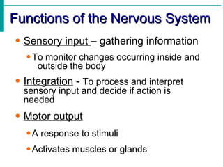
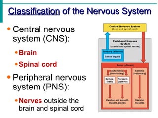

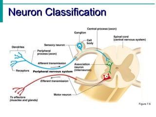
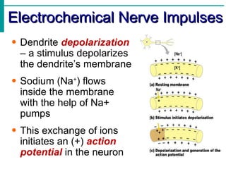
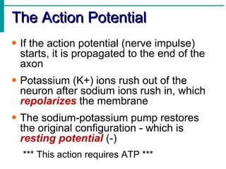
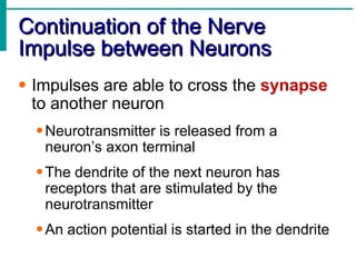
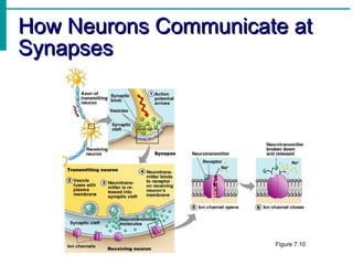
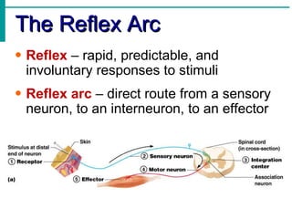
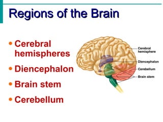
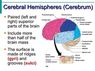

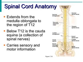
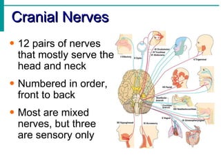
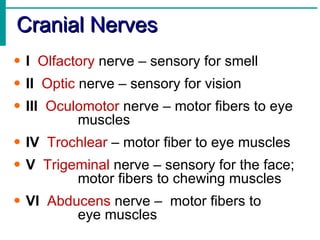
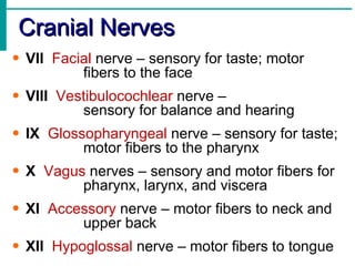


The document provides an overview of the nervous system, including its main functions and classifications. It describes the central nervous system (brain and spinal cord) and peripheral nervous system. It also discusses the structure and function of neurons, how impulses are transmitted, and the main regions and components of the brain and spinal cord.


