Downloaded 574 times

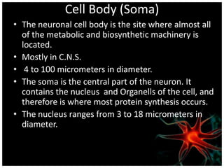
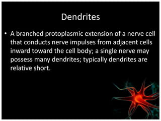
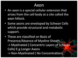
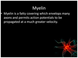
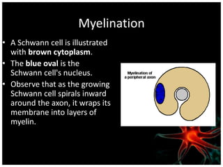

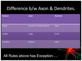
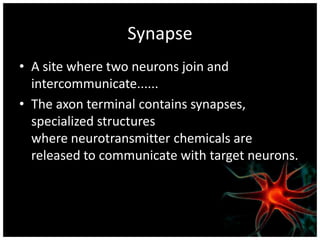
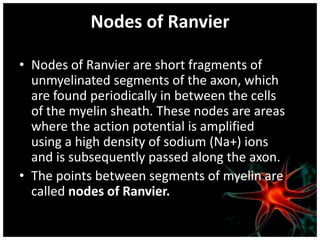
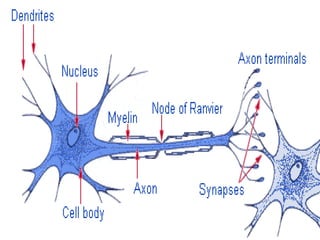
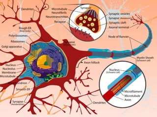


Nervous tissue is composed of neurons and supporting cells. Neurons have a cell body containing the nucleus, dendrites which receive signals, and an axon which transmits signals. The axon may be myelinated by Schwann cells to increase signal transmission speed. At synapses, the axon terminal releases neurotransmitters which allow communication with other neurons. Supporting glial cells aid neurons structurally and metabolically.