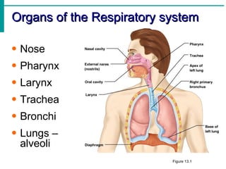
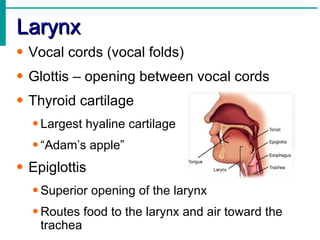
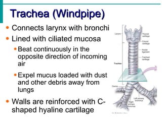
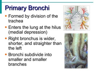
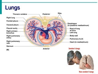
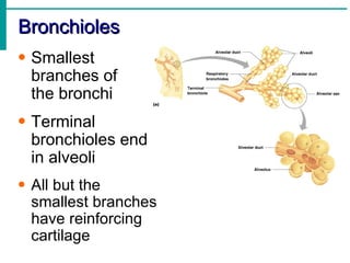
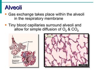
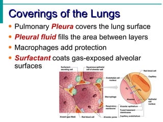
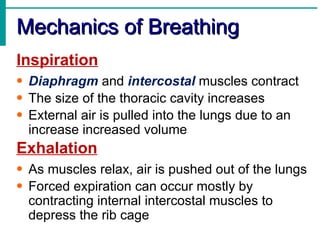
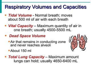
Essentials of Human Anatomy & Physiology


The Respiratory System
   Anatomy & Physiology II


           Mr. Hildebrandt
Respiratory System Functions
• Gas exchange between the
  blood and external environment
• Exchange of Oxygen & Carbon
  Dioxide takes place within the
  lungs in the alveoli
• Purify, warm, and humidify the
  incoming air
• Produce sound for
  communication
Nonrespiratory Functions
• Actions can be caused by reflexes or
    voluntary actions
• Examples:
  • Cough – clears lungs of debris
  • Sneeze – clears upper respiratory tract
  • Yawn – deep inspiration triggered by ???
  • Hiccup – phrenic nerve is irratated which
     leads to rhythmic spasms.
Organs of the Respiratory system

• Nose
• Pharynx
• Larynx
• Trachea
• Bronchi
• Lungs –
  alveoli
                           Figure 13.1
The Nose
• The only externally visible
  part of the respiratory
  system
• Air enters the nose
  through the external nares
  (nostrils)
• The interior of the nose
  consists of a nasal cavity
  divided by a nasal septum
Upper Respiratory Tract
Nasal Cavity
• Lateral walls have projections called
  conchae
• Nasal cavity is separated from the oral
     cavity by the hard & soft palates
• Olfactory receptors are located in the
  mucosa on the superior surface
• Cavity is lined with respiratory mucosa
  • Moistens air
  • Traps incoming foreign particles
Pharynx (Throat)
• Muscular passage from nasal cavity to
  larynx
• Three regions of the pharynx:
  • Nasopharynx – superior region behind nasal
    cavity
  • Oropharynx – middle region behind mouth
  • Laryngopharynx – inferior region attached to
    larynx
• Oropharynx & laryngopharynx are the
  passageways for air & food
Larynx
• Vocal cords (vocal folds)
• Glottis – opening between vocal cords
• Thyroid cartilage
  • Largest hyaline cartilage
  • “Adam’s apple”
• Epiglottis
  • Superior opening of the larynx
  • Routes food to the larynx and air toward the
    trachea
Trachea (Windpipe)
• Connects larynx with bronchi
• Lined with ciliated mucosa
  • Beat continuously in the
    opposite direction of incoming
    air
  • Expel mucus loaded with dust
    and other debris away from
    lungs
• Walls are reinforced with C-
  shaped hyaline cartilage
Primary Bronchi
• Formed by division of the
  trachea
• Enters the lung at the hilus
  (medial depression)
• Right bronchus is wider,
  shorter, and straighter than
  the left
• Bronchi subdivide into
  smaller and smaller
  branches
Lungs
• Site of gas exchange
• Occupy most of the thoracic cavity
  • Apex is near the clavicle (superior portion)
    • Base rests on the diaphragm (inferior
      portion)
  • Each lung is divided into lobes
    • Left lung – two lobes
    • Right lung – three lobes
Lungs
Bronchioles
• Smallest
  branches of
  the bronchi
• Terminal
  bronchioles end
  in alveoli
• All but the
  smallest branches
  have reinforcing
  cartilage
Alveoli
• Gas exchange takes place within the alveoli
    in the respiratory membrane
• Tiny blood capillaries surround alveoli and
     allow for simple diffusion of O2 & CO2
Coverings of the Lungs
•   Pulmonary Pleura covers the lung surface
•   Pleural fluid fills the area between layers
•   Macrophages add protection
•   Surfactant coats gas-exposed alveolar
    surfaces
Mechanics of Breathing
Inspiration
• Diaphragm and intercostal muscles contract
• The size of the thoracic cavity increases
• External air is pulled into the lungs due to an
  increase increased volume
Exhalation
• As muscles relax, air is pushed out of the lungs
• Forced expiration can occur mostly by
  contracting internal intercostal muscles to
  depress the rib cage
Respiratory Volumes and Capacities
• Tidal Volume - Normal breath; moves
  about 500 ml of air with each breath
• Vital Capacity – Maximum quantity of air in
     one breath; usually 4500-5500 mL
•   Dead Space Volume
    • Air that remains in conducting zone
     and never reaches alveoli
    • About 150 ml
• Total Lung Capacity – Maximum amount
     lungs can hold; usually 4650-6400 mL

Respiratory System Notes

  • 1.
    Essentials of HumanAnatomy & Physiology The Respiratory System Anatomy & Physiology II Mr. Hildebrandt
  • 2.
    Respiratory System Functions •Gas exchange between the blood and external environment • Exchange of Oxygen & Carbon Dioxide takes place within the lungs in the alveoli • Purify, warm, and humidify the incoming air • Produce sound for communication
  • 3.
    Nonrespiratory Functions • Actionscan be caused by reflexes or voluntary actions • Examples: • Cough – clears lungs of debris • Sneeze – clears upper respiratory tract • Yawn – deep inspiration triggered by ??? • Hiccup – phrenic nerve is irratated which leads to rhythmic spasms.
  • 4.
    Organs of theRespiratory system • Nose • Pharynx • Larynx • Trachea • Bronchi • Lungs – alveoli Figure 13.1
  • 5.
    The Nose • Theonly externally visible part of the respiratory system • Air enters the nose through the external nares (nostrils) • The interior of the nose consists of a nasal cavity divided by a nasal septum
  • 6.
  • 7.
    Nasal Cavity • Lateralwalls have projections called conchae • Nasal cavity is separated from the oral cavity by the hard & soft palates • Olfactory receptors are located in the mucosa on the superior surface • Cavity is lined with respiratory mucosa • Moistens air • Traps incoming foreign particles
  • 8.
    Pharynx (Throat) • Muscularpassage from nasal cavity to larynx • Three regions of the pharynx: • Nasopharynx – superior region behind nasal cavity • Oropharynx – middle region behind mouth • Laryngopharynx – inferior region attached to larynx • Oropharynx & laryngopharynx are the passageways for air & food
  • 9.
    Larynx • Vocal cords(vocal folds) • Glottis – opening between vocal cords • Thyroid cartilage • Largest hyaline cartilage • “Adam’s apple” • Epiglottis • Superior opening of the larynx • Routes food to the larynx and air toward the trachea
  • 10.
    Trachea (Windpipe) • Connectslarynx with bronchi • Lined with ciliated mucosa • Beat continuously in the opposite direction of incoming air • Expel mucus loaded with dust and other debris away from lungs • Walls are reinforced with C- shaped hyaline cartilage
  • 11.
    Primary Bronchi • Formedby division of the trachea • Enters the lung at the hilus (medial depression) • Right bronchus is wider, shorter, and straighter than the left • Bronchi subdivide into smaller and smaller branches
  • 12.
    Lungs • Site ofgas exchange • Occupy most of the thoracic cavity • Apex is near the clavicle (superior portion) • Base rests on the diaphragm (inferior portion) • Each lung is divided into lobes • Left lung – two lobes • Right lung – three lobes
  • 13.
  • 14.
    Bronchioles • Smallest branches of the bronchi • Terminal bronchioles end in alveoli • All but the smallest branches have reinforcing cartilage
  • 15.
    Alveoli • Gas exchangetakes place within the alveoli in the respiratory membrane • Tiny blood capillaries surround alveoli and allow for simple diffusion of O2 & CO2
  • 16.
    Coverings of theLungs • Pulmonary Pleura covers the lung surface • Pleural fluid fills the area between layers • Macrophages add protection • Surfactant coats gas-exposed alveolar surfaces
  • 17.
    Mechanics of Breathing Inspiration •Diaphragm and intercostal muscles contract • The size of the thoracic cavity increases • External air is pulled into the lungs due to an increase increased volume Exhalation • As muscles relax, air is pushed out of the lungs • Forced expiration can occur mostly by contracting internal intercostal muscles to depress the rib cage
  • 18.
    Respiratory Volumes andCapacities • Tidal Volume - Normal breath; moves about 500 ml of air with each breath • Vital Capacity – Maximum quantity of air in one breath; usually 4500-5500 mL • Dead Space Volume • Air that remains in conducting zone and never reaches alveoli • About 150 ml • Total Lung Capacity – Maximum amount lungs can hold; usually 4650-6400 mL