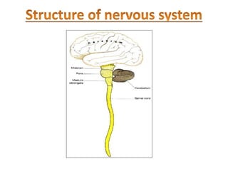
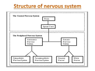
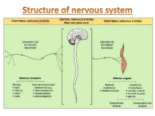
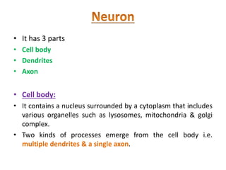
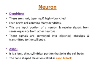
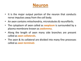
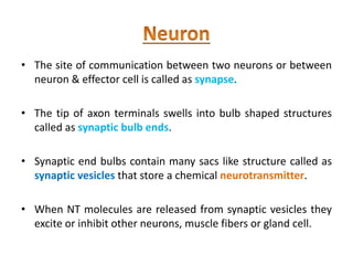
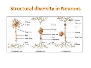
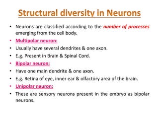
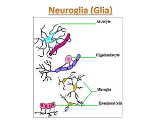
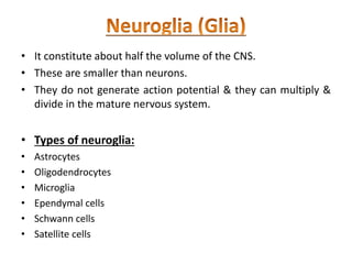
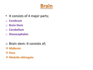
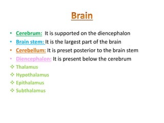
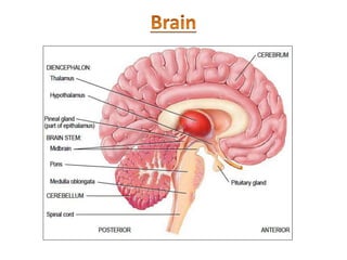
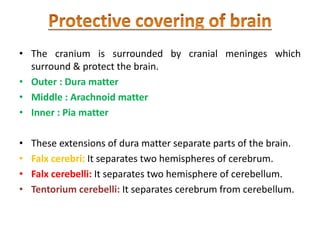
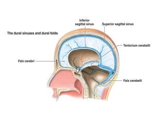
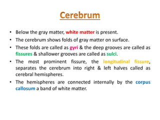
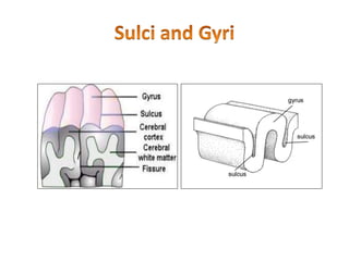
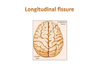
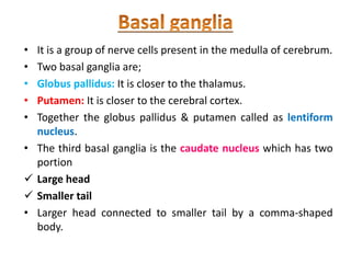
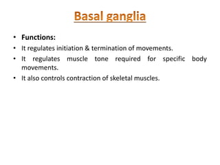
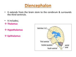
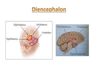
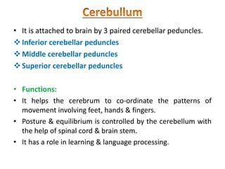
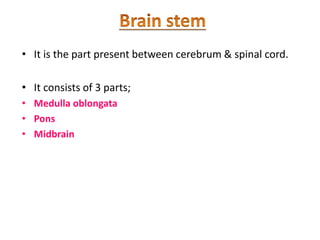
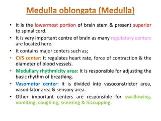
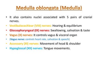
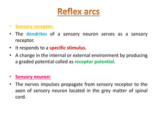
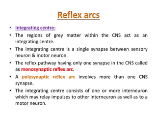
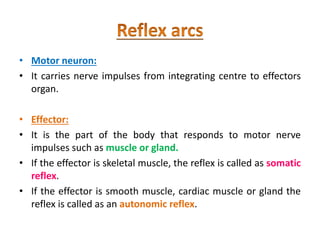
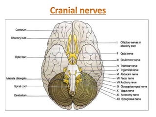
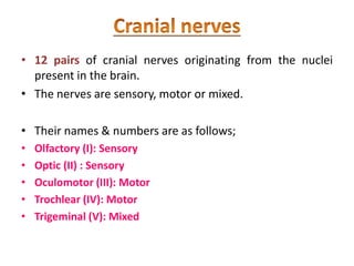
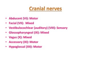
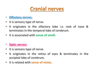
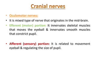
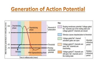
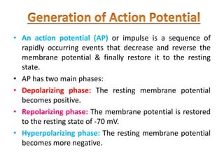
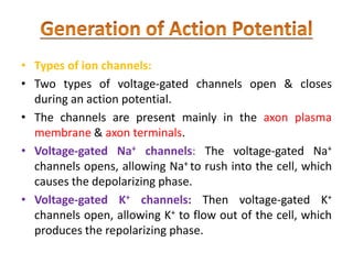
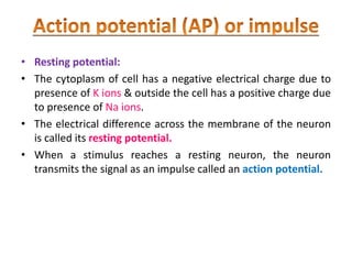
SlideShare a Scribd company logo
• To describe the functions of nervous system
• To describe the structure of neuron
• To explain the formation & circulation of CSF
• To describe the structure & function of cerebrum
• To describe the lobes of cerebrum
• To describe the structure & function of basal ganglia
• To describe the structure & functions of limbic system
• To describe the locations & functions of sensory, association,
and motor areas of cerebral cortex
• To describe the structures & functions of brain stem
• To describe the structure & functions of cerebellum
• To describe the structure & functions of diencephalon
• To identify the cranial nerves by name, number, type & give
functions of each nerve
• To describe preganglionic & postganglionic neurons of the
ANS
• To compare the sympathetic & parasympathetic divisions of
ANS
• To describe the events that generates an action potential
• To enlist excitatory and inhibitory neurotransmitters
• To describe the protective covering, internal and external
anatomy of spinal cord
• To describe the functional components of a reflex arc
• Neurology:
• The branch of science that deals with the structure,
functions & diseases of nervous system (brain &
spinal cord).
• The CNS consists of brain & spinal cord, integrates &
correlates many different kinds of incoming sensory
information.
• CNS is also the source of thoughts, emotions & memories.
• NS is divided into two main parts. CNS & PNS
• The components of PNS are subdivided into a somatic
nervous system (SNS) and autonomic nervous system (ANS).
• The SNS consist of sensory neuron & motor neuron.
• Sensory neurons: They convey information from somatic
receptors in the head, body wall, limbs, eyes, tongue, ear to
the CNS.
• Motor neurons: They conduct impulses from the CNS to
skeletal muscles (The PNS is voluntary).
• The ANS consists of sympathetic & parasympathetic division.
• Sensory neuron: They convey information from autonomic
sensory receptors, located primarily in visceral organs such as
stomach & lungs to the CNS.
• Motor neuron: They conduct nerve impulses from CNS to
smooth muscles, cardiac muscles & glands (The ANS is
involuntary)
• Sensory function:
• Sensory receptors detect internal stimuli such as increase in blood
acidity & external stimuli such as raindrop falling on your arm.
• The neurons that carry sensory information from brain & spinal
cord are sensory or afferent neurons.
• Integrative function:
• The nervous system integrates the sensory information by analyzing
& storing some of it & making decisions of appropriate responses.
• The neurons involved in the processes called as interneurons.
• Motor function:
• The neurons that carry information away from brain & spinal cord
are known motor or efferent neurons.
• It is the fundamental unit of nervous system.
• Nervous system consists of 2 types of cells.
 Neurons
 Neuroglia
• Neurons perform the function of sensing, thinking,
remembering, controlling muscle activity & regulating
glandular secretions.
• Neuroglia supports, nourish & protect the neurons &
maintain homeostasis in the interstitial fluid that bathes
neurons.
• It has 3 parts
• Cell body
• Dendrites
• Axon
• Cell body:
• It contains a nucleus surrounded by a cytoplasm that includes
various organelles such as lysosomes, mitochondria & golgi
complex.
• Two kinds of processes emerge from the cell body i.e.
multiple dendrites & a single axon.
• Dendrites:
• These are short, tapering & highly branched.
• Each nerve cell contains many dendrites.
• This are input portion of a neuron & receive signals from
sense organs or from other neurons.
• These signals are converted into electrical impulses &
transmitted to the cell body.
• Axon:
• It is a long, thin, cylindrical portion that joins the cell body.
• The cone shaped elevation called as axon hillock.
• It is the major output portion of the neuron that conducts
nerve impulses away from the cell body.
• An axon contains mitochondria, microtubules & neurofibrils.
• The cytoplasm of axon called as axoplasm is surrounded by a
plasma membrane known as axolemma.
• Along the length of axon many side branches are present
called as axon collaterals.
• The axon & its collateral are divided into many fine processes
called as axon terminals
• The site of communication between two neurons or between
neuron & effector cell is called as synapse.
• The tip of axon terminals swells into bulb shaped structures
called as synaptic bulb ends.
• Synaptic end bulbs contain many sacs like structure called as
synaptic vesicles that store a chemical neurotransmitter.
• When NT molecules are released from synaptic vesicles they
excite or inhibit other neurons, muscle fibers or gland cell.
• Neurons are classified according to the number of processes
emerging from the cell body.
• Multipolar neuron:
• Usually have several dendrites & one axon.
• E.g. Present in Brain & Spinal Cord.
• Bipolar neuron:
• Have one main dendrite & one axon.
• E.g. Retina of eye, inner ear & olfactory area of the brain.
• Unipolar neuron:
• These are sensory neurons present in the embryo as bipolar
neurons.
• It constitute about half the volume of the CNS.
• These are smaller than neurons.
• They do not generate action potential & they can multiply &
divide in the mature nervous system.
• Types of neuroglia:
• Astrocytes
• Oligodendrocytes
• Microglia
• Ependymal cells
• Schwann cells
• Satellite cells
• Astrocytes:
• These are star shaped with many processes.
• Functions:
• Maintains appropriate environment for generation of nerve
impulses, provide nutrients to neurons, take up excess NT,
helps to form the BBB.
• Oligodendrocytes:
• These are smaller than astrocytes, with fewer processes,
round or oval body.
• Function:
• Forms supporting network around neurons; produce myelin
sheath around the axons.
• Microglia:
• These are small cells with few processes & give rise to
monocytes & macrophages.
• Functions:
• Protect neurons from disease by engulfing invading microbes
• Ependymal cell:
• These are arranged in a single layer, ranges in shape from
cuboidal to columnar, many are ciliated.
• Functions:
• Lines ventricles of the brain & central canal of spinal cord,
form CSF & assists in its circulation.
• Schwann cells:
• These are flat cells that encircle PNS axons.
• Functions:
• They produces myelin sheath around the axon of neuron.
• They participate in regeneration of PNS axons.
• Satellite cells:
• These are flat cells arranged around the cell bodies of neurons
in ganglia.
• Functions:
• They support neurons in PNS ganglia.
• It consists of 4 major parts;
o Cerebrum
o Brain Stem
o Cerebellum
o Dineacephalon
o Brain stem: It consists of;
 Midbrain
 Pons
 Medulla oblongata
• Cerebrum: It is supported on the diencephalon
• Brain stem: It is the largest part of the brain
• Cerebellum: It is preset posterior to the brain stem
• Diencephalon: It is present below the cerebrum
 Thalamus
 Hypothalamus
 Epithalamus
 Subthalamus
• The cranium is surrounded by cranial meninges which
surround & protect the brain.
• Outer : Dura matter
• Middle : Arachnoid matter
• Inner : Pia matter
• These extensions of dura matter separate parts of the brain.
• Falx cerebri: It separates two hemispheres of cerebrum.
• Falx cerebelli: It separates two hemisphere of cerebellum.
• Tentorium cerebelli: It separates cerebrum from cerebellum.
• It is a clear, colorless liquid that protects the brain & spinal
cord against chemical & physical injuries.
• It also carries oxygen, glucose & other chemicals from the
blood to neurons & neuroglia.
• Approximately 500 ml of CSF is formed every day.
• It is present in ventricles of brain, in cisterns round the brain
& in the sub-arachnoid space around both the brain & spinal
cord.
• The total volume of CSF is 80 to 150 ml in an adult.
• CSF contains glucose, proteins, lactic acid, urea, cation (Na⁺,
K⁺, Ca⁺, Mg⁺) & anions (Cl⁻ & HCOз⁻).
• It also contains some white blood cells.
• Functions of CSF:
• It provides support & protects the delicate structure of brain
& spinal cord.
• It acts as a shock absorber providing cushioning to brain &
spinal cord.
• It maintains uniform pressure around delicate structures.
• It provides chemical protection to brain & spinal cord.
• CSF is a medium for exchange of nutrients & waste products
between the blood & nervous tissue.
• It is the seat of intelligence.
• It provides us the ability to read, write & speak, to make
calculations & compose music, to remember the past, plan for
the future & imagine the things that have never existed
before.
• It is the largest part of brain.
• The right & left halves of cerebrum called as cerebral
hemispheres.
• The superficial layer of the cerebrum is gray matter.
• This layer is 2 - 4 mm thick contains billons of neurons.
• Below the gray matter, white matter is present.
• The cerebrum shows folds of gray matter on surface.
• These folds are called as gyri & the deep grooves are called as
fissures & shallower grooves are called as sulci.
• The most prominent fissure, the longitudinal fissure,
separates the cerebrum into right & left halves called as
cerebral hemispheres.
• The hemispheres are connected internally by the corpus
callosum a band of white matter.
• Each cerebral hemisphere is divided into 4 lobes.
 Frontal lobes
 Parietal lobe
 Occipital lobe
 Temporal lobe
• Central sulcus separates the frontal lobe from the parietal
lobe.
• The precentral gyrus located immediately anterior to the
central sulcus.
• The Post central gyrus is located immediately posterior to
central sulcus.
• The lateral sulcus separates the frontal lobe from the
temporal lobe.
• The parietooccipital sulcus separates the parietal lobe
from the occipital lobe.
• Major area present in the frontal lobe are as follows.
• Motor area: Also known as 1⁰ motor cortex. The area control
muscles of speech & fine movements of fingers & hands.
• Premotor areas: It lies anterior to motor area. It controls
involuntary movements that perform specific tasks.
• Supplimental motor area: It is present above the premotor
area.
• Broca′s area: It is a speech related area causing activation of
vocal cords with movements of mouth & tongue.
• Voluntary eye field: It is located just above broca’s area & is
responsible for voluntary eye movements.
• Prefrontal area: It lies anterior to voluntary eye field. It helps
in concentrating & elaboration of thoughts. It also helps in
planning future.
• Area of hand skills: It is located anterior to 1⁰ motor cortex. It
co-ordinates the skillful hand movements.
• The major areas of parietal lobe include;
• Primary sensory area:
• This area receives information from sensations like
temperature, touch & pain.
• This area can judge the texture of material, shapes & forms of
objects, weights, pressure, etc.
• Sensory association area:
• It lies behind primary sensory area.
• Recognition of complex objects lies with this area.
• It contains 1⁰ visual area & visual association areas.
• 1⁰ visual area:
• This area detects the size, shapes & colors of various
objects.
• Visual association areas:
• It is associated with detection of more complex visual
patterns.
• The primary auditory areas & auditory association
areas.
• The areas helps to identify the different pitches of sound,
judge the intensity & analyzes different properties of
sound.
• It performs 3 main activities.
• Mental activity: It involves in memory, intelligence, sense of
responsibility, thinking, reasoning, moral sense, learning,
word formation & interpretation.
• Sensory activity: This receives perception of pain,
temperature, touch, sight, hearing, taste & smell.
• Motor activity: It is associated with control of voluntary
muscle contraction.
• It consist of,
Basal ganglia
Thalamus
Hypothalamus
• It is a group of nerve cells present in the medulla of cerebrum.
• Two basal ganglia are;
• Globus pallidus: It is closer to the thalamus.
• Putamen: It is closer to the cerebral cortex.
• Together the globus pallidus & putamen called as lentiform
nucleus.
• The third basal ganglia is the caudate nucleus which has two
portion
 Large head
 Smaller tail
• Larger head connected to smaller tail by a comma-shaped
body.
• Functions:
• It regulates initiation & termination of movements.
• It regulates muscle tone required for specific body
movements.
• It also controls contraction of skeletal muscles.
• It extends from the brain stem to the cerebrum & surrounds
the third ventricle.
• It includes;
 Thalamus
 Hypothalamus
 Epithalamus
• It measures about 3 cm in length & makes up 80 % of the
diencephalon.
• It consists of paired oval masses of gray matter present on
each side of 3 ventricle.
• Functions:
• Sensory input from various sensory organs are given to the
thalamus before redistribution to the cerebrum.
• This information further conveyed to the cerebrum.
• It plays important role in maintenance of consciousness.
• It is a small part of the diencephalon located just below
the thalamus.
• It is divided into 4 main regions.
• Mammillary region
• Tuberal region
• Supryoptic region
• Preoptic region
• Functions:
• Control of ANS:
• It regulates the contraction of smooth & cardiac muscle,
secretion of many glands.
• It regulates visceral activities such as regulation of HR,
movement of food through GIT & contraction of urinary
bladder.
• Production of hormones:
• The hypothalamus produces several hormones.
• Regulation of eating & drinking:
• It regulates eating & drinking habits.
• Regulation of emotional & behavioral patterns:
• Together with limbic system, the hypothalamus participates in
expressions of aggression, pain, pleasure, behavioral patterns
related to sexual arousal.
• Control of body temperature:
• It controls the body temperature.
• Regulation of circadian rhythms & states of consciousness:
• It regulates the patterns of awakening & sleep.
• It is a small region present posterior to the thalamus that
consists of pineal gland
• Pineal gland is having a size of small pea.
• It is a part of endocrine system as it secretes the
hormone melatonin.
• More amount of melatonin is liberated during darkness
than in light.
• This hormone is thought to promote sleepiness.
• It is second largest part of brain.
• It is present posterior to brain stem.
• The shape is like a butterfly.
• The central constricted area is the vermis & the lateral lobes
are cerebral hemispheres.
• Each hemisphere consists of 3 lobes,
 Anterior lobe
 Posterior lobe
 Flocculonodular lobe
• The superficial layer of cerebellum called as cerebellar cortex,
consists of gray matter in the form of folia (leaves).
• Deep to the gray matter are tracts called as arbor vitae that
look like branches of tree.
• In deep white matter, cerebellar nuclei are present.
• It is attached to brain by 3 paired cerebellar peduncles.
Inferior cerebellar peduncles
Middle cerebellar peduncles
Superior cerebellar peduncles
• Functions:
• It helps the cerebrum to co-ordinate the patterns of
movement involving feet, hands & fingers.
• Posture & equilibrium is controlled by the cerebellum with
the help of spinal cord & brain stem.
• It has a role in learning & language processing.
• It is the part present between cerebrum & spinal cord.
• It consists of 3 parts;
• Medulla oblongata
• Pons
• Midbrain
• It is the lowermost portion of brain stem & present superior
to spinal cord.
• It is very important centre of brain as many regulatory centers
are located here.
• It contains major centers such as;
• CVS center: It regulates heart rate, force of contraction & the
diameter of blood vessels.
• Medullary rhythmicity area: It is responsible for adjusting the
basic rhythm of breathing.
• Vasomotor center: It is divided into vasoconstrictor area,
vasodilator area & sensory area.
• Other important centers are responsible for swallowing,
vomiting, coughing, sneezing & hiccupping.
• It also contains nuclei associated with 5 pairs of cranial
nerves.
• Vestibulocochlear (VIII) nerves: Hearing & equilibrium
• Glossopharyngeal (IX) nerves: Swallowing, salivation & taste
• Vagus (X) nerves: It controls vagus & visceral organ
• (Vagus nerve: controls heart rate, salivation & speech)
• Accessory (XI) nerves: Movement of head & shoulder
• Hypoglossal (XII) nerves: Tongue movements.
• It is the first part of brain stem, about 2.5 cm long.
• It contains nuclei for 4 pairs of cranial nerves.
• Trigeminal nerves (V): Chewing & sensation of head & face.
• Abducens nerves (VI): It regulates eyeball movements.
• Facial nerves (VII): Sensations of taste, salivation & facial
expressions.
• Vestibulocochlear nerves: Related to hearing & equilibrium.
• Respiratory center is also located in pons.
• It is the middle part of brain stem, 2.5 cm long.
• It is associated with nuclei for 2 pairs of cranial nerves.
• Oculomotor (III) nerves: Movement of eyeball & regulating
the size of pupil.
• Trochlear (IV) nerves: Responsible for movement of eyeball.
• It is a complex set of brain structures that lies on both sides of
the thalamus, under the cerebrum
• The main components are as follows;
• Cingulate gyrus: This lies above the corpus callosum.
• Parahippocompal gyrus: This lies in the temporal lobe.
• Hippocampus: It is present in the temporal lobe.
• Dentate gyrus: It lies between the hippocampus &
parahippocompal gyrus.
• Amygdala: It is composed of neurons located close to the tail
of caudate nucleus.
• Septal nuclei: It is present under the corpus callosum.
• Mammillary bodies of the hypothalamus: These are 2 round
masses.
• Olfactory bulbs: These are flat bodies that rest on the
cribiform plate.
• Fornix, stria termilanlis, stria medullarius &
mammilothalamic tract are other parts of limbic system.
• Functions:
• It plays primary role in various emotions including pain,
pleasure, affection & anger.
• It is also involved in the olfaction and memory.
• People with damage to limbic system structures forget recent
events & cannot recall the past events.
• The spinal cord is located within the vertebral column.
• Meninges of spinal cord:
• These are 3 connective tissues coverings that encircle the
spinal cord & brain.
• The spinal meninges surrounds the spinal cord &
continuous with cranial meninges that encircle the brain.
• Outermost: Dura mater
• Middlemost: Arachanoid mater
• Innermost: Pia mater
• It is roughly cylindrical in shape & its thickness is about little
finger.
• In adults, it extends from the medulla oblongata and to the
second lumbar vertebrae.
• The length is 42-45 cm & diameter is 2 cm.
• Externally it shows 2 enlargements.
• Cervical enlargement: Superior enlargement, nerves to &
from the upper limbs arises from the cervical enlargement.
• Lumbar enlargement: Inferior enlargement, nerves to & from
lower limbs arises from lumbar enlargement.
• CSF is present in the central canal of subarachnoid space.
• 31 pairs of spinal nerves originate from the spinal cord.
• The spinal nerve is connected to the spinal cord by roots.
• 2 types of roots are present:
 Posterior root or dorsal root
 Anterior root or ventral root
• The dorsal or sensory root contains sensory nerve fibers
which conducts the nerve impulses from periphery to the
spinal cord.
• The ventral or motor root contains motor nerve fibers
conducting the nerve impulses from spinal cord to the
periphery.
• There are 31 pairs of spinal nerves originating from the spinal
cord.
• According to the region they are divided into;
• Cervical nerves: 8 pairs
• Thoracic nerves: 12 pairs
• Lumbar nerves: 5 pairs
• Sacral nerves: 5 pairs
• Coccygeal nerve: 1 pair
• The T.S. shows grey matter in the center & white matter
surrounds it.
• The grooves penetrate the white matter of spinal cord &
divide it into right & left sides.
 Anterior median fissure: Deep groove on anterior side
 Posterior median sulcus: Shallower groove on posterior side.
• The grey matter of spinal cord is shaped like letter ‘H’ or
butterfly & is surrounded by white matter.
• In the centre of grey matter is space called as central canal.
• The grey matter consists of dendrites, cell bodies of neurons,
unmyelinated neurons & neuroglia.
• The white matter consists of bundles of myelinated axons of
neurons.
• The grey matter on each side of the spinal cord is divided into
regions called as horns.
• Anterior or ventral horns:
• Posterior or dorsal horns:
• The anterior & posterior grey horns divide the white matter
on each side into parts called as columns:
 Anterior (ventral) white columns
 Posterior (dorsal) white columns
 Lateral white columns
• A reflex arc is a flat, autonomic, unplanned sequence of
actions that occurs in response to a particular stimulus.
• When integration takes place in the spinal cord grey matter,
the reflex is called as spinal reflex.
• If integration occurs in the brain stem called as cranial reflex.
• When there is contraction of skeletal muscles called as
somatic reflexes.
• When there is contraction of smooth muscles, cardiac muscles
& glands called as autonomic (visceral) reflex.
• It consists of 5 functional components.
Sensory receptors
Sensory neuron
Integrating centre
Motor neuron
Effector organ
• Sensory receptor:
• The dendrites of a sensory neuron serves as a sensory
receptor.
• It responds to a specific stimulus.
• A change in the internal or external environment by producing
a graded potential called as receptor potential.
• Sensory neuron:
• The nerves impulses propagate from sensory receptor to the
axon of sensory neuron located in the grey matter of spinal
cord.
• Integrating centre:
• The regions of grey matter within the CNS act as an
integrating centre.
• The integrating centre is a single synapse between sensory
neuron & motor neuron.
• The reflex pathway having only one synapse in the CNS called
as monosynaptic reflex arc.
• A polysynaptic reflex arc involves more than one CNS
synapse.
• The integrating centre consists of one or more interneuron
which may relay impulses to other interneuron as well as to a
motor neuron.
• Motor neuron:
• It carries nerve impulses from integrating centre to effectors
organ.
• Effector:
• It is the part of the body that responds to motor nerve
impulses such as muscle or gland.
• If the effector is skeletal muscle, the reflex is called as somatic
reflex.
• If the effector is smooth muscle, cardiac muscle or gland the
reflex is called as an autonomic reflex.
• 12 pairs of cranial nerves originating from the nuclei
present in the brain.
• The nerves are sensory, motor or mixed.
• Their names & numbers are as follows;
• Olfactory (I): Sensory
• Optic (II) : Sensory
• Oculomotor (III): Motor
• Trochlear (IV): Motor
• Trigeminal (V): Mixed
• Abducent (VI): Motor
• Facial (VII): Mixed
• Vestibulocochlear (auditory) (VIII): Sensory
• Glossopharyngeal (IX): Mixed
• Vagus (X): Mixed
• Accessory (XI): Motor
• Hypoglossal (XII): Motor
• Olfactory nerves:
• It is sensory type of nerve.
• It originates in the olfactory lobe i.e. root of nose &
terminates in the temporal lobe of cerebrum.
• It is associated with sense of smell.
• Optic nerves:
• It is sensory type of nerve.
• It originates in the retina of eyes & terminates in the
occipital lobe of cerebrum.
• It is related with sense of vision.
• Oculomotor nerves:
• It is mixed type of nerve that originates in the mid-brain.
• Efferent (motor) portion: It innervates skeletal muscles
that moves the eyeball & innervates smooth muscles
that constrict pupil.
• Afferent (sensory) portion: It is related to movement
eyeball & regulating the size of pupil.
• Trochlear nerve:
• It is mixed type of nerve & originates in midbrain.
• It is the smallest of the 12 cranial nerves.
• Motor portion is related to movement of eyeball &
sensory portion carries information from eye to
midbrain.
• Trigeminal nerve:
• Mixed type of nerve.
• Largest among all the cranial nerves.
• Motor portion originates from pons & innervates the
muscles of mastication (skeletal chewing muscles).
• Sensory portion consists of 3 branches;
• Ophthalmic nerve: It contains nerves from eyelids, eyeball,
lacrimal glands, nasal cavity, nose and forehead.
• Maxillary nerve: It contains nerve from the mucosa of
nose, pharynx, teeth, upper lip & lower eyelid.
• Mandibular nerve: It contains nerve from tongue, teeth,
skin, mandible & cheek.
• Motor function: Chewing
• Sensory function: Conveys impulses for touch, pain &
temperature.
• Abducens nerve:
• A mixed type of nerve & originates in the pons.
• Motor portion: It innervates the skeletal muscles that
moves eyeball.
• Sensory portion: It is related to movement of eyeball &
muscles sense (proprioception)
• Motor function: Movement of eyeball
• Sensory function: Proprioception
• Facial nerve:
• It is a mixed type of nerve.
• Motor nerve originates from pons & innervates skeletal
muscle of face, nose, palate, lacrimal & salivary gland.
• Sensory nerve transmits information from taste buds in
the tongue & mouth.
• Motor function: Facial expression
• Sensory function: Taste & Proprioception
• Vestibulocochlear nerve:
• Sensory type of nerve.
• It consists of two nerves,
• Vestibular nerve: It arises from semicircular canals of the
inner ear & conveys impulses to the cerebellum. They are
associated with maintenance of posture & balance.
• Cochlear nerve: It originates in the spiral organ of the
inner ear & conveys impulses to the hearing area of
cerebrum. It is responsible for hearing.
• Glossopharyngeal nerve:
• Mixed type of nerve.
• Motor nerves originate from medulla oblongata &
innervate the tongue & pharynx.
• Sensory nerves originate from salivary glands &
terminates in medulla oblongata.
• Motor function: Movement of pharynx during
swallowing & speech.
• Sensory function: Taste, touch, pain & temperature
sensations, monitoring of blood pressure.
• Vagus nerve:
• Mixed type of nerve.
• Motor fibers: originates in the medulla & innervates the
smooth muscles of pharynx, larynx, trachea, heart
esophagus, stomach, intestine, pancreas, gall bladder,
bile duct, spleen, kidney, ureter, blood vessels in thoracic
& abdominal cavities.
• Sensory fibers: convey impulses from same organs to
brain.
• Motor function: Swallowing, coughing & voice
production.
• Sensory function: Taste, touch, pain, temperature
regulation & monitoring of blood pressure.
• Accessory nerve:
• It is mixed type of nerve.
• It originates from medulla oblongata & innervates the
muscles of pharynx & skeletal muscle of neck.
• Motor function: Swallowing & movement of head &
shoulders.
• Hypoglossal nerve:
• It is mixed type of nerve.
• It originates in the medulla & innervates the muscle of
tongue.
• Sensory function: Proprioception.
• Motor function: Movement of tongue during speech &
swallowing.
• The peripheral nervous system consists of somatic
nervous system (SNS) and autonomic nervous system
(ANS).
• Somatic nervous system consists of sensory neuron &
motor neuron.
• Sensory neurons convey message from periphery to the
CNS.
• These include sensations of pain, temperature, taste,
smell, hearing & vision, etc.
• Motor neurons conveys information from brain to
periphery.
• The autonomic nervous system which controls the
autonomic function of the body i.e. initiates in the brain.
• The effects of autonomic activity are rapid and effector
organs are;
• Smooth muscle i.e. changes in bronchioles & blood
vessels diameter.
• Cardiac muscle i.e. changes in rate & force of the
heartbeat.
• Glands i.e. increase or decrease in GIT secretions.
• It consists of two types of neurons.
• Autonomic sensory neuron
• Autonomic motor neuron
• Autonomic sensory neuron (afferent):
• These neurons are associated with sensory receptors
located in blood vessels, visceral organs and muscles.
• Autonomic motor neuron (efferent):
• These regulates visceral activities by either increase or
decrease ongoing activities in the effector tissues
(cardiac muscle, smooth muscles or glands)
• E.g. Change in pupil diameter, dilation or constriction of
blood vessels, adjusting the heart rate & force of
contraction.
• The autonomic nervous system is separated into two
divisions.
• Sympathetic (Thoracolumbar outflow) division
• Parasympathetic (Carniosacral outflow) division
• The two divisions have both structural & functional
differences.
• They normally work in opposite manner.
• Each division has two motor neurons, autonomic ganglia
and effector organs.
• These are;
• Pre-ganglionic neurons
• Post-ganglionic neurons
• The autonomic ganglion is the collection of cell bodies
outside the CNS.
• The motor neurons which lies before the ganglion is
called as preganglionic neuron.
• The myelinated axon is called as preganglionic fiber.
• The motor neuron which lies after the ganglion and
terminates in the effector organ called as postganglionic
neuron.
• The myelinated axon is called as postganglionic fibers.
• It is called as thoracolumbar division.
• It consists of two types of autonomic ganglia.
• Sympathetic trunk ganglia
• Prevertebral ganglia
• Sympathetic trunk ganglia:
• These are the ganglia lie in a vertical row on either side
of the vertebral column.
• These lies close to the spinal cord and therefore the
preganglionic fibers are short.
• Prevertebral ganglia:
• These are 3 types of ganglion;
• Coeliac ganglion
• Superior mesenteric ganglion
• Inferior mesenteric ganglion
• These are the ganglion situated close to the abdominal
cavity.
• The preganglionic nerve fibers are shorter.
• The postganglionic nerve fibers are longer.
• The preganglionic neurotransmitter is acetylcholine.
• Most of the postganglionic nerve fibers are adrenergic.
• The targeted receptors are mostly adrenergic.
• Distributed throughout the body.
• It is called as craniosacral division.
• It contains parasympathetic ganglia.
• The parasympathetic ganglia are dispersed.
• The ganglia are near or within the wall of the effector
organs.
• The preganglionic nerve fibers are large.
• The post ganglionic nerve fibers are shorter.
• The preganglionic nerve fibers are acetylcholine.
• Most of the postganglionic nerve fibers are cholinergic.
• The targeted receptors are mostly cholinergic.
• The distribution is limited, particularly to heart, viscera of
thorax, abdomen & pelvis.
• The NT is synthesized in the nerve endings.
• Enzymes & precursor molecules are required for NT.
• Once synthesized, the NT is stored in storage vesicles in
the nerve endings.
• A nerve action potential causes release of NT into the
synaptic space.
• The liberated NT then attaches to the receptors on the
effector organ, resulting in depolarization & generation
of action potential.
• Two basic NT involved are acetylcholine & nor-
epinephrine (Nor-adrenaline).
• Small molecules NT
• Neuropeptides
• Small molecules NT:
• These are rapidly acting produces acute responses.
• Acetylcholine
• Different amines
• Norepinephrine
• Epinephrine
• Dopamine
• Serotonin
• Different amino acids
• Glycin
• Gamma-amino-butyric acid (GABA)
• Aspartate
• Glutamate
• Slowly acting NT having prolonged effects.
• These are hormones releasing from hypothalamus. E.g.
TRH, LHRH, Somatostatin, etc.
• Pituitary peptides: E.g. ACTH, Vasopressin, Oxytocin, etc.
• Peptides acting on gut & brain: E.g. Substance P, Insulin,
Glucagon, Enkephalin, etc.
• Neuropeptides from other tissues: E.g. Bradykinin,
Angiotensin-II, etc.
• It is a NT which is released by PNS & CNS neurons.
• It is an excitatory NT at NMJ, where it acts directly on
open ligand-gated cation channels.
• The enzyme acetyl cholinesterase (AchE) inactivates Ach
by splitting it into acetate & choline.
• Certain amino acids are modified & decarboxylated to
produce biogenic amines.
• Norepinephrine (NE) plays role in arousal (awakening
from deep sleep), dreaming & regulating mood.
• Neurons containing dopamine NT are active during
emotional responses & pleasurable responses.
• Dopamine helps to regulate the skeletal muscle tone.
• Schizophrenia is due to accumulation of excess
dopamine.
• Muscular stiffness is due to degeneration of
dopamienrgic neurons in parkinson’s disease.
• Serotonin plays important role in sensory perception,
temperature regulation, control of mood, appetite &
induction of a sleep.
• An action potential (AP) or impulse is a sequence of
rapidly occurring events that decrease and reverse the
membrane potential & finally restore it to the resting
state.
• AP has two main phases:
• Depolarizing phase: The resting membrane potential
becomes positive.
• Repolarizing phase: The membrane potential is restored
to the resting state of -70 mV.
• Hyperpolarizing phase: The resting membrane potential
becomes more negative.
• Types of ion channels:
• Two types of voltage-gated channels open & closes
during an action potential.
• The channels are present mainly in the axon plasma
membrane & axon terminals.
• Voltage-gated Na+ channels: The voltage-gated Na+
channels opens, allowing Na+ to rush into the cell, which
causes the depolarizing phase.
• Voltage-gated K+ channels: Then voltage-gated K+
channels open, allowing K+ to flow out of the cell, which
produces the repolarizing phase.
• Resting potential:
• The cytoplasm of cell has a negative electrical charge due to
presence of K ions & outside the cell has a positive charge due
to presence of Na ions.
• The electrical difference across the membrane of the neuron
is called its resting potential.
• When a stimulus reaches a resting neuron, the neuron
transmits the signal as an impulse called an action potential.
• The voltage-gated Na+ channels open rapidly & causes
inward movement of Na+ causes depolarizing phase of
the action potential.
• The inflow of Na+ changes the membrane potential from -
-70 mV to 30 mV.
• Each voltage gated Na+ channel has two separate gates,
an activation gate & an inactivation gate.
• After a short period, the voltage gated Na+ channels gates
closed.
• It causes opening of voltage-gated K+ channels so, K ions
get moves out of the cell.
• The membrane potential restores to -70 mv.
• When the voltage-gated K+ channels are open, outflow of
K+ may be large enough to cause an after-hyperpolarizing
phase of the action potential.
• During this phase, the membrane potential becomes
even more negative (about - 90 mV).
• The sodium-potassium pump goes back to work, moving
Na+ ions to the outside of the cell and K+ ions to the
inside, returning the neuron to its normal polarized state.
• Epilepsy:
• It is characterized by short, recurrent attacks of motor,
sensory or psychological malfunctions although it never
affects intelligence.
• The attacks are called as epileptic seizures, affects about
1% of the world population.
• Multiple sclerosis (MS):
• It is a disease that causes a progressive destruction of
myelin sheaths of neurons.
• It usually appears between the age of 20 to 40 affecting
females twice as often as males.
• Cerebrovascular accident (CVA):
• It is also called as stroke or brain attack.
• It is characterized by various neurological symptoms such
as paralysis, loss of sensations that arises from destruction
of brain tissue.
• The common causes are intracerebral hemorrhage, emboli
(blood clot) & atherosclerosis of the cerebral arteries.
• Transient Ischemic Attacks (TIA):
• It is an episode of temporary cerebral dysfunction caused
by impaired blood flow to the brain.
• The symptoms include dizziness, weakness, numbness, or
paralysis in a limb or in one side of the body, drooping of
one side of the face, headache, slurred speech and a partial
loss of vision or double vision.
• Alzheimer Disease (AD):
• It is neurodegenerative disorder characterized by
progressive dementia, primarily affecting cholinergic
neurons in the brain.
• In this loss of reasoning and ability to care for oneself
that affects 11 % of population over 65 ages.
• The cause for AD is still unknown, but it may be due to
combination of genetic factors, environmental factors or
life style factors.
• Brain tumor:
• A brain tumor is an abnormal growth of tissue in the
brain that may be malignant or benign.
• The symptoms of a brain tumor depend on its size,
location, and rate of growth.
• The symptoms are headache, poor balance,
coordination, dizziness, double vision, slurred speech,
nausea and vomiting, fever, abnormal pulse and
breathing rates, personality changes, numbness,
weakness of limbs, and seizures.

More Related Content

What's hot

Nervous system introduction
Nervous system  introductionNervous system  introduction
Nervous system introduction
Idris Siddiqui
 
Nervous System
Nervous SystemNervous System
Nervous System
jeremyschriner
 
PPT on Nervous System
PPT on Nervous SystemPPT on Nervous System
PPT on Nervous System
Dr Ashok dhaka Bishnoi
 
Diencephalon 1
Diencephalon 1Diencephalon 1
Diencephalon 1
Dr. sana yaseen
 
Unit 11 nervous system
Unit 11 nervous systemUnit 11 nervous system
Unit 11 nervous system
CHANDAN260
 
Central and peripheral nervous system
Central and peripheral nervous systemCentral and peripheral nervous system
Central and peripheral nervous system
Fawad Bukhari
 
Classification of Nervous System
Classification of Nervous SystemClassification of Nervous System
Classification of Nervous System
SalmaAsghar4
 
Structure, function and classification of neuron
Structure, function and classification of neuronStructure, function and classification of neuron
Structure, function and classification of neuron
Dr Sara Sadiq
 
NERVE PHYSIOLOGY- NEURON & NEUROGLIA
NERVE PHYSIOLOGY- NEURON & NEUROGLIANERVE PHYSIOLOGY- NEURON & NEUROGLIA
NERVE PHYSIOLOGY- NEURON & NEUROGLIA
Nabeel Beeran Abdul Rahiman
 
NERVOUS SYSTEM ANATOMY
NERVOUS SYSTEM ANATOMYNERVOUS SYSTEM ANATOMY
NERVOUS SYSTEM ANATOMY
Fasama H. Kollie
 
Nervous system
Nervous systemNervous system
Nervous system
Ishfaq Ahmad
 
Central Nervous System.ppt
Central Nervous System.pptCentral Nervous System.ppt
Central Nervous System.pptShama
 
Nervous system
Nervous systemNervous system
Nervous system
DR .PALLAVI PATHANIA
 
Neuron & its structural & functional type by Murtaza Syed
Neuron & its structural & functional type by Murtaza SyedNeuron & its structural & functional type by Murtaza Syed
Neuron & its structural & functional type by Murtaza Syed
Murtaza Syed
 
Chapter 14: The Nervous System Part 3 - Reflex Actions
Chapter 14: The Nervous System Part 3 - Reflex ActionsChapter 14: The Nervous System Part 3 - Reflex Actions
Chapter 14: The Nervous System Part 3 - Reflex Actionsj3di79
 

What's hot (20)

Nervous system introduction
Nervous system  introductionNervous system  introduction
Nervous system introduction
 
Nervous System
Nervous SystemNervous System
Nervous System
 
PPT on Nervous System
PPT on Nervous SystemPPT on Nervous System
PPT on Nervous System
 
Diencephalon 1
Diencephalon 1Diencephalon 1
Diencephalon 1
 
Unit 11 nervous system
Unit 11 nervous systemUnit 11 nervous system
Unit 11 nervous system
 
Central and peripheral nervous system
Central and peripheral nervous systemCentral and peripheral nervous system
Central and peripheral nervous system
 
Neurons
NeuronsNeurons
Neurons
 
NERVOUS SYSTEM
NERVOUS SYSTEMNERVOUS SYSTEM
NERVOUS SYSTEM
 
Classification of Nervous System
Classification of Nervous SystemClassification of Nervous System
Classification of Nervous System
 
Structure, function and classification of neuron
Structure, function and classification of neuronStructure, function and classification of neuron
Structure, function and classification of neuron
 
NERVE PHYSIOLOGY- NEURON & NEUROGLIA
NERVE PHYSIOLOGY- NEURON & NEUROGLIANERVE PHYSIOLOGY- NEURON & NEUROGLIA
NERVE PHYSIOLOGY- NEURON & NEUROGLIA
 
NERVOUS SYSTEM ANATOMY
NERVOUS SYSTEM ANATOMYNERVOUS SYSTEM ANATOMY
NERVOUS SYSTEM ANATOMY
 
Nervous system
Nervous systemNervous system
Nervous system
 
Central Nervous System.ppt
Central Nervous System.pptCentral Nervous System.ppt
Central Nervous System.ppt
 
Neuron physiology
Neuron physiologyNeuron physiology
Neuron physiology
 
Nervous system
Nervous systemNervous system
Nervous system
 
Neuron & its structural & functional type by Murtaza Syed
Neuron & its structural & functional type by Murtaza SyedNeuron & its structural & functional type by Murtaza Syed
Neuron & its structural & functional type by Murtaza Syed
 
The Brain
The BrainThe Brain
The Brain
 
Chapter 14: The Nervous System Part 3 - Reflex Actions
Chapter 14: The Nervous System Part 3 - Reflex ActionsChapter 14: The Nervous System Part 3 - Reflex Actions
Chapter 14: The Nervous System Part 3 - Reflex Actions
 
Spinal cord
Spinal cordSpinal cord
Spinal cord
 

Similar to Nervous system

Nervous system
Nervous system Nervous system
Nervous system
Revathi Boyina
 
Nervous System.pdf
Nervous System.pdfNervous System.pdf
Nervous System.pdf
CITY NURSING SCHOOL
 
Nervous system unit iii stds
Nervous system unit iii stdsNervous system unit iii stds
Nervous system unit iii stds
shafaatullahkhatt
 
Nervous system
Nervous systemNervous system
Nervous system
Poonam Beniwal
 
nervous system
 nervous system nervous system
nervous system
mubashar iqbal
 
Anatomy and physiology of brain ppt, its organization
Anatomy and physiology of brain ppt,  its organizationAnatomy and physiology of brain ppt,  its organization
Anatomy and physiology of brain ppt, its organization
Jyotirmayee Mandal
 
Central Nervous System.pptx
Central Nervous System.pptxCentral Nervous System.pptx
Central Nervous System.pptx
MariamMahmood28
 
nervous-system gollis.pptx
nervous-system gollis.pptxnervous-system gollis.pptx
nervous-system gollis.pptx
KalitMohamedAbdulahi
 
Nervous System-class 6.pdf
Nervous System-class 6.pdfNervous System-class 6.pdf
Nervous System-class 6.pdf
RachaelAdeleke
 
Ns3
Ns3Ns3
Nervous System 1 (63).pptx
Nervous System 1 (63).pptxNervous System 1 (63).pptx
Nervous System 1 (63).pptx
syedumair76
 
Central nervous system 1
Central nervous system 1Central nervous system 1
Central nervous system 1
Dr. Arpit Gohel
 
Brain anatomy
Brain anatomyBrain anatomy
Brain anatomy
Ayshah Hashimi
 
Brain anatomy
Brain anatomyBrain anatomy
Brain anatomy
Ayshah Hashimi
 
central nervous system
central nervous systemcentral nervous system
central nervous system
laraib jameel
 
Response&coordination
Response&coordinationResponse&coordination
Response&coordination
Khadijaparekh93
 
Unit 1 introduction to physiology
Unit 1 introduction to physiologyUnit 1 introduction to physiology
Unit 1 introduction to physiology
menwar
 
Unit 7 nervous system nrs 237
Unit 7 nervous system nrs 237Unit 7 nervous system nrs 237
Unit 7 nervous system nrs 237
menwar
 
Spinal cord and spinal nerves
Spinal cord and spinal nervesSpinal cord and spinal nerves
Spinal cord and spinal nerves
Ravish Yadav
 
Biophysio nervous system
Biophysio nervous systemBiophysio nervous system
Biophysio nervous system
Enoch Taclan
 

Similar to Nervous system (20)

Nervous system
Nervous system Nervous system
Nervous system
 
Nervous System.pdf
Nervous System.pdfNervous System.pdf
Nervous System.pdf
 
Nervous system unit iii stds
Nervous system unit iii stdsNervous system unit iii stds
Nervous system unit iii stds
 
Nervous system
Nervous systemNervous system
Nervous system
 
nervous system
 nervous system nervous system
nervous system
 
Anatomy and physiology of brain ppt, its organization
Anatomy and physiology of brain ppt,  its organizationAnatomy and physiology of brain ppt,  its organization
Anatomy and physiology of brain ppt, its organization
 
Central Nervous System.pptx
Central Nervous System.pptxCentral Nervous System.pptx
Central Nervous System.pptx
 
nervous-system gollis.pptx
nervous-system gollis.pptxnervous-system gollis.pptx
nervous-system gollis.pptx
 
Nervous System-class 6.pdf
Nervous System-class 6.pdfNervous System-class 6.pdf
Nervous System-class 6.pdf
 
Ns3
Ns3Ns3
Ns3
 
Nervous System 1 (63).pptx
Nervous System 1 (63).pptxNervous System 1 (63).pptx
Nervous System 1 (63).pptx
 
Central nervous system 1
Central nervous system 1Central nervous system 1
Central nervous system 1
 
Brain anatomy
Brain anatomyBrain anatomy
Brain anatomy
 
Brain anatomy
Brain anatomyBrain anatomy
Brain anatomy
 
central nervous system
central nervous systemcentral nervous system
central nervous system
 
Response&coordination
Response&coordinationResponse&coordination
Response&coordination
 
Unit 1 introduction to physiology
Unit 1 introduction to physiologyUnit 1 introduction to physiology
Unit 1 introduction to physiology
 
Unit 7 nervous system nrs 237
Unit 7 nervous system nrs 237Unit 7 nervous system nrs 237
Unit 7 nervous system nrs 237
 
Spinal cord and spinal nerves
Spinal cord and spinal nervesSpinal cord and spinal nerves
Spinal cord and spinal nerves
 
Biophysio nervous system
Biophysio nervous systemBiophysio nervous system
Biophysio nervous system
 

More from deepaingawale21

Introduction to hap
Introduction to hapIntroduction to hap
Introduction to hap
deepaingawale21
 
Urinary system
Urinary systemUrinary system
Urinary system
deepaingawale21
 
Special sense organs
Special sense organsSpecial sense organs
Special sense organs
deepaingawale21
 
Respiratory system
Respiratory systemRespiratory system
Respiratory system
deepaingawale21
 
Reproductive system
Reproductive systemReproductive system
Reproductive system
deepaingawale21
 
Lymphatic system
Lymphatic systemLymphatic system
Lymphatic system
deepaingawale21
 
Cell and tissue
Cell and tissueCell and tissue
Cell and tissue
deepaingawale21
 
Cardiovascular system
Cardiovascular systemCardiovascular system
Cardiovascular system
deepaingawale21
 
Muscular system
Muscular systemMuscular system
Muscular system
deepaingawale21
 
Health education ppt
Health education pptHealth education ppt
Health education ppt
deepaingawale21
 
Digestive system
Digestive systemDigestive system
Digestive system
deepaingawale21
 
Blood
BloodBlood

More from deepaingawale21 (12)

Introduction to hap
Introduction to hapIntroduction to hap
Introduction to hap
 
Urinary system
Urinary systemUrinary system
Urinary system
 
Special sense organs
Special sense organsSpecial sense organs
Special sense organs
 
Respiratory system
Respiratory systemRespiratory system
Respiratory system
 
Reproductive system
Reproductive systemReproductive system
Reproductive system
 
Lymphatic system
Lymphatic systemLymphatic system
Lymphatic system
 
Cell and tissue
Cell and tissueCell and tissue
Cell and tissue
 
Cardiovascular system
Cardiovascular systemCardiovascular system
Cardiovascular system
 
Muscular system
Muscular systemMuscular system
Muscular system
 
Health education ppt
Health education pptHealth education ppt
Health education ppt
 
Digestive system
Digestive systemDigestive system
Digestive system
 
Blood
BloodBlood
Blood
 

Recently uploaded

New Drug Discovery and Development .....
New Drug Discovery and Development .....New Drug Discovery and Development .....
New Drug Discovery and Development .....
NEHA GUPTA
 
Ophthalmology Clinical Tests for OSCE exam
Ophthalmology Clinical Tests for OSCE examOphthalmology Clinical Tests for OSCE exam
Ophthalmology Clinical Tests for OSCE exam
KafrELShiekh University
 
Hemodialysis: Chapter 3, Dialysis Water Unit - Dr.Gawad
Hemodialysis: Chapter 3, Dialysis Water Unit - Dr.GawadHemodialysis: Chapter 3, Dialysis Water Unit - Dr.Gawad
Hemodialysis: Chapter 3, Dialysis Water Unit - Dr.Gawad
NephroTube - Dr.Gawad
 
KDIGO 2024 guidelines for diabetologists
KDIGO 2024 guidelines for diabetologistsKDIGO 2024 guidelines for diabetologists
KDIGO 2024 guidelines for diabetologists
د.محمود نجيب
 
Lung Cancer: Artificial Intelligence, Synergetics, Complex System Analysis, S...
Lung Cancer: Artificial Intelligence, Synergetics, Complex System Analysis, S...Lung Cancer: Artificial Intelligence, Synergetics, Complex System Analysis, S...
Lung Cancer: Artificial Intelligence, Synergetics, Complex System Analysis, S...
Oleg Kshivets
 
NVBDCP.pptx Nation vector borne disease control program
NVBDCP.pptx Nation vector borne disease control programNVBDCP.pptx Nation vector borne disease control program
NVBDCP.pptx Nation vector borne disease control program
Sapna Thakur
 
ACUTE SCROTUM.....pdf. ACUTE SCROTAL CONDITIOND
ACUTE SCROTUM.....pdf. ACUTE SCROTAL CONDITIONDACUTE SCROTUM.....pdf. ACUTE SCROTAL CONDITIOND
ACUTE SCROTUM.....pdf. ACUTE SCROTAL CONDITIOND
DR SETH JOTHAM
 
24 Upakrama.pptx class ppt useful in all
24 Upakrama.pptx class ppt useful in all24 Upakrama.pptx class ppt useful in all
24 Upakrama.pptx class ppt useful in all
DrSathishMS1
 
ARTIFICIAL INTELLIGENCE IN HEALTHCARE.pdf
ARTIFICIAL INTELLIGENCE IN  HEALTHCARE.pdfARTIFICIAL INTELLIGENCE IN  HEALTHCARE.pdf
ARTIFICIAL INTELLIGENCE IN HEALTHCARE.pdf
Anujkumaranit
 
ANATOMY AND PHYSIOLOGY OF URINARY SYSTEM.pptx
ANATOMY AND PHYSIOLOGY OF URINARY SYSTEM.pptxANATOMY AND PHYSIOLOGY OF URINARY SYSTEM.pptx
ANATOMY AND PHYSIOLOGY OF URINARY SYSTEM.pptx
Swetaba Besh
 
micro teaching on communication m.sc nursing.pdf
micro teaching on communication m.sc nursing.pdfmicro teaching on communication m.sc nursing.pdf
micro teaching on communication m.sc nursing.pdf
Anurag Sharma
 
Cervical & Brachial Plexus By Dr. RIG.pptx
Cervical & Brachial Plexus By Dr. RIG.pptxCervical & Brachial Plexus By Dr. RIG.pptx
Cervical & Brachial Plexus By Dr. RIG.pptx
Dr. Rabia Inam Gandapore
 
Couples presenting to the infertility clinic- Do they really have infertility...
Couples presenting to the infertility clinic- Do they really have infertility...Couples presenting to the infertility clinic- Do they really have infertility...
Couples presenting to the infertility clinic- Do they really have infertility...
Sujoy Dasgupta
 
Novas diretrizes da OMS para os cuidados perinatais de mais qualidade
Novas diretrizes da OMS para os cuidados perinatais de mais qualidadeNovas diretrizes da OMS para os cuidados perinatais de mais qualidade
Novas diretrizes da OMS para os cuidados perinatais de mais qualidade
Prof. Marcus Renato de Carvalho
 
Triangles of Neck and Clinical Correlation by Dr. RIG.pptx
Triangles of Neck and Clinical Correlation by Dr. RIG.pptxTriangles of Neck and Clinical Correlation by Dr. RIG.pptx
Triangles of Neck and Clinical Correlation by Dr. RIG.pptx
Dr. Rabia Inam Gandapore
 
The Normal Electrocardiogram - Part I of II
The Normal Electrocardiogram - Part I of IIThe Normal Electrocardiogram - Part I of II
The Normal Electrocardiogram - Part I of II
MedicoseAcademics
 
How STIs Influence the Development of Pelvic Inflammatory Disease.pptx
How STIs Influence the Development of Pelvic Inflammatory Disease.pptxHow STIs Influence the Development of Pelvic Inflammatory Disease.pptx
How STIs Influence the Development of Pelvic Inflammatory Disease.pptx
FFragrant
 
BENIGN PROSTATIC HYPERPLASIA.BPH. BPHpdf
BENIGN PROSTATIC HYPERPLASIA.BPH. BPHpdfBENIGN PROSTATIC HYPERPLASIA.BPH. BPHpdf
BENIGN PROSTATIC HYPERPLASIA.BPH. BPHpdf
DR SETH JOTHAM
 
Phone Us ❤85270-49040❤ #ℂall #gIRLS In Surat By Surat @ℂall @Girls Hotel With...
Phone Us ❤85270-49040❤ #ℂall #gIRLS In Surat By Surat @ℂall @Girls Hotel With...Phone Us ❤85270-49040❤ #ℂall #gIRLS In Surat By Surat @ℂall @Girls Hotel With...
Phone Us ❤85270-49040❤ #ℂall #gIRLS In Surat By Surat @ℂall @Girls Hotel With...
Savita Shen $i11
 
POST OPERATIVE OLIGURIA and its management
POST OPERATIVE OLIGURIA and its managementPOST OPERATIVE OLIGURIA and its management
POST OPERATIVE OLIGURIA and its management
touseefaziz1
 

Recently uploaded (20)

New Drug Discovery and Development .....
New Drug Discovery and Development .....New Drug Discovery and Development .....
New Drug Discovery and Development .....
 
Ophthalmology Clinical Tests for OSCE exam
Ophthalmology Clinical Tests for OSCE examOphthalmology Clinical Tests for OSCE exam
Ophthalmology Clinical Tests for OSCE exam
 
Hemodialysis: Chapter 3, Dialysis Water Unit - Dr.Gawad
Hemodialysis: Chapter 3, Dialysis Water Unit - Dr.GawadHemodialysis: Chapter 3, Dialysis Water Unit - Dr.Gawad
Hemodialysis: Chapter 3, Dialysis Water Unit - Dr.Gawad
 
KDIGO 2024 guidelines for diabetologists
KDIGO 2024 guidelines for diabetologistsKDIGO 2024 guidelines for diabetologists
KDIGO 2024 guidelines for diabetologists
 
Lung Cancer: Artificial Intelligence, Synergetics, Complex System Analysis, S...
Lung Cancer: Artificial Intelligence, Synergetics, Complex System Analysis, S...Lung Cancer: Artificial Intelligence, Synergetics, Complex System Analysis, S...
Lung Cancer: Artificial Intelligence, Synergetics, Complex System Analysis, S...
 
NVBDCP.pptx Nation vector borne disease control program
NVBDCP.pptx Nation vector borne disease control programNVBDCP.pptx Nation vector borne disease control program
NVBDCP.pptx Nation vector borne disease control program
 
ACUTE SCROTUM.....pdf. ACUTE SCROTAL CONDITIOND
ACUTE SCROTUM.....pdf. ACUTE SCROTAL CONDITIONDACUTE SCROTUM.....pdf. ACUTE SCROTAL CONDITIOND
ACUTE SCROTUM.....pdf. ACUTE SCROTAL CONDITIOND
 
24 Upakrama.pptx class ppt useful in all
24 Upakrama.pptx class ppt useful in all24 Upakrama.pptx class ppt useful in all
24 Upakrama.pptx class ppt useful in all
 
ARTIFICIAL INTELLIGENCE IN HEALTHCARE.pdf
ARTIFICIAL INTELLIGENCE IN  HEALTHCARE.pdfARTIFICIAL INTELLIGENCE IN  HEALTHCARE.pdf
ARTIFICIAL INTELLIGENCE IN HEALTHCARE.pdf
 
ANATOMY AND PHYSIOLOGY OF URINARY SYSTEM.pptx
ANATOMY AND PHYSIOLOGY OF URINARY SYSTEM.pptxANATOMY AND PHYSIOLOGY OF URINARY SYSTEM.pptx
ANATOMY AND PHYSIOLOGY OF URINARY SYSTEM.pptx
 
micro teaching on communication m.sc nursing.pdf
micro teaching on communication m.sc nursing.pdfmicro teaching on communication m.sc nursing.pdf
micro teaching on communication m.sc nursing.pdf
 
Cervical & Brachial Plexus By Dr. RIG.pptx
Cervical & Brachial Plexus By Dr. RIG.pptxCervical & Brachial Plexus By Dr. RIG.pptx
Cervical & Brachial Plexus By Dr. RIG.pptx
 
Couples presenting to the infertility clinic- Do they really have infertility...
Couples presenting to the infertility clinic- Do they really have infertility...Couples presenting to the infertility clinic- Do they really have infertility...
Couples presenting to the infertility clinic- Do they really have infertility...
 
Novas diretrizes da OMS para os cuidados perinatais de mais qualidade
Novas diretrizes da OMS para os cuidados perinatais de mais qualidadeNovas diretrizes da OMS para os cuidados perinatais de mais qualidade
Novas diretrizes da OMS para os cuidados perinatais de mais qualidade
 
Triangles of Neck and Clinical Correlation by Dr. RIG.pptx
Triangles of Neck and Clinical Correlation by Dr. RIG.pptxTriangles of Neck and Clinical Correlation by Dr. RIG.pptx
Triangles of Neck and Clinical Correlation by Dr. RIG.pptx
 
The Normal Electrocardiogram - Part I of II
The Normal Electrocardiogram - Part I of IIThe Normal Electrocardiogram - Part I of II
The Normal Electrocardiogram - Part I of II
 
How STIs Influence the Development of Pelvic Inflammatory Disease.pptx
How STIs Influence the Development of Pelvic Inflammatory Disease.pptxHow STIs Influence the Development of Pelvic Inflammatory Disease.pptx
How STIs Influence the Development of Pelvic Inflammatory Disease.pptx
 
BENIGN PROSTATIC HYPERPLASIA.BPH. BPHpdf
BENIGN PROSTATIC HYPERPLASIA.BPH. BPHpdfBENIGN PROSTATIC HYPERPLASIA.BPH. BPHpdf
BENIGN PROSTATIC HYPERPLASIA.BPH. BPHpdf
 
Phone Us ❤85270-49040❤ #ℂall #gIRLS In Surat By Surat @ℂall @Girls Hotel With...
Phone Us ❤85270-49040❤ #ℂall #gIRLS In Surat By Surat @ℂall @Girls Hotel With...Phone Us ❤85270-49040❤ #ℂall #gIRLS In Surat By Surat @ℂall @Girls Hotel With...
Phone Us ❤85270-49040❤ #ℂall #gIRLS In Surat By Surat @ℂall @Girls Hotel With...
 
POST OPERATIVE OLIGURIA and its management
POST OPERATIVE OLIGURIA and its managementPOST OPERATIVE OLIGURIA and its management
POST OPERATIVE OLIGURIA and its management
 

Nervous system

  • 1.
  • 2. • To describe the functions of nervous system • To describe the structure of neuron • To explain the formation & circulation of CSF • To describe the structure & function of cerebrum • To describe the lobes of cerebrum • To describe the structure & function of basal ganglia • To describe the structure & functions of limbic system • To describe the locations & functions of sensory, association, and motor areas of cerebral cortex • To describe the structures & functions of brain stem • To describe the structure & functions of cerebellum
  • 3. • To describe the structure & functions of diencephalon • To identify the cranial nerves by name, number, type & give functions of each nerve • To describe preganglionic & postganglionic neurons of the ANS • To compare the sympathetic & parasympathetic divisions of ANS • To describe the events that generates an action potential • To enlist excitatory and inhibitory neurotransmitters • To describe the protective covering, internal and external anatomy of spinal cord • To describe the functional components of a reflex arc
  • 4. • Neurology: • The branch of science that deals with the structure, functions & diseases of nervous system (brain & spinal cord).
  • 5.
  • 6.
  • 7.
  • 8. • The CNS consists of brain & spinal cord, integrates & correlates many different kinds of incoming sensory information. • CNS is also the source of thoughts, emotions & memories. • NS is divided into two main parts. CNS & PNS • The components of PNS are subdivided into a somatic nervous system (SNS) and autonomic nervous system (ANS). • The SNS consist of sensory neuron & motor neuron. • Sensory neurons: They convey information from somatic receptors in the head, body wall, limbs, eyes, tongue, ear to the CNS.
  • 9. • Motor neurons: They conduct impulses from the CNS to skeletal muscles (The PNS is voluntary). • The ANS consists of sympathetic & parasympathetic division. • Sensory neuron: They convey information from autonomic sensory receptors, located primarily in visceral organs such as stomach & lungs to the CNS. • Motor neuron: They conduct nerve impulses from CNS to smooth muscles, cardiac muscles & glands (The ANS is involuntary)
  • 10. • Sensory function: • Sensory receptors detect internal stimuli such as increase in blood acidity & external stimuli such as raindrop falling on your arm. • The neurons that carry sensory information from brain & spinal cord are sensory or afferent neurons. • Integrative function: • The nervous system integrates the sensory information by analyzing & storing some of it & making decisions of appropriate responses. • The neurons involved in the processes called as interneurons. • Motor function: • The neurons that carry information away from brain & spinal cord are known motor or efferent neurons.
  • 11.
  • 12. • It is the fundamental unit of nervous system. • Nervous system consists of 2 types of cells.  Neurons  Neuroglia • Neurons perform the function of sensing, thinking, remembering, controlling muscle activity & regulating glandular secretions. • Neuroglia supports, nourish & protect the neurons & maintain homeostasis in the interstitial fluid that bathes neurons.
  • 13. • It has 3 parts • Cell body • Dendrites • Axon • Cell body: • It contains a nucleus surrounded by a cytoplasm that includes various organelles such as lysosomes, mitochondria & golgi complex. • Two kinds of processes emerge from the cell body i.e. multiple dendrites & a single axon.
  • 14. • Dendrites: • These are short, tapering & highly branched. • Each nerve cell contains many dendrites. • This are input portion of a neuron & receive signals from sense organs or from other neurons. • These signals are converted into electrical impulses & transmitted to the cell body. • Axon: • It is a long, thin, cylindrical portion that joins the cell body. • The cone shaped elevation called as axon hillock.
  • 15. • It is the major output portion of the neuron that conducts nerve impulses away from the cell body. • An axon contains mitochondria, microtubules & neurofibrils. • The cytoplasm of axon called as axoplasm is surrounded by a plasma membrane known as axolemma. • Along the length of axon many side branches are present called as axon collaterals. • The axon & its collateral are divided into many fine processes called as axon terminals
  • 16. • The site of communication between two neurons or between neuron & effector cell is called as synapse. • The tip of axon terminals swells into bulb shaped structures called as synaptic bulb ends. • Synaptic end bulbs contain many sacs like structure called as synaptic vesicles that store a chemical neurotransmitter. • When NT molecules are released from synaptic vesicles they excite or inhibit other neurons, muscle fibers or gland cell.
  • 17.
  • 18. • Neurons are classified according to the number of processes emerging from the cell body. • Multipolar neuron: • Usually have several dendrites & one axon. • E.g. Present in Brain & Spinal Cord. • Bipolar neuron: • Have one main dendrite & one axon. • E.g. Retina of eye, inner ear & olfactory area of the brain. • Unipolar neuron: • These are sensory neurons present in the embryo as bipolar neurons.
  • 19.
  • 20. • It constitute about half the volume of the CNS. • These are smaller than neurons. • They do not generate action potential & they can multiply & divide in the mature nervous system. • Types of neuroglia: • Astrocytes • Oligodendrocytes • Microglia • Ependymal cells • Schwann cells • Satellite cells
  • 21. • Astrocytes: • These are star shaped with many processes. • Functions: • Maintains appropriate environment for generation of nerve impulses, provide nutrients to neurons, take up excess NT, helps to form the BBB. • Oligodendrocytes: • These are smaller than astrocytes, with fewer processes, round or oval body. • Function: • Forms supporting network around neurons; produce myelin sheath around the axons.
  • 22. • Microglia: • These are small cells with few processes & give rise to monocytes & macrophages. • Functions: • Protect neurons from disease by engulfing invading microbes • Ependymal cell: • These are arranged in a single layer, ranges in shape from cuboidal to columnar, many are ciliated. • Functions: • Lines ventricles of the brain & central canal of spinal cord, form CSF & assists in its circulation.
  • 23. • Schwann cells: • These are flat cells that encircle PNS axons. • Functions: • They produces myelin sheath around the axon of neuron. • They participate in regeneration of PNS axons. • Satellite cells: • These are flat cells arranged around the cell bodies of neurons in ganglia. • Functions: • They support neurons in PNS ganglia.
  • 24. • It consists of 4 major parts; o Cerebrum o Brain Stem o Cerebellum o Dineacephalon o Brain stem: It consists of;  Midbrain  Pons  Medulla oblongata
  • 25. • Cerebrum: It is supported on the diencephalon • Brain stem: It is the largest part of the brain • Cerebellum: It is preset posterior to the brain stem • Diencephalon: It is present below the cerebrum  Thalamus  Hypothalamus  Epithalamus  Subthalamus
  • 26.
  • 27. • The cranium is surrounded by cranial meninges which surround & protect the brain. • Outer : Dura matter • Middle : Arachnoid matter • Inner : Pia matter • These extensions of dura matter separate parts of the brain. • Falx cerebri: It separates two hemispheres of cerebrum. • Falx cerebelli: It separates two hemisphere of cerebellum. • Tentorium cerebelli: It separates cerebrum from cerebellum.
  • 28.
  • 29. • It is a clear, colorless liquid that protects the brain & spinal cord against chemical & physical injuries. • It also carries oxygen, glucose & other chemicals from the blood to neurons & neuroglia. • Approximately 500 ml of CSF is formed every day. • It is present in ventricles of brain, in cisterns round the brain & in the sub-arachnoid space around both the brain & spinal cord. • The total volume of CSF is 80 to 150 ml in an adult. • CSF contains glucose, proteins, lactic acid, urea, cation (Na⁺, K⁺, Ca⁺, Mg⁺) & anions (Cl⁻ & HCOз⁻). • It also contains some white blood cells.
  • 30.
  • 31. • Functions of CSF: • It provides support & protects the delicate structure of brain & spinal cord. • It acts as a shock absorber providing cushioning to brain & spinal cord. • It maintains uniform pressure around delicate structures. • It provides chemical protection to brain & spinal cord. • CSF is a medium for exchange of nutrients & waste products between the blood & nervous tissue.
  • 32. • It is the seat of intelligence. • It provides us the ability to read, write & speak, to make calculations & compose music, to remember the past, plan for the future & imagine the things that have never existed before. • It is the largest part of brain. • The right & left halves of cerebrum called as cerebral hemispheres. • The superficial layer of the cerebrum is gray matter. • This layer is 2 - 4 mm thick contains billons of neurons.
  • 33. • Below the gray matter, white matter is present. • The cerebrum shows folds of gray matter on surface. • These folds are called as gyri & the deep grooves are called as fissures & shallower grooves are called as sulci. • The most prominent fissure, the longitudinal fissure, separates the cerebrum into right & left halves called as cerebral hemispheres. • The hemispheres are connected internally by the corpus callosum a band of white matter.
  • 34.
  • 35.
  • 36.
  • 37.
  • 38.
  • 39. • Each cerebral hemisphere is divided into 4 lobes.  Frontal lobes  Parietal lobe  Occipital lobe  Temporal lobe • Central sulcus separates the frontal lobe from the parietal lobe. • The precentral gyrus located immediately anterior to the central sulcus. • The Post central gyrus is located immediately posterior to central sulcus. • The lateral sulcus separates the frontal lobe from the temporal lobe. • The parietooccipital sulcus separates the parietal lobe from the occipital lobe.
  • 40. • Major area present in the frontal lobe are as follows. • Motor area: Also known as 1⁰ motor cortex. The area control muscles of speech & fine movements of fingers & hands. • Premotor areas: It lies anterior to motor area. It controls involuntary movements that perform specific tasks. • Supplimental motor area: It is present above the premotor area.
  • 41.
  • 42. • Broca′s area: It is a speech related area causing activation of vocal cords with movements of mouth & tongue. • Voluntary eye field: It is located just above broca’s area & is responsible for voluntary eye movements. • Prefrontal area: It lies anterior to voluntary eye field. It helps in concentrating & elaboration of thoughts. It also helps in planning future. • Area of hand skills: It is located anterior to 1⁰ motor cortex. It co-ordinates the skillful hand movements.
  • 43. • The major areas of parietal lobe include; • Primary sensory area: • This area receives information from sensations like temperature, touch & pain. • This area can judge the texture of material, shapes & forms of objects, weights, pressure, etc. • Sensory association area: • It lies behind primary sensory area. • Recognition of complex objects lies with this area.
  • 44. • It contains 1⁰ visual area & visual association areas. • 1⁰ visual area: • This area detects the size, shapes & colors of various objects. • Visual association areas: • It is associated with detection of more complex visual patterns.
  • 45. • The primary auditory areas & auditory association areas. • The areas helps to identify the different pitches of sound, judge the intensity & analyzes different properties of sound.
  • 46. • It performs 3 main activities. • Mental activity: It involves in memory, intelligence, sense of responsibility, thinking, reasoning, moral sense, learning, word formation & interpretation. • Sensory activity: This receives perception of pain, temperature, touch, sight, hearing, taste & smell. • Motor activity: It is associated with control of voluntary muscle contraction.
  • 47. • It consist of, Basal ganglia Thalamus Hypothalamus
  • 48.
  • 49. • It is a group of nerve cells present in the medulla of cerebrum. • Two basal ganglia are; • Globus pallidus: It is closer to the thalamus. • Putamen: It is closer to the cerebral cortex. • Together the globus pallidus & putamen called as lentiform nucleus. • The third basal ganglia is the caudate nucleus which has two portion  Large head  Smaller tail • Larger head connected to smaller tail by a comma-shaped body.
  • 50. • Functions: • It regulates initiation & termination of movements. • It regulates muscle tone required for specific body movements. • It also controls contraction of skeletal muscles.
  • 51. • It extends from the brain stem to the cerebrum & surrounds the third ventricle. • It includes;  Thalamus  Hypothalamus  Epithalamus
  • 52.
  • 53. • It measures about 3 cm in length & makes up 80 % of the diencephalon. • It consists of paired oval masses of gray matter present on each side of 3 ventricle. • Functions: • Sensory input from various sensory organs are given to the thalamus before redistribution to the cerebrum. • This information further conveyed to the cerebrum. • It plays important role in maintenance of consciousness.
  • 54. • It is a small part of the diencephalon located just below the thalamus. • It is divided into 4 main regions. • Mammillary region • Tuberal region • Supryoptic region • Preoptic region
  • 55. • Functions: • Control of ANS: • It regulates the contraction of smooth & cardiac muscle, secretion of many glands. • It regulates visceral activities such as regulation of HR, movement of food through GIT & contraction of urinary bladder. • Production of hormones: • The hypothalamus produces several hormones. • Regulation of eating & drinking: • It regulates eating & drinking habits.
  • 56. • Regulation of emotional & behavioral patterns: • Together with limbic system, the hypothalamus participates in expressions of aggression, pain, pleasure, behavioral patterns related to sexual arousal. • Control of body temperature: • It controls the body temperature. • Regulation of circadian rhythms & states of consciousness: • It regulates the patterns of awakening & sleep.
  • 57. • It is a small region present posterior to the thalamus that consists of pineal gland • Pineal gland is having a size of small pea. • It is a part of endocrine system as it secretes the hormone melatonin. • More amount of melatonin is liberated during darkness than in light. • This hormone is thought to promote sleepiness.
  • 58.
  • 59.
  • 60. • It is second largest part of brain. • It is present posterior to brain stem. • The shape is like a butterfly. • The central constricted area is the vermis & the lateral lobes are cerebral hemispheres. • Each hemisphere consists of 3 lobes,  Anterior lobe  Posterior lobe  Flocculonodular lobe • The superficial layer of cerebellum called as cerebellar cortex, consists of gray matter in the form of folia (leaves). • Deep to the gray matter are tracts called as arbor vitae that look like branches of tree. • In deep white matter, cerebellar nuclei are present.
  • 61. • It is attached to brain by 3 paired cerebellar peduncles. Inferior cerebellar peduncles Middle cerebellar peduncles Superior cerebellar peduncles • Functions: • It helps the cerebrum to co-ordinate the patterns of movement involving feet, hands & fingers. • Posture & equilibrium is controlled by the cerebellum with the help of spinal cord & brain stem. • It has a role in learning & language processing.
  • 62. • It is the part present between cerebrum & spinal cord. • It consists of 3 parts; • Medulla oblongata • Pons • Midbrain
  • 63. • It is the lowermost portion of brain stem & present superior to spinal cord. • It is very important centre of brain as many regulatory centers are located here. • It contains major centers such as; • CVS center: It regulates heart rate, force of contraction & the diameter of blood vessels. • Medullary rhythmicity area: It is responsible for adjusting the basic rhythm of breathing. • Vasomotor center: It is divided into vasoconstrictor area, vasodilator area & sensory area. • Other important centers are responsible for swallowing, vomiting, coughing, sneezing & hiccupping.
  • 64. • It also contains nuclei associated with 5 pairs of cranial nerves. • Vestibulocochlear (VIII) nerves: Hearing & equilibrium • Glossopharyngeal (IX) nerves: Swallowing, salivation & taste • Vagus (X) nerves: It controls vagus & visceral organ • (Vagus nerve: controls heart rate, salivation & speech) • Accessory (XI) nerves: Movement of head & shoulder • Hypoglossal (XII) nerves: Tongue movements.
  • 65. • It is the first part of brain stem, about 2.5 cm long. • It contains nuclei for 4 pairs of cranial nerves. • Trigeminal nerves (V): Chewing & sensation of head & face. • Abducens nerves (VI): It regulates eyeball movements. • Facial nerves (VII): Sensations of taste, salivation & facial expressions. • Vestibulocochlear nerves: Related to hearing & equilibrium. • Respiratory center is also located in pons.
  • 66. • It is the middle part of brain stem, 2.5 cm long. • It is associated with nuclei for 2 pairs of cranial nerves. • Oculomotor (III) nerves: Movement of eyeball & regulating the size of pupil. • Trochlear (IV) nerves: Responsible for movement of eyeball.
  • 67.
  • 68. • It is a complex set of brain structures that lies on both sides of the thalamus, under the cerebrum • The main components are as follows; • Cingulate gyrus: This lies above the corpus callosum. • Parahippocompal gyrus: This lies in the temporal lobe. • Hippocampus: It is present in the temporal lobe. • Dentate gyrus: It lies between the hippocampus & parahippocompal gyrus. • Amygdala: It is composed of neurons located close to the tail of caudate nucleus. • Septal nuclei: It is present under the corpus callosum. • Mammillary bodies of the hypothalamus: These are 2 round masses.
  • 69. • Olfactory bulbs: These are flat bodies that rest on the cribiform plate. • Fornix, stria termilanlis, stria medullarius & mammilothalamic tract are other parts of limbic system. • Functions: • It plays primary role in various emotions including pain, pleasure, affection & anger. • It is also involved in the olfaction and memory. • People with damage to limbic system structures forget recent events & cannot recall the past events.
  • 70. • The spinal cord is located within the vertebral column. • Meninges of spinal cord: • These are 3 connective tissues coverings that encircle the spinal cord & brain. • The spinal meninges surrounds the spinal cord & continuous with cranial meninges that encircle the brain. • Outermost: Dura mater • Middlemost: Arachanoid mater • Innermost: Pia mater
  • 71.
  • 72. • It is roughly cylindrical in shape & its thickness is about little finger. • In adults, it extends from the medulla oblongata and to the second lumbar vertebrae. • The length is 42-45 cm & diameter is 2 cm. • Externally it shows 2 enlargements. • Cervical enlargement: Superior enlargement, nerves to & from the upper limbs arises from the cervical enlargement. • Lumbar enlargement: Inferior enlargement, nerves to & from lower limbs arises from lumbar enlargement.
  • 73. • CSF is present in the central canal of subarachnoid space. • 31 pairs of spinal nerves originate from the spinal cord. • The spinal nerve is connected to the spinal cord by roots. • 2 types of roots are present:  Posterior root or dorsal root  Anterior root or ventral root • The dorsal or sensory root contains sensory nerve fibers which conducts the nerve impulses from periphery to the spinal cord. • The ventral or motor root contains motor nerve fibers conducting the nerve impulses from spinal cord to the periphery.
  • 74. • There are 31 pairs of spinal nerves originating from the spinal cord. • According to the region they are divided into; • Cervical nerves: 8 pairs • Thoracic nerves: 12 pairs • Lumbar nerves: 5 pairs • Sacral nerves: 5 pairs • Coccygeal nerve: 1 pair
  • 75.
  • 76. • The T.S. shows grey matter in the center & white matter surrounds it. • The grooves penetrate the white matter of spinal cord & divide it into right & left sides.  Anterior median fissure: Deep groove on anterior side  Posterior median sulcus: Shallower groove on posterior side. • The grey matter of spinal cord is shaped like letter ‘H’ or butterfly & is surrounded by white matter. • In the centre of grey matter is space called as central canal. • The grey matter consists of dendrites, cell bodies of neurons, unmyelinated neurons & neuroglia.
  • 77. • The white matter consists of bundles of myelinated axons of neurons. • The grey matter on each side of the spinal cord is divided into regions called as horns. • Anterior or ventral horns: • Posterior or dorsal horns: • The anterior & posterior grey horns divide the white matter on each side into parts called as columns:  Anterior (ventral) white columns  Posterior (dorsal) white columns  Lateral white columns
  • 78.
  • 79. • A reflex arc is a flat, autonomic, unplanned sequence of actions that occurs in response to a particular stimulus. • When integration takes place in the spinal cord grey matter, the reflex is called as spinal reflex. • If integration occurs in the brain stem called as cranial reflex. • When there is contraction of skeletal muscles called as somatic reflexes. • When there is contraction of smooth muscles, cardiac muscles & glands called as autonomic (visceral) reflex.
  • 80. • It consists of 5 functional components. Sensory receptors Sensory neuron Integrating centre Motor neuron Effector organ
  • 81. • Sensory receptor: • The dendrites of a sensory neuron serves as a sensory receptor. • It responds to a specific stimulus. • A change in the internal or external environment by producing a graded potential called as receptor potential. • Sensory neuron: • The nerves impulses propagate from sensory receptor to the axon of sensory neuron located in the grey matter of spinal cord.
  • 82. • Integrating centre: • The regions of grey matter within the CNS act as an integrating centre. • The integrating centre is a single synapse between sensory neuron & motor neuron. • The reflex pathway having only one synapse in the CNS called as monosynaptic reflex arc. • A polysynaptic reflex arc involves more than one CNS synapse. • The integrating centre consists of one or more interneuron which may relay impulses to other interneuron as well as to a motor neuron.
  • 83. • Motor neuron: • It carries nerve impulses from integrating centre to effectors organ. • Effector: • It is the part of the body that responds to motor nerve impulses such as muscle or gland. • If the effector is skeletal muscle, the reflex is called as somatic reflex. • If the effector is smooth muscle, cardiac muscle or gland the reflex is called as an autonomic reflex.
  • 84.
  • 85. • 12 pairs of cranial nerves originating from the nuclei present in the brain. • The nerves are sensory, motor or mixed. • Their names & numbers are as follows; • Olfactory (I): Sensory • Optic (II) : Sensory • Oculomotor (III): Motor • Trochlear (IV): Motor • Trigeminal (V): Mixed
  • 86. • Abducent (VI): Motor • Facial (VII): Mixed • Vestibulocochlear (auditory) (VIII): Sensory • Glossopharyngeal (IX): Mixed • Vagus (X): Mixed • Accessory (XI): Motor • Hypoglossal (XII): Motor
  • 87. • Olfactory nerves: • It is sensory type of nerve. • It originates in the olfactory lobe i.e. root of nose & terminates in the temporal lobe of cerebrum. • It is associated with sense of smell. • Optic nerves: • It is sensory type of nerve. • It originates in the retina of eyes & terminates in the occipital lobe of cerebrum. • It is related with sense of vision.
  • 88. • Oculomotor nerves: • It is mixed type of nerve that originates in the mid-brain. • Efferent (motor) portion: It innervates skeletal muscles that moves the eyeball & innervates smooth muscles that constrict pupil. • Afferent (sensory) portion: It is related to movement eyeball & regulating the size of pupil.
  • 89. • Trochlear nerve: • It is mixed type of nerve & originates in midbrain. • It is the smallest of the 12 cranial nerves. • Motor portion is related to movement of eyeball & sensory portion carries information from eye to midbrain.
  • 90. • Trigeminal nerve: • Mixed type of nerve. • Largest among all the cranial nerves. • Motor portion originates from pons & innervates the muscles of mastication (skeletal chewing muscles). • Sensory portion consists of 3 branches; • Ophthalmic nerve: It contains nerves from eyelids, eyeball, lacrimal glands, nasal cavity, nose and forehead.
  • 91. • Maxillary nerve: It contains nerve from the mucosa of nose, pharynx, teeth, upper lip & lower eyelid. • Mandibular nerve: It contains nerve from tongue, teeth, skin, mandible & cheek. • Motor function: Chewing • Sensory function: Conveys impulses for touch, pain & temperature.
  • 92. • Abducens nerve: • A mixed type of nerve & originates in the pons. • Motor portion: It innervates the skeletal muscles that moves eyeball. • Sensory portion: It is related to movement of eyeball & muscles sense (proprioception) • Motor function: Movement of eyeball • Sensory function: Proprioception
  • 93. • Facial nerve: • It is a mixed type of nerve. • Motor nerve originates from pons & innervates skeletal muscle of face, nose, palate, lacrimal & salivary gland. • Sensory nerve transmits information from taste buds in the tongue & mouth. • Motor function: Facial expression • Sensory function: Taste & Proprioception
  • 94. • Vestibulocochlear nerve: • Sensory type of nerve. • It consists of two nerves, • Vestibular nerve: It arises from semicircular canals of the inner ear & conveys impulses to the cerebellum. They are associated with maintenance of posture & balance. • Cochlear nerve: It originates in the spiral organ of the inner ear & conveys impulses to the hearing area of cerebrum. It is responsible for hearing.
  • 95. • Glossopharyngeal nerve: • Mixed type of nerve. • Motor nerves originate from medulla oblongata & innervate the tongue & pharynx. • Sensory nerves originate from salivary glands & terminates in medulla oblongata. • Motor function: Movement of pharynx during swallowing & speech. • Sensory function: Taste, touch, pain & temperature sensations, monitoring of blood pressure.
  • 96. • Vagus nerve: • Mixed type of nerve. • Motor fibers: originates in the medulla & innervates the smooth muscles of pharynx, larynx, trachea, heart esophagus, stomach, intestine, pancreas, gall bladder, bile duct, spleen, kidney, ureter, blood vessels in thoracic & abdominal cavities. • Sensory fibers: convey impulses from same organs to brain. • Motor function: Swallowing, coughing & voice production. • Sensory function: Taste, touch, pain, temperature regulation & monitoring of blood pressure.
  • 97. • Accessory nerve: • It is mixed type of nerve. • It originates from medulla oblongata & innervates the muscles of pharynx & skeletal muscle of neck. • Motor function: Swallowing & movement of head & shoulders.
  • 98. • Hypoglossal nerve: • It is mixed type of nerve. • It originates in the medulla & innervates the muscle of tongue. • Sensory function: Proprioception. • Motor function: Movement of tongue during speech & swallowing.
  • 99. • The peripheral nervous system consists of somatic nervous system (SNS) and autonomic nervous system (ANS). • Somatic nervous system consists of sensory neuron & motor neuron. • Sensory neurons convey message from periphery to the CNS. • These include sensations of pain, temperature, taste, smell, hearing & vision, etc.
  • 100. • Motor neurons conveys information from brain to periphery. • The autonomic nervous system which controls the autonomic function of the body i.e. initiates in the brain. • The effects of autonomic activity are rapid and effector organs are; • Smooth muscle i.e. changes in bronchioles & blood vessels diameter. • Cardiac muscle i.e. changes in rate & force of the heartbeat. • Glands i.e. increase or decrease in GIT secretions.
  • 101. • It consists of two types of neurons. • Autonomic sensory neuron • Autonomic motor neuron • Autonomic sensory neuron (afferent): • These neurons are associated with sensory receptors located in blood vessels, visceral organs and muscles. • Autonomic motor neuron (efferent): • These regulates visceral activities by either increase or decrease ongoing activities in the effector tissues (cardiac muscle, smooth muscles or glands)
  • 102. • E.g. Change in pupil diameter, dilation or constriction of blood vessels, adjusting the heart rate & force of contraction. • The autonomic nervous system is separated into two divisions. • Sympathetic (Thoracolumbar outflow) division • Parasympathetic (Carniosacral outflow) division • The two divisions have both structural & functional differences. • They normally work in opposite manner. • Each division has two motor neurons, autonomic ganglia and effector organs.
  • 103. • These are; • Pre-ganglionic neurons • Post-ganglionic neurons • The autonomic ganglion is the collection of cell bodies outside the CNS. • The motor neurons which lies before the ganglion is called as preganglionic neuron. • The myelinated axon is called as preganglionic fiber. • The motor neuron which lies after the ganglion and terminates in the effector organ called as postganglionic neuron. • The myelinated axon is called as postganglionic fibers.
  • 104.
  • 105.
  • 106.
  • 107. • It is called as thoracolumbar division. • It consists of two types of autonomic ganglia. • Sympathetic trunk ganglia • Prevertebral ganglia • Sympathetic trunk ganglia: • These are the ganglia lie in a vertical row on either side of the vertebral column. • These lies close to the spinal cord and therefore the preganglionic fibers are short.
  • 108. • Prevertebral ganglia: • These are 3 types of ganglion; • Coeliac ganglion • Superior mesenteric ganglion • Inferior mesenteric ganglion • These are the ganglion situated close to the abdominal cavity.
  • 109.
  • 110. • The preganglionic nerve fibers are shorter. • The postganglionic nerve fibers are longer. • The preganglionic neurotransmitter is acetylcholine. • Most of the postganglionic nerve fibers are adrenergic. • The targeted receptors are mostly adrenergic. • Distributed throughout the body.
  • 111. • It is called as craniosacral division. • It contains parasympathetic ganglia. • The parasympathetic ganglia are dispersed. • The ganglia are near or within the wall of the effector organs. • The preganglionic nerve fibers are large. • The post ganglionic nerve fibers are shorter.
  • 112. • The preganglionic nerve fibers are acetylcholine. • Most of the postganglionic nerve fibers are cholinergic. • The targeted receptors are mostly cholinergic. • The distribution is limited, particularly to heart, viscera of thorax, abdomen & pelvis.
  • 113. • The NT is synthesized in the nerve endings. • Enzymes & precursor molecules are required for NT. • Once synthesized, the NT is stored in storage vesicles in the nerve endings. • A nerve action potential causes release of NT into the synaptic space. • The liberated NT then attaches to the receptors on the effector organ, resulting in depolarization & generation of action potential. • Two basic NT involved are acetylcholine & nor- epinephrine (Nor-adrenaline).
  • 114. • Small molecules NT • Neuropeptides
  • 115. • Small molecules NT: • These are rapidly acting produces acute responses. • Acetylcholine • Different amines • Norepinephrine • Epinephrine • Dopamine • Serotonin
  • 116. • Different amino acids • Glycin • Gamma-amino-butyric acid (GABA) • Aspartate • Glutamate
  • 117. • Slowly acting NT having prolonged effects. • These are hormones releasing from hypothalamus. E.g. TRH, LHRH, Somatostatin, etc. • Pituitary peptides: E.g. ACTH, Vasopressin, Oxytocin, etc. • Peptides acting on gut & brain: E.g. Substance P, Insulin, Glucagon, Enkephalin, etc. • Neuropeptides from other tissues: E.g. Bradykinin, Angiotensin-II, etc.
  • 118. • It is a NT which is released by PNS & CNS neurons. • It is an excitatory NT at NMJ, where it acts directly on open ligand-gated cation channels. • The enzyme acetyl cholinesterase (AchE) inactivates Ach by splitting it into acetate & choline.
  • 119. • Certain amino acids are modified & decarboxylated to produce biogenic amines. • Norepinephrine (NE) plays role in arousal (awakening from deep sleep), dreaming & regulating mood. • Neurons containing dopamine NT are active during emotional responses & pleasurable responses.
  • 120. • Dopamine helps to regulate the skeletal muscle tone. • Schizophrenia is due to accumulation of excess dopamine. • Muscular stiffness is due to degeneration of dopamienrgic neurons in parkinson’s disease. • Serotonin plays important role in sensory perception, temperature regulation, control of mood, appetite & induction of a sleep.
  • 121.
  • 122. • An action potential (AP) or impulse is a sequence of rapidly occurring events that decrease and reverse the membrane potential & finally restore it to the resting state. • AP has two main phases: • Depolarizing phase: The resting membrane potential becomes positive. • Repolarizing phase: The membrane potential is restored to the resting state of -70 mV. • Hyperpolarizing phase: The resting membrane potential becomes more negative.
  • 123. • Types of ion channels: • Two types of voltage-gated channels open & closes during an action potential. • The channels are present mainly in the axon plasma membrane & axon terminals. • Voltage-gated Na+ channels: The voltage-gated Na+ channels opens, allowing Na+ to rush into the cell, which causes the depolarizing phase. • Voltage-gated K+ channels: Then voltage-gated K+ channels open, allowing K+ to flow out of the cell, which produces the repolarizing phase.
  • 124. • Resting potential: • The cytoplasm of cell has a negative electrical charge due to presence of K ions & outside the cell has a positive charge due to presence of Na ions. • The electrical difference across the membrane of the neuron is called its resting potential. • When a stimulus reaches a resting neuron, the neuron transmits the signal as an impulse called an action potential.
  • 125. • The voltage-gated Na+ channels open rapidly & causes inward movement of Na+ causes depolarizing phase of the action potential. • The inflow of Na+ changes the membrane potential from - -70 mV to 30 mV. • Each voltage gated Na+ channel has two separate gates, an activation gate & an inactivation gate.
  • 126. • After a short period, the voltage gated Na+ channels gates closed. • It causes opening of voltage-gated K+ channels so, K ions get moves out of the cell. • The membrane potential restores to -70 mv.
  • 127. • When the voltage-gated K+ channels are open, outflow of K+ may be large enough to cause an after-hyperpolarizing phase of the action potential. • During this phase, the membrane potential becomes even more negative (about - 90 mV).
  • 128. • The sodium-potassium pump goes back to work, moving Na+ ions to the outside of the cell and K+ ions to the inside, returning the neuron to its normal polarized state.
  • 129. • Epilepsy: • It is characterized by short, recurrent attacks of motor, sensory or psychological malfunctions although it never affects intelligence. • The attacks are called as epileptic seizures, affects about 1% of the world population.
  • 130. • Multiple sclerosis (MS): • It is a disease that causes a progressive destruction of myelin sheaths of neurons. • It usually appears between the age of 20 to 40 affecting females twice as often as males. • Cerebrovascular accident (CVA): • It is also called as stroke or brain attack. • It is characterized by various neurological symptoms such as paralysis, loss of sensations that arises from destruction of brain tissue. • The common causes are intracerebral hemorrhage, emboli (blood clot) & atherosclerosis of the cerebral arteries.
  • 131. • Transient Ischemic Attacks (TIA): • It is an episode of temporary cerebral dysfunction caused by impaired blood flow to the brain. • The symptoms include dizziness, weakness, numbness, or paralysis in a limb or in one side of the body, drooping of one side of the face, headache, slurred speech and a partial loss of vision or double vision.
  • 132. • Alzheimer Disease (AD): • It is neurodegenerative disorder characterized by progressive dementia, primarily affecting cholinergic neurons in the brain. • In this loss of reasoning and ability to care for oneself that affects 11 % of population over 65 ages. • The cause for AD is still unknown, but it may be due to combination of genetic factors, environmental factors or life style factors.
  • 133. • Brain tumor: • A brain tumor is an abnormal growth of tissue in the brain that may be malignant or benign. • The symptoms of a brain tumor depend on its size, location, and rate of growth. • The symptoms are headache, poor balance, coordination, dizziness, double vision, slurred speech, nausea and vomiting, fever, abnormal pulse and breathing rates, personality changes, numbness, weakness of limbs, and seizures.