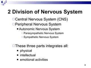
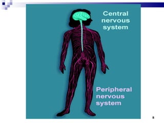
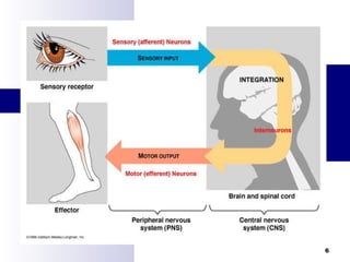
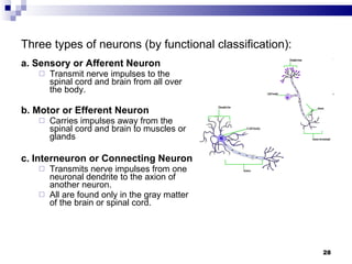
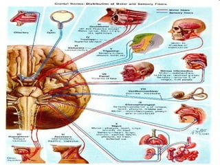
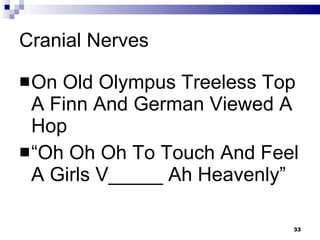
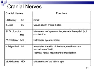
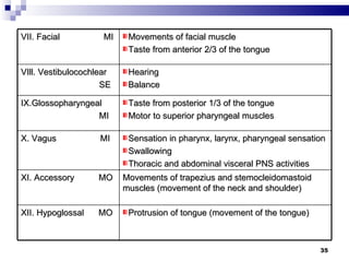
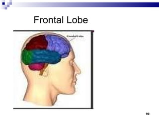
DISTURBANCE IN PERCEPTION & COORDINATION LUDY MAE B. NALZARO, RN, MN
The Nervous System
The Nervous System Master controlling and communicating system of the body 3 Overlapping Functions: Uses  sensory receptors  to interpret stimulus Uses  integration  to process the input Uses  motor output  to elicit a response to the stimulus
2 Division of Nervous System Central Nervous System (CNS) Peripheral Nervous System Autonomic Nervous System Parasympathetic Nervous System Sympathetic Nervous System These three parts integrates all: physical intellectual  emotional activities
 
 
Central Nervous System It includes the brain and spinal cord; occupies the dorsal body cavity INTEGRATOR COMMAND CENTERS
Brain Collects integrates and interprets all stimuli Composition: Cerebrum (Cerebral Cortex),  Brain stem Cerebellum.
Cerebrum It gives us the ability to think and reason It is enclosed in three membrane layers called  meninges It has four lobes and two hemispheres, these lobes controls specific functions
The Lobes of the Cerebrum The brain is composed of lobes: Frontal lobe personality, memory and motor function Broca’s area (speech) Parietal lobe sensory function (tactile) Temporal lobe hearing and olfaction and emotion by the limbic system Occipital lobe vision
Anatomy and Physiology The cerebellum  is involved in coordination and equilibrium The diencephalon consists of the : Thalamus- the relay center of all sensory input Hypothalamus- center for endocrine regulation, sleep, temperature, thirst, sexual arousal and emotional response
Brain Stem the midbrain, pons, and medulla. It relays messages between the cerebrum and diencephalon and the spinal cord It regulates automatic body functions such as heart rate, breathing, swallowing and coughing
Anatomy and Physiology The brainstem is composed of the: MIDBRAIN for visual and auditory reflexes Pons respiratory apneustic center, nucleus of cranial nerves- 5,6,7,8 Medulla oblongata respiratory and cardiovascular centers, nucleus of cranial nerves 9,10,11,12
Spinal Cord The primary path for nerve impulses traveling between peripheral areas of the body and the brain. It contains the sensory – to – motor pathway known as the  reflex arc It is made up of an H shaped mass of gray matter, divided into the dorsal and ventral horns
Peripheral Nervous System (PNS )  a. Cranial nerves:  (12 pairs) b. Spinal nerves: (31 pairs)   Carry nerve impulses to and from the spinal cord to body parts not served by the cranial nerves.  c.  Somatic nervous system  Carries nerve impulses to the skeletal muscles, joints and skin      d.  Autonomic nervous system (ANS)   Carries nerve impulses to the smooth muscles of internal organs and to glands without conscious thought
The Autonomic Nervous System Composed of a special group of neurons that regulates cardiac muscle, smooth muscles of the visceral organs and glands Critical to the stability of our internal environment (homeostasis) Controls adjustments needed to best support body activities; fine-tuning occurs without our awareness or attention
Autonomic nervous system (ANS)   1.   Sympathetic system   Controls "fight or flight" responses Neurotransmitter is norepinephrine (or noradrenaline). Mobilizes the body during extreme situations (fear, exercise, rage, stress) Enables the body to cope rapidly and vigorously with situations that threaten homeostasis Functions to provide the best conditions for responding to some threat
Autonomic nervous system (ANS)   2.  Parasympathetic system   Controls those responses associated with a  relaxed state. Neurotransmitter is acetylcholine (Ach). Chiefly concerned  with promoting normal digestion and elimination and with conserving body energy Decreases demands on the cardiovascular system
Things to know: Broca’s area  ability to speak;  found in front of the Central Sulcus of Rolando  in the left hemisphere Higher intellectual reasoning  found in the anterior part of the frontal lobes;  language comprehension;  memory in both temporal and frontal lobes
Wernicke’s area  Located at the junction of the temporal, parietal and occipital lobes;  allows one to sound out words;  COMPREHENSION  found in the right hemisphere Corpus Callosum  connects the 2 hemispheres
Synapse   A place where nerve impulses are transmitted from one neuron to another (from presynaptic neuron to the postsynaptic neuron) Three structures of a synapse:  1.  Synaptic knob  (on presynaptic neuron's axon)         Contains vessicles filled with a chemical(s) called a neurotransmitter(s) 2.  Synaptic cleft   Space between synaptic knob and plasma membrane of postsynaptic neuron 3.  Plasma membrane  (of  postsynaptic neuron )  Has receptor site (proteins) in its membrane for the neurotransmitter
    Neurotransmitters   1.  Acetylcholine   a. associated with nerve impulse conduction at synapse. b. belongs to a chemical group called  cholinergics . 2.  Norepinepherine                   a.  belong to a chemical group called  adrenergics and catecholamines. b. associated with sleep, mood, motor function and pleasure recognition 3.  Dopamine   a.   also a  catecholamine . b.   associated with sleep, mood, motor function and pleasure recognition 4.   Serotonin   a.   also a  catecholamine. b.   associated with sleep, mood, motor function and pleasure recognition 5.  Endorphins   Inhibits pain- type nerve impulses. 6.  Enkephalins   Inhibits pain- type nerve impulses
Cells of Nervous System: Neuroglia (glial cells) Provide support for the activity and nutrition of neurons Neurons composes of cell body, axon and dendrites
Types of Neuralgia in CNS Astrocytes  Nutrition, attached to blood vessels Oligodendrites like Schwann cells of PNS Microglia Phagocytic cells Ependymal cells Lines the ventricles in regulating diffusion of substances like interstitial fluid and CSF
Types of Neuralgia Found in PNS Schwann cells        Form myelin sheaths around peripheral axons and are composed of:  Myelin: A white fatty substance Provides insulation  Forms  myelin sheath  around some axons in the PNS Nodes of Ranvier:   Nonmyelinated gaps on axons that lie between adjacent Schwann cells. Neurilemma:   Outer membrane of a Schwann cell Plays an important role in the regeneration of cut or injured axons. Note:    Since Schwann cells are only found in the PNS, the CNS (brain & spinal cord) has far less potential for regeneration.
 
Neurons  a.   Cell Body   Part that contains the nucleus b.  Dendrite(s)   Carries a nerve impulse  towards  the cell body c.  Axon(s)   Carries a nerve impulse  away  from the cell body (and towards the dendrite of the next neuron) d.   Axons are also called  nerve fibers . e.   A nerve is a bundle of axons  outside  the CNS They are the basic  structural and functional unit  of the nervous system.   They conduct nerve impulses.  Note:  Definition of a   nerve impulse   :  A self-propagating wave of electrical disturbance that travels along the surface of a neuron's plasma membrane.
Three types of neurons (by functional classification): a. Sensory or Afferent Neuron  Transmit nerve impulses to the spinal cord and brain from all over the body. b. Motor or Efferent Neuron Carries impulses away from the spinal cord and brain to muscles or glands c.   Interneuron or Connecting   Neuron   Transmits nerve impulses from one neuronal dendrite to the axion of another neuron. All are found only in the gray matter of the brain or spinal cord.
The Peripheral Nervous System Nerves are classified as Afferents  carry impulses towards the CNS Efferents  carry impulses towards the muscles Mixed  Carries both sensory and motor fibers
Cranial Nerves – 12 pairs - nerves serving the head and neck - VAGUS NERVE – is the only pair of Cranial Nerves that extends to the thoracic and abdominal cavities - described by name, number, course and major function - most cranial nerves are mixed nerves except for the Optic, Olfactory and Acoustic Nerve (pure sensory)
CRANIAL NERVES
 
Cranial Nerves On Old Olympus Treeless Top A Finn And German Viewed A Hop “ Oh Oh Oh To Touch And Feel A Girls V_____ Ah Heavenly”
Cranial Nerves Cranial Nerves Functions I.Olfactory  SE Smell II.Optic  SE Visual acuity, Visual Fields III. Oculomotor  MO Movements of eye muscles, elevate the eyelid, [upil constriction IV.Trochlear  MO Extrocular eye movement V.Trigeminal  MI Innervates the skin of the face, nasal mucosa; sensations of teeth  *corneal reflex; Movement of mastication VI.Abducens  MO Movements of the lateral eye
VII. Facial  MI Movements of facial muscle Taste from anterior 2/3 of the tongue VIll. Vestibulocochlear SE Hearing Balance IX.Glossopharyngeal MI Taste from posterior 1/3 of the tongue Motor to superior pharyngeal muscles X. Vagus  MI Sensation in pharynx, larynx, pharyngeal sensation Swallowing Thoracic and abdominal visceral PNS activities XI. Accessory  MO Movements of trapezius and stemocleidomastoid muscles (movement of the neck and shoulder) XII. Hypoglossal  MO Protrusion of tongue (movement of the tongue)
Effects of Injury
Neurologic Assessment
History Taking Current Health Status Previous Health Status Family Health Lifestyle
Current health status Discover the patient’s chief complaint by asking this sample questions: Why have you come to the hospital? What has been bothering you lately? Do you have headaches? If so, how often? What precipitates them? Do you ever feel a tingling or numbness? If so, Where? How’s your memory and ability to concentrate? Do you have trouble urinating? Walking? Do you have trouble reading or writing?
Current health status Using the patient’s own words, document his reasons for seeking care If the patient is suffering from neurologic disorder you can expect reports of: Headaches motor disturbance Seizures sensory deviations or an altered level of consciousness (LOC).
Previous health status Many chronic diseases can affect neurologic system. Ask if he has had any: - major illnesses - recurrent minor illnesses - accidents , injuries - surgical procedures , allergies  - birth (full/premature)
Family health status Information about the patient’s family may reveal a hereditary disorder. epilepsy Huntington’s disease Amyotrophic lateral sclerosis Diabetes  Cardio or renal disease HPN Cancer bleeding disorder mental disorder  stroke
Lifestyle Cultural and social background  Educational level Occupation Drug use
Dietary habits Review 24 hrs day’s diet Vit. B12 deficiency Damage to CNS –polyneuritis and weakness Vit B complex deficiency Lead to peripheral nerve damage
Physical Assessment Mental Status Cranial nerve function Sensory function Motor function Reflexes
Mental Status Listen and watch for clues to the patients orientation and memory. It involves evaluating the patients: - LOC  –  most sensitive indicator - Appearance and behavior - mood swings - Speech - ability to express, fluency of language, comprehension - Cognitive function  - long & short term memories - Constructional ability
QUICK CHECK OF MENTAL STATUS Question Function screened What’s your name? Orientation to person What’s today’s date? Orientation to time What year is it? Orientation to time Where are you now? Orientation to place How old are you? Memory Where were you born? Remote memory What did you have for breakfast? Recent memory Who’s the Phil. President? General knowledge Can you count back from 20 to 1? Attention and calculation  Why are you here? Judgement
Glasgow Coma Scale
GLASGOW COMA SCALE - An objective measure to describe LOC in terms of:  Eye Opening Motor Response Verbal Response Result:  3 - indicating deep unconsciousness  7 – candidate for intubation 14 - minor
Glasgow Coma Scale
 
GCS VALUES **Should not be considered a complete assessment tool **Not a sensitive tool for evaluation of altered sensorium
GCS VALUES Best possible score  = 15 Moderate disability  =  11 Coma   =  7  Lowest possible score = 3
Level of Consciousness - Most sensitive indicator of changes in neurologic status of the patient. I. Alert – conscious, coherent, cognitive   - alert, awake, responsive to  stimuli     - follows commands and responds completely and appropriately to stimuli
II. Lethargic, somnolence (sleepy) , drowsiness or obtunded (slowed psychomotor responses ) - can be aroused with little difficulty
III. Stupor Physical and mental activities are minimal.  Person inaccessible to many stimuli. Requires vigorous stimulation for a response. Can not be fully aroused IV. Light Coma - does not respond to ordinary stimuli but may respond to painful stimuli.
V. Deep Coma Limbs flaccid and motionless, muscles, tendon, plantar reflexes absent. No reaction to painful stimuli, abnormal motor responses, incontinence present. Pupils constricted or dilated and unresponsive to light; corneal and pharyngeal reflexes are minimal or absent.
Cranial Nerves It provides valuable information about the status of the CNS, particularly the brain stem The optic, oculomotor, trochlear and abducens are more vulnerable to an increase in intracranial pressure than other cranial nerves.
Cranial Nerve Function: Cranial Nerve 1- Olfactory Check first for the patency of the nose Instruct to close the eyes Occlude one nostrils at a time Hold familiar substance and asks for the identification Repeat with the other nostrils PROBLEM- ANOSMIA- “loss of smell”
CN II Optic - Sensory Visual acuity Snellen’s chart-distance vision Visual fields confrontation test Check of peripheral vision; 11/2 -2 feet Your own visual field must be normal
Visual Field Confrontation test
Nystagmus
Myopia -  far away objects appear  blurred  and near objects appear clearly  Hyperopia -  farsightedness   Presbyopia Astigmatism Cataracts  Glaucoma
Cataract
Cranial Nerve Function: Cranial Nerve 2- Optic Check the visual acuity with the use of the Snellen chart Check for visual field by confrontation test Check for pupillary reflex- direct and consensual Fundoscopy to check for papilledema
Cranial Nerve Function: Cranial Nerve 3, 4 and 6 Assess simultaneously the movement of the extra-ocular muscles Deviations: Opthalmoplegia- inability to move the eye in a direction Diplopia- complaint of double vision
Rev. Nov 2006
 
 
Mydriasis –blown pupil Miosis  (constriction of the pupil ) Anisocoria (Unequal size of pupils ) Ptosis
Cranial Nerve Function: Cranial Nerve 5 -trigeminal Sensory portion  assess for sensation of the facial skin Motor portion assess the muscles of mastication Assess corneal reflex
CN VII. Facial mixed Sensory and Motor Motor Function of Face Taste sensation of Tongue Ask client to smile, close eyes, raise eyebrows, pout mouth Test anterior 2/3 of tongue for diff. taste sensation  Bell’s Palsy
 
CN VIII – Auditory  Hearing Gross Hearing acuity test – Normal tone of voice and Whispered voice Watch tick Test Weber’s test – bone conduction test Rinne’s test – comparison of air and bone conduction
Hearing Weber’s test positive – if sound is louder in impaired ear –  bone conductive hearing   loss ; if sound is louder in normal ear –  sensorineural hearing loss Weber’s negative : normal  = if sound felt on both ears are equal
Weber’s Test
Rinne’s Test
Hearing Rinne’s test positive –  normal  = Air conduction > Bone conduction Rinne’s negative – Abnormal =  Bone conduction = Air conduction Bone conduction > Air conduction
CN IX – Glossopharyngeal – Mixed Sensory and motor Test – gag reflex Tongue movement – CN IX and CN XII Hypoglossal
Cranial Nerve Function: Cranial Nerve 9- glossopharyngeal Together with Cranial nerve 10 –vagus Assess for gag reflex Watch the soft palate rising after instructing the client to say “AH” The posterior one-third of the tongue is supplied by the glossopharyngeal nerve
CN X – Vagus – Mixed Sensory and motor Together with assessment of CN IX- test patient for hoarseness of voice
CN XI– Accessory Spinal Nerve motor Ask Client to shrug shoulders against resistance Move head against resistance
Cranial Nerve Function: Cranial Nerve 12- hypoglossal Ask patient to protrude the tongue and note for symmetry
Sensory Perception Stereognosis – ability to perceive sensory stimuli. Agnosia – inability to perceive stimuli. Global aphasia  Aphasia - difficulty in producing or comprehending spoken or written language severe  Broca's  aphasia  and  Wernicke's  aphasia  combined  Receptive aphasia  neurological damage to  Wernicke’s  area  in the brain Expressive aphasia the loss of the ability to produce language (spoken or written)  5 Areas of Sensation: - Pain  - Position - Light Touch   - Discrimination - Vibration
Motor Function Regulating mechanisms: Frontal lobe – motor center; responsible for voluntary, purposeful, coordinated movement. Apraxia  inability to perform fine motor activities. Agraphia  inability to write. Cerebellum  (center for balance) equilibrium, sense of posture, direction. Ataxia  uncoordinated movement.
Coordination Finger to nose – eyes open then closed Distance – 18 inches Rapid alternating movements; thumb to finger position Heel to shin Romberg test – with eyes closed; no swaying for 5 seconds Coordination – heel to toe fashion and then stand on each foot
Finger to Nose test
Alternating Supination & Pronation of Hands to Knees
Heel to Shin test
Heel to Toe walk
Romberg’s  Test
  Frontal Lobe
General Appearance - involuntary, unpurposeful, uncoordinated movement, asymmetry of face, muscle dystrophy. Muscle Power - weakness (“paresis”) - paralysis (“plegia”) Muscle Tone - flaccidity (hypotonicity) - rigidity (hypertonicity)
Plegia(Paralysis)
Muscle Volume Atrophy  Loss of muscle movement. Hypertrophy  Increase in muscle volume. Muscle Movement  Bradykinesia  Slow muscle movement not associated with weakness. Akinesia  Absence of muscle movement.
  Atrophy
Bradykinesia
Muscle Visual scanning of Symmetry Contour Size (e.g. atrophy) Involuntary movement Palpation Tone tension present in a resting muscle Testing of strength
Muscle Strength Functional level Lovett Scale Grade % of normal No evidence of contractility 0 0 0 Evidence of slight contractility Trace 1 10 Complete ROM minus gravity Poor 2 25 Complete ROM with gravity Fair 3 50 Complete ROM with some resistance Good 4 75 Complete ROM vs. gravity with full resistance Normal 5 100
Muscle strength test
Reflexes Defect in sensory pathways from tendons & muscles or the motor component Plantar reflex – start from the heel Babinski reflex – normal in children before they can walk (lesions of the pyramidal tract or motor nerves Grasp reflex – normal in infants < 4 mo old Present if with widespread brain damage
Deep Tendon Reflexes Grading of reflexes 4+ - brisk, hyperactive 3+ - more brisk than normal 2+ - normal 1+ - low normal, slightly diminished 0 – no response
Reflexes Function of reflex arcs and the spinal cord segment Biceps - C5,6 Brachioradialis - C5,6 Triceps - C6, 7, 8 Patellar - L2, 3, 4 Ankle - S1, 2 Plantar - L4, 5; S1, 2
Meningeal Brudzinski
Meningeal Kernig
Biceps reflex
Triceps reflex
Patellar reflex
Achilles reflex
Babinski Reflex
Diagnostic Assessment
Imaging Studies Computed Tomography (CT Scan) Isotope Brain Scan Magnetic Resonance Imaging (MRI) Positron Emission Tomography (PET) Skull and Spinal X- rays
DIAGNOSTIC TESTS Lumbar puncture Insertion of a needle into the lumbar subarachnoid space and withdrawal of CSF for diagnostic and therapeutic purposes. Ensure consent, determine ability to lie still Contraindicated in patients with increased ICP Keep flat on bed after procedure Increase fluid intake after procedure
Computed Tomography Scan It combines radiology and computer analysis of tissue density (with the use of dye) to study the intracranial structures. Highly informative diagnostic test using the computer to analyze data. Purpose is to detect intracranial bleeding, space occupying lesions, cerebral edema and shifts of brain structures. Hydrocephalus and cerebral atrophy can also be identified. The head is scanned by a narrow beam of x-ray, performed quickly within about 30 mins not including data analysis.
 
CT Scan Spine scan will diagnose: - Herniated disk - Spinal cord tumors - Spinal stenosis
CT Scan Brain scan can detect: - Brain contusion - Brain calcifications - Cerebral Atrophy - Hydrocephalus - Inflammation - Space – occupying lesions  - Vascular anomalies
CT Scan Nursing Consideration: - Confirm that the patient isn’t allergic to iodine or shellfish - If the test calls for a contrast medium, explain that an I.V. catheter will be inserted - Explain to the patient that he may feel flushed or notice a metallic taste in his mouth when the contrast is injected - The procedure will last for 10 to 30 mins - Encourage the patient to resume normal activities and a regular diet after the test - The contrast medium may discolor his urine for 24 hours, and suggest that he drink more fluids to help flush the medium out of his system
Isotope Brain Scan A scanning device monitors the brain’s uptake of a radioactive isotope It will detect cerebral lesions, neoplasms, brain abscess, cerebral edema, hematoma, infarction
Isotope Brain Scan Nursing Consideration: - Withhold medications, as ordered - Confirm that the patient isn’t allergic to iodine or shellfish - If the test calls for a contrast medium, explain that an I.V. catheter will be inserted
Isotope Brain Scan Nursing Consideration: Tell the patient that he’ll be asked to change the position several times during the procedure while the technician takes picture of his brain Unless contraindicated, suggest that he drink more fluids to help flush the contrast medium out of his system
Uses magnetic waves Provides more anatomically detailed pictures and does not use radiation exposure Extremely sensitive in detecting abnormalities in the brain, especially chemical changes within cells and for delineating the extent of IC tumors. Patients with pacemakers, orthopedic metal prosthesis and implanted metal devices cannot undergo this procedure Magnetic Resonance Imaging
 
MRI Nursing Consideration: - Explain that the procedure can take up to 1 ½ hours  and that he’ll be remain still for intervals of 5 to 20 minutes - Have the patient remove all metallic items - The test is painless but the machinery may seem loud and frightening - Provide sedation, as ordered, to promote relaxation
SKULL & SPINAL X-RAYS Skull X-ray is taken from two angles: Anteroposterior (AP) and Lateral. Waters’ view to examine frontal and maxillary sinuses, facial bones and eye orbits. Towne’s view to examine the occipital bone
Skull x-rays help detect: - fractures - bony tumors or calcifications - space occupying lesions - vascular abnormalities
 
Spinal X-rays It detects: - Spinal fractures - Displacement & Subluxation - Destructive lesions - Arthritic changes - Structural abnormalities
Cerebral Angiography It detects: Stenosis or occlusion associated w/ thrombi Aneurysms Locate vessel displacement associated w/ tumors, abscesses, cerebral edema, hematoma, or herniation
Nursing Consideration: - Confirm that he is not allergic to iodine or shellfish - Feeling of flushed sensation in his face as the dye is injected - Monitor the catheter injection site for signs of bleeding - Monitor the peripheral pulse in the arm or leg used for catheter insertion
- Monitor the patient for neurologic changes and such complications as hemiparesis, hemiplegia, aphasia, and impaired LOC - Monitor for adverse reaction to the contrast medium
DIAGNOSTIC TESTS EEG Records by means of electrodes applied on the scalp surface. The electrical activity that is generated in the brain. It is useful in diagnosing epilepsies & as screening for coma and organic brain syndrome; also serves as indicator of brain death. Nursing Responsibilities: Withhold medications that may interfere with the results- anticonvulsants, sedatives and stimulants Wash hair thoroughly before procedure
Electroencephalography It is recording of the brains continuous electrical activity. It will identify: - seizure disorder - head injuries - intracranial lesions - TIA’s - stroke - brain death
Nursing Consideration: - Explain that the physician will apply paste and attached the electrodes to areas of skin of the head and neck after these areas have been lightly abraded to ensure good contact
- Discuss any specific activity that the patient will be asked to perform, such as hyperventilating for 3 minutes or sleeping depending on the purpose of EEG - Use acetone to remove any remaining paste from the patient’s skin - Resume normal activities, as ordered
ASSIGNMENT SUBMISSION = DECEMBER  1,2011 (THURSDAY) NEKS WEK!  Description and Nursing Management of the following diagnostic examinations: Skull x-ray CT Scan MRI EEG PET Scan Lumbar puncture Myelography Cerebral angiography Prothrombin Time (PT) Partial Thrombo-plastin Time (PTT) International Normalized Ratio (INR) Electrolytes and Blood Glucose Coagulation studies
END
TREATMENTS Drug Therapy Adrenergic blockers (Ergotamine Tartrate, Dihydroergotamine mesylate) Anticonvulsants (Carbamazepine, clonazepam, diazepam, valproate)
Antiparkinson agents  (Benztropine, levodopa, tolcapone, carbidopa – levodopa) Calcium Channel Blockers (Nimodipine) Cortocosteroids (Dexametahsone, Prednisone) Diuretics (Mannitol) Opiod Analgesics (Codeine, Morphine)
SURGERY The only viable intervention when a neurologic disorder is life – threatening Cerebellar stimulator implantation, cerebral aneurysm repair, craniotomy and intracranial hematoma aspiration
Cerebellar Stimulator Implantation 2 electrodes are position in the patients cerebellum. The electrodes are connected to a power source and pulse generator. The generator sends electrical impulses to the electrodes which stimulates nerve fibers in cerebellar cortex
These electrical impulses help regulate uncoordinated neuromuscular activity. Benefits for the patient may include a reduction in spasticity and abnormal movements, improved muscle, clearer speech and decrease number of seizures
Patient Preparation: - The patient will be in general anesthesia - The patient’s head will be shaved and positioned in a special headrest - The surgeon will have four incisions for implanting the electrodes ( 2 on head, one on neck & one in abdomen)
Monitoring and aftercare Assess the neurologic status and vital signs every hour for the first 24 hours WOF signs of complications, such as increase ICP, infection & fluid imbalance
Provide IV fluids if the patient experiences excessive nausea & vomiting Observe for seizures or spasticity, and record occurrences to help doctor evaluate the effectiveness of the device Keep the patient on bed rest until the 2 nd  post operative day.
Elevate the head 15 to 30 degrees, and turn the patient every 2 hours Slowly increase the patients activity, but make sure he avoids overexertion Check the dressing regularly for excessive bleeding or drainage. Assess suture line for signs of infection Administer antibiotics and analgesic
Home Care Instruction Teach the patient and family how to change a dressing and recommend to change it every 2 days until the surgeon removes the sutures Describe the signs of infection Record any seizure activity for the doctor to evaluate the effectiveness of the operation
Cerebral Aneurysm Repair it clamp the affected artery, wrap the aneurysm wall with synthetic material or clip or ligate the aneurysm
Patient Preparation Tell the patient and family that he’ll be monitored in the ICU before and after the surgery, where he will be observed for signs of bleeding, vasopasm and increase ICP
Monitoring and aftercare Gradually increase the patients level of activity Monitor the incision site Monitor neurologic status and V/S Provide patient and family with emotional support
Home care instruction Continue take the prescribed anticonvulsant to minimize seizure Emphasize the importance of returning for scheduled follow up Refer the patient of family to a support group
Craniotomy Involves creation of incision into the skull to expose the brain. Potential complications, include infection, hemorrhage, respiratory compromise, Increase ICP
Preparation Patients head will be shaved Discuss the recovery period Tell the patient to expect headache and facial swelling for 2 – 3 days Performed & document a baseline neuro assessment ICU after surgery
Monitoring & Aftercare Gradually increase level of activity Monitor incision site Monitor neurologic status and VS Provide emotional support
Home Care Instruction Teach proper wound care Remind the patient to continue taking prescribed anticonvulsant medication to minimize seizure Emphasize on returning for scheduled follow up
Nursing Diagnosis Impaired Physical Mobility Expected outcomes ( Will show no evidence of complications, Will achiev the highest level of mobility & Will maintain muscle strength and joint ROM)
Impaired Skin Integrity Expected outcome ( Will maintain intact skin integrity, Will not develop complications & will maintain the optimal nutrition needed)
Impaired urinary elimination Expected outcomes ( Will empty his bladder completely & regularly & Patient won’t develop UTI
Impaired Gas Exchange Expected outcomes ( Patient will not develop a respiratory infection, will maintain optimal oxygen saturation level)
Thank YOU !

INTRODUCTION TO NEURO NURSING

  • 1.
    DISTURBANCE IN PERCEPTION& COORDINATION LUDY MAE B. NALZARO, RN, MN
  • 2.
  • 3.
    The Nervous SystemMaster controlling and communicating system of the body 3 Overlapping Functions: Uses sensory receptors to interpret stimulus Uses integration to process the input Uses motor output to elicit a response to the stimulus
  • 4.
    2 Division ofNervous System Central Nervous System (CNS) Peripheral Nervous System Autonomic Nervous System Parasympathetic Nervous System Sympathetic Nervous System These three parts integrates all: physical intellectual emotional activities
  • 5.
  • 6.
  • 7.
    Central Nervous SystemIt includes the brain and spinal cord; occupies the dorsal body cavity INTEGRATOR COMMAND CENTERS
  • 8.
    Brain Collects integratesand interprets all stimuli Composition: Cerebrum (Cerebral Cortex), Brain stem Cerebellum.
  • 9.
    Cerebrum It givesus the ability to think and reason It is enclosed in three membrane layers called meninges It has four lobes and two hemispheres, these lobes controls specific functions
  • 10.
    The Lobes ofthe Cerebrum The brain is composed of lobes: Frontal lobe personality, memory and motor function Broca’s area (speech) Parietal lobe sensory function (tactile) Temporal lobe hearing and olfaction and emotion by the limbic system Occipital lobe vision
  • 11.
    Anatomy and PhysiologyThe cerebellum is involved in coordination and equilibrium The diencephalon consists of the : Thalamus- the relay center of all sensory input Hypothalamus- center for endocrine regulation, sleep, temperature, thirst, sexual arousal and emotional response
  • 12.
    Brain Stem themidbrain, pons, and medulla. It relays messages between the cerebrum and diencephalon and the spinal cord It regulates automatic body functions such as heart rate, breathing, swallowing and coughing
  • 13.
    Anatomy and PhysiologyThe brainstem is composed of the: MIDBRAIN for visual and auditory reflexes Pons respiratory apneustic center, nucleus of cranial nerves- 5,6,7,8 Medulla oblongata respiratory and cardiovascular centers, nucleus of cranial nerves 9,10,11,12
  • 14.
    Spinal Cord Theprimary path for nerve impulses traveling between peripheral areas of the body and the brain. It contains the sensory – to – motor pathway known as the reflex arc It is made up of an H shaped mass of gray matter, divided into the dorsal and ventral horns
  • 15.
    Peripheral Nervous System(PNS ) a. Cranial nerves:  (12 pairs) b. Spinal nerves: (31 pairs) Carry nerve impulses to and from the spinal cord to body parts not served by the cranial nerves. c. Somatic nervous system Carries nerve impulses to the skeletal muscles, joints and skin     d. Autonomic nervous system (ANS) Carries nerve impulses to the smooth muscles of internal organs and to glands without conscious thought
  • 16.
    The Autonomic NervousSystem Composed of a special group of neurons that regulates cardiac muscle, smooth muscles of the visceral organs and glands Critical to the stability of our internal environment (homeostasis) Controls adjustments needed to best support body activities; fine-tuning occurs without our awareness or attention
  • 17.
    Autonomic nervous system(ANS) 1.   Sympathetic system Controls &quot;fight or flight&quot; responses Neurotransmitter is norepinephrine (or noradrenaline). Mobilizes the body during extreme situations (fear, exercise, rage, stress) Enables the body to cope rapidly and vigorously with situations that threaten homeostasis Functions to provide the best conditions for responding to some threat
  • 18.
    Autonomic nervous system(ANS) 2.  Parasympathetic system Controls those responses associated with a relaxed state. Neurotransmitter is acetylcholine (Ach). Chiefly concerned with promoting normal digestion and elimination and with conserving body energy Decreases demands on the cardiovascular system
  • 19.
    Things to know:Broca’s area ability to speak; found in front of the Central Sulcus of Rolando in the left hemisphere Higher intellectual reasoning found in the anterior part of the frontal lobes; language comprehension; memory in both temporal and frontal lobes
  • 20.
    Wernicke’s area Located at the junction of the temporal, parietal and occipital lobes; allows one to sound out words; COMPREHENSION found in the right hemisphere Corpus Callosum connects the 2 hemispheres
  • 21.
    Synapse A place where nerve impulses are transmitted from one neuron to another (from presynaptic neuron to the postsynaptic neuron) Three structures of a synapse: 1. Synaptic knob (on presynaptic neuron's axon)        Contains vessicles filled with a chemical(s) called a neurotransmitter(s) 2. Synaptic cleft Space between synaptic knob and plasma membrane of postsynaptic neuron 3. Plasma membrane (of postsynaptic neuron ) Has receptor site (proteins) in its membrane for the neurotransmitter
  • 22.
       Neurotransmitters 1.  Acetylcholine a. associated with nerve impulse conduction at synapse. b. belongs to a chemical group called cholinergics . 2.  Norepinepherine                  a.  belong to a chemical group called adrenergics and catecholamines. b. associated with sleep, mood, motor function and pleasure recognition 3. Dopamine a.   also a catecholamine . b.   associated with sleep, mood, motor function and pleasure recognition 4.  Serotonin a.   also a catecholamine. b.   associated with sleep, mood, motor function and pleasure recognition 5. Endorphins Inhibits pain- type nerve impulses. 6. Enkephalins Inhibits pain- type nerve impulses
  • 23.
    Cells of NervousSystem: Neuroglia (glial cells) Provide support for the activity and nutrition of neurons Neurons composes of cell body, axon and dendrites
  • 24.
    Types of Neuralgiain CNS Astrocytes Nutrition, attached to blood vessels Oligodendrites like Schwann cells of PNS Microglia Phagocytic cells Ependymal cells Lines the ventricles in regulating diffusion of substances like interstitial fluid and CSF
  • 25.
    Types of NeuralgiaFound in PNS Schwann cells       Form myelin sheaths around peripheral axons and are composed of: Myelin: A white fatty substance Provides insulation Forms myelin sheath around some axons in the PNS Nodes of Ranvier: Nonmyelinated gaps on axons that lie between adjacent Schwann cells. Neurilemma: Outer membrane of a Schwann cell Plays an important role in the regeneration of cut or injured axons. Note:   Since Schwann cells are only found in the PNS, the CNS (brain & spinal cord) has far less potential for regeneration.
  • 26.
  • 27.
    Neurons a.  Cell Body Part that contains the nucleus b.  Dendrite(s) Carries a nerve impulse towards the cell body c.  Axon(s) Carries a nerve impulse away from the cell body (and towards the dendrite of the next neuron) d.   Axons are also called nerve fibers . e.   A nerve is a bundle of axons outside the CNS They are the basic structural and functional unit of the nervous system.   They conduct nerve impulses. Note: Definition of a  nerve impulse : A self-propagating wave of electrical disturbance that travels along the surface of a neuron's plasma membrane.
  • 28.
    Three types ofneurons (by functional classification): a. Sensory or Afferent Neuron  Transmit nerve impulses to the spinal cord and brain from all over the body. b. Motor or Efferent Neuron Carries impulses away from the spinal cord and brain to muscles or glands c.   Interneuron or Connecting Neuron Transmits nerve impulses from one neuronal dendrite to the axion of another neuron. All are found only in the gray matter of the brain or spinal cord.
  • 29.
    The Peripheral NervousSystem Nerves are classified as Afferents carry impulses towards the CNS Efferents carry impulses towards the muscles Mixed Carries both sensory and motor fibers
  • 30.
    Cranial Nerves –12 pairs - nerves serving the head and neck - VAGUS NERVE – is the only pair of Cranial Nerves that extends to the thoracic and abdominal cavities - described by name, number, course and major function - most cranial nerves are mixed nerves except for the Optic, Olfactory and Acoustic Nerve (pure sensory)
  • 31.
  • 32.
  • 33.
    Cranial Nerves OnOld Olympus Treeless Top A Finn And German Viewed A Hop “ Oh Oh Oh To Touch And Feel A Girls V_____ Ah Heavenly”
  • 34.
    Cranial Nerves CranialNerves Functions I.Olfactory SE Smell II.Optic SE Visual acuity, Visual Fields III. Oculomotor MO Movements of eye muscles, elevate the eyelid, [upil constriction IV.Trochlear MO Extrocular eye movement V.Trigeminal MI Innervates the skin of the face, nasal mucosa; sensations of teeth *corneal reflex; Movement of mastication VI.Abducens MO Movements of the lateral eye
  • 35.
    VII. Facial MI Movements of facial muscle Taste from anterior 2/3 of the tongue VIll. Vestibulocochlear SE Hearing Balance IX.Glossopharyngeal MI Taste from posterior 1/3 of the tongue Motor to superior pharyngeal muscles X. Vagus MI Sensation in pharynx, larynx, pharyngeal sensation Swallowing Thoracic and abdominal visceral PNS activities XI. Accessory MO Movements of trapezius and stemocleidomastoid muscles (movement of the neck and shoulder) XII. Hypoglossal MO Protrusion of tongue (movement of the tongue)
  • 36.
  • 37.
  • 38.
    History Taking CurrentHealth Status Previous Health Status Family Health Lifestyle
  • 39.
    Current health statusDiscover the patient’s chief complaint by asking this sample questions: Why have you come to the hospital? What has been bothering you lately? Do you have headaches? If so, how often? What precipitates them? Do you ever feel a tingling or numbness? If so, Where? How’s your memory and ability to concentrate? Do you have trouble urinating? Walking? Do you have trouble reading or writing?
  • 40.
    Current health statusUsing the patient’s own words, document his reasons for seeking care If the patient is suffering from neurologic disorder you can expect reports of: Headaches motor disturbance Seizures sensory deviations or an altered level of consciousness (LOC).
  • 41.
    Previous health statusMany chronic diseases can affect neurologic system. Ask if he has had any: - major illnesses - recurrent minor illnesses - accidents , injuries - surgical procedures , allergies - birth (full/premature)
  • 42.
    Family health statusInformation about the patient’s family may reveal a hereditary disorder. epilepsy Huntington’s disease Amyotrophic lateral sclerosis Diabetes Cardio or renal disease HPN Cancer bleeding disorder mental disorder stroke
  • 43.
    Lifestyle Cultural andsocial background Educational level Occupation Drug use
  • 44.
    Dietary habits Review24 hrs day’s diet Vit. B12 deficiency Damage to CNS –polyneuritis and weakness Vit B complex deficiency Lead to peripheral nerve damage
  • 45.
    Physical Assessment MentalStatus Cranial nerve function Sensory function Motor function Reflexes
  • 46.
    Mental Status Listenand watch for clues to the patients orientation and memory. It involves evaluating the patients: - LOC – most sensitive indicator - Appearance and behavior - mood swings - Speech - ability to express, fluency of language, comprehension - Cognitive function - long & short term memories - Constructional ability
  • 47.
    QUICK CHECK OFMENTAL STATUS Question Function screened What’s your name? Orientation to person What’s today’s date? Orientation to time What year is it? Orientation to time Where are you now? Orientation to place How old are you? Memory Where were you born? Remote memory What did you have for breakfast? Recent memory Who’s the Phil. President? General knowledge Can you count back from 20 to 1? Attention and calculation Why are you here? Judgement
  • 48.
  • 49.
    GLASGOW COMA SCALE- An objective measure to describe LOC in terms of: Eye Opening Motor Response Verbal Response Result: 3 - indicating deep unconsciousness 7 – candidate for intubation 14 - minor
  • 50.
  • 51.
  • 52.
    GCS VALUES **Shouldnot be considered a complete assessment tool **Not a sensitive tool for evaluation of altered sensorium
  • 53.
    GCS VALUES Bestpossible score = 15 Moderate disability = 11 Coma = 7 Lowest possible score = 3
  • 54.
    Level of Consciousness- Most sensitive indicator of changes in neurologic status of the patient. I. Alert – conscious, coherent, cognitive - alert, awake, responsive to stimuli - follows commands and responds completely and appropriately to stimuli
  • 55.
    II. Lethargic, somnolence(sleepy) , drowsiness or obtunded (slowed psychomotor responses ) - can be aroused with little difficulty
  • 56.
    III. Stupor Physicaland mental activities are minimal. Person inaccessible to many stimuli. Requires vigorous stimulation for a response. Can not be fully aroused IV. Light Coma - does not respond to ordinary stimuli but may respond to painful stimuli.
  • 57.
    V. Deep ComaLimbs flaccid and motionless, muscles, tendon, plantar reflexes absent. No reaction to painful stimuli, abnormal motor responses, incontinence present. Pupils constricted or dilated and unresponsive to light; corneal and pharyngeal reflexes are minimal or absent.
  • 58.
    Cranial Nerves Itprovides valuable information about the status of the CNS, particularly the brain stem The optic, oculomotor, trochlear and abducens are more vulnerable to an increase in intracranial pressure than other cranial nerves.
  • 59.
    Cranial Nerve Function:Cranial Nerve 1- Olfactory Check first for the patency of the nose Instruct to close the eyes Occlude one nostrils at a time Hold familiar substance and asks for the identification Repeat with the other nostrils PROBLEM- ANOSMIA- “loss of smell”
  • 60.
    CN II Optic- Sensory Visual acuity Snellen’s chart-distance vision Visual fields confrontation test Check of peripheral vision; 11/2 -2 feet Your own visual field must be normal
  • 61.
  • 62.
  • 63.
    Myopia - far away objects appear blurred and near objects appear clearly Hyperopia - farsightedness Presbyopia Astigmatism Cataracts Glaucoma
  • 64.
  • 65.
    Cranial Nerve Function:Cranial Nerve 2- Optic Check the visual acuity with the use of the Snellen chart Check for visual field by confrontation test Check for pupillary reflex- direct and consensual Fundoscopy to check for papilledema
  • 66.
    Cranial Nerve Function:Cranial Nerve 3, 4 and 6 Assess simultaneously the movement of the extra-ocular muscles Deviations: Opthalmoplegia- inability to move the eye in a direction Diplopia- complaint of double vision
  • 67.
  • 68.
  • 69.
  • 70.
    Mydriasis –blown pupilMiosis (constriction of the pupil ) Anisocoria (Unequal size of pupils ) Ptosis
  • 71.
    Cranial Nerve Function:Cranial Nerve 5 -trigeminal Sensory portion assess for sensation of the facial skin Motor portion assess the muscles of mastication Assess corneal reflex
  • 72.
    CN VII. Facialmixed Sensory and Motor Motor Function of Face Taste sensation of Tongue Ask client to smile, close eyes, raise eyebrows, pout mouth Test anterior 2/3 of tongue for diff. taste sensation Bell’s Palsy
  • 73.
  • 74.
    CN VIII –Auditory Hearing Gross Hearing acuity test – Normal tone of voice and Whispered voice Watch tick Test Weber’s test – bone conduction test Rinne’s test – comparison of air and bone conduction
  • 75.
    Hearing Weber’s testpositive – if sound is louder in impaired ear – bone conductive hearing loss ; if sound is louder in normal ear – sensorineural hearing loss Weber’s negative : normal = if sound felt on both ears are equal
  • 76.
  • 77.
  • 78.
    Hearing Rinne’s testpositive – normal = Air conduction > Bone conduction Rinne’s negative – Abnormal = Bone conduction = Air conduction Bone conduction > Air conduction
  • 79.
    CN IX –Glossopharyngeal – Mixed Sensory and motor Test – gag reflex Tongue movement – CN IX and CN XII Hypoglossal
  • 80.
    Cranial Nerve Function:Cranial Nerve 9- glossopharyngeal Together with Cranial nerve 10 –vagus Assess for gag reflex Watch the soft palate rising after instructing the client to say “AH” The posterior one-third of the tongue is supplied by the glossopharyngeal nerve
  • 81.
    CN X –Vagus – Mixed Sensory and motor Together with assessment of CN IX- test patient for hoarseness of voice
  • 82.
    CN XI– AccessorySpinal Nerve motor Ask Client to shrug shoulders against resistance Move head against resistance
  • 83.
    Cranial Nerve Function:Cranial Nerve 12- hypoglossal Ask patient to protrude the tongue and note for symmetry
  • 84.
    Sensory Perception Stereognosis– ability to perceive sensory stimuli. Agnosia – inability to perceive stimuli. Global aphasia Aphasia - difficulty in producing or comprehending spoken or written language severe Broca's aphasia and Wernicke's aphasia combined Receptive aphasia neurological damage to Wernicke’s area in the brain Expressive aphasia the loss of the ability to produce language (spoken or written) 5 Areas of Sensation: - Pain - Position - Light Touch - Discrimination - Vibration
  • 85.
    Motor Function Regulatingmechanisms: Frontal lobe – motor center; responsible for voluntary, purposeful, coordinated movement. Apraxia inability to perform fine motor activities. Agraphia inability to write. Cerebellum (center for balance) equilibrium, sense of posture, direction. Ataxia uncoordinated movement.
  • 86.
    Coordination Finger tonose – eyes open then closed Distance – 18 inches Rapid alternating movements; thumb to finger position Heel to shin Romberg test – with eyes closed; no swaying for 5 seconds Coordination – heel to toe fashion and then stand on each foot
  • 87.
  • 88.
    Alternating Supination &Pronation of Hands to Knees
  • 89.
  • 90.
  • 91.
  • 92.
  • 93.
    General Appearance -involuntary, unpurposeful, uncoordinated movement, asymmetry of face, muscle dystrophy. Muscle Power - weakness (“paresis”) - paralysis (“plegia”) Muscle Tone - flaccidity (hypotonicity) - rigidity (hypertonicity)
  • 94.
  • 95.
    Muscle Volume Atrophy Loss of muscle movement. Hypertrophy Increase in muscle volume. Muscle Movement Bradykinesia Slow muscle movement not associated with weakness. Akinesia Absence of muscle movement.
  • 96.
  • 97.
  • 98.
    Muscle Visual scanningof Symmetry Contour Size (e.g. atrophy) Involuntary movement Palpation Tone tension present in a resting muscle Testing of strength
  • 99.
    Muscle Strength Functionallevel Lovett Scale Grade % of normal No evidence of contractility 0 0 0 Evidence of slight contractility Trace 1 10 Complete ROM minus gravity Poor 2 25 Complete ROM with gravity Fair 3 50 Complete ROM with some resistance Good 4 75 Complete ROM vs. gravity with full resistance Normal 5 100
  • 100.
  • 101.
    Reflexes Defect insensory pathways from tendons & muscles or the motor component Plantar reflex – start from the heel Babinski reflex – normal in children before they can walk (lesions of the pyramidal tract or motor nerves Grasp reflex – normal in infants < 4 mo old Present if with widespread brain damage
  • 102.
    Deep Tendon ReflexesGrading of reflexes 4+ - brisk, hyperactive 3+ - more brisk than normal 2+ - normal 1+ - low normal, slightly diminished 0 – no response
  • 103.
    Reflexes Function ofreflex arcs and the spinal cord segment Biceps - C5,6 Brachioradialis - C5,6 Triceps - C6, 7, 8 Patellar - L2, 3, 4 Ankle - S1, 2 Plantar - L4, 5; S1, 2
  • 104.
  • 105.
  • 106.
  • 107.
  • 108.
  • 109.
  • 110.
  • 111.
  • 112.
    Imaging Studies ComputedTomography (CT Scan) Isotope Brain Scan Magnetic Resonance Imaging (MRI) Positron Emission Tomography (PET) Skull and Spinal X- rays
  • 113.
    DIAGNOSTIC TESTS Lumbarpuncture Insertion of a needle into the lumbar subarachnoid space and withdrawal of CSF for diagnostic and therapeutic purposes. Ensure consent, determine ability to lie still Contraindicated in patients with increased ICP Keep flat on bed after procedure Increase fluid intake after procedure
  • 114.
    Computed Tomography ScanIt combines radiology and computer analysis of tissue density (with the use of dye) to study the intracranial structures. Highly informative diagnostic test using the computer to analyze data. Purpose is to detect intracranial bleeding, space occupying lesions, cerebral edema and shifts of brain structures. Hydrocephalus and cerebral atrophy can also be identified. The head is scanned by a narrow beam of x-ray, performed quickly within about 30 mins not including data analysis.
  • 115.
  • 116.
    CT Scan Spinescan will diagnose: - Herniated disk - Spinal cord tumors - Spinal stenosis
  • 117.
    CT Scan Brainscan can detect: - Brain contusion - Brain calcifications - Cerebral Atrophy - Hydrocephalus - Inflammation - Space – occupying lesions - Vascular anomalies
  • 118.
    CT Scan NursingConsideration: - Confirm that the patient isn’t allergic to iodine or shellfish - If the test calls for a contrast medium, explain that an I.V. catheter will be inserted - Explain to the patient that he may feel flushed or notice a metallic taste in his mouth when the contrast is injected - The procedure will last for 10 to 30 mins - Encourage the patient to resume normal activities and a regular diet after the test - The contrast medium may discolor his urine for 24 hours, and suggest that he drink more fluids to help flush the medium out of his system
  • 119.
    Isotope Brain ScanA scanning device monitors the brain’s uptake of a radioactive isotope It will detect cerebral lesions, neoplasms, brain abscess, cerebral edema, hematoma, infarction
  • 120.
    Isotope Brain ScanNursing Consideration: - Withhold medications, as ordered - Confirm that the patient isn’t allergic to iodine or shellfish - If the test calls for a contrast medium, explain that an I.V. catheter will be inserted
  • 121.
    Isotope Brain ScanNursing Consideration: Tell the patient that he’ll be asked to change the position several times during the procedure while the technician takes picture of his brain Unless contraindicated, suggest that he drink more fluids to help flush the contrast medium out of his system
  • 122.
    Uses magnetic wavesProvides more anatomically detailed pictures and does not use radiation exposure Extremely sensitive in detecting abnormalities in the brain, especially chemical changes within cells and for delineating the extent of IC tumors. Patients with pacemakers, orthopedic metal prosthesis and implanted metal devices cannot undergo this procedure Magnetic Resonance Imaging
  • 123.
  • 124.
    MRI Nursing Consideration:- Explain that the procedure can take up to 1 ½ hours and that he’ll be remain still for intervals of 5 to 20 minutes - Have the patient remove all metallic items - The test is painless but the machinery may seem loud and frightening - Provide sedation, as ordered, to promote relaxation
  • 125.
    SKULL & SPINALX-RAYS Skull X-ray is taken from two angles: Anteroposterior (AP) and Lateral. Waters’ view to examine frontal and maxillary sinuses, facial bones and eye orbits. Towne’s view to examine the occipital bone
  • 126.
    Skull x-rays helpdetect: - fractures - bony tumors or calcifications - space occupying lesions - vascular abnormalities
  • 127.
  • 128.
    Spinal X-rays Itdetects: - Spinal fractures - Displacement & Subluxation - Destructive lesions - Arthritic changes - Structural abnormalities
  • 129.
    Cerebral Angiography Itdetects: Stenosis or occlusion associated w/ thrombi Aneurysms Locate vessel displacement associated w/ tumors, abscesses, cerebral edema, hematoma, or herniation
  • 130.
    Nursing Consideration: -Confirm that he is not allergic to iodine or shellfish - Feeling of flushed sensation in his face as the dye is injected - Monitor the catheter injection site for signs of bleeding - Monitor the peripheral pulse in the arm or leg used for catheter insertion
  • 131.
    - Monitor thepatient for neurologic changes and such complications as hemiparesis, hemiplegia, aphasia, and impaired LOC - Monitor for adverse reaction to the contrast medium
  • 132.
    DIAGNOSTIC TESTS EEGRecords by means of electrodes applied on the scalp surface. The electrical activity that is generated in the brain. It is useful in diagnosing epilepsies & as screening for coma and organic brain syndrome; also serves as indicator of brain death. Nursing Responsibilities: Withhold medications that may interfere with the results- anticonvulsants, sedatives and stimulants Wash hair thoroughly before procedure
  • 133.
    Electroencephalography It isrecording of the brains continuous electrical activity. It will identify: - seizure disorder - head injuries - intracranial lesions - TIA’s - stroke - brain death
  • 134.
    Nursing Consideration: -Explain that the physician will apply paste and attached the electrodes to areas of skin of the head and neck after these areas have been lightly abraded to ensure good contact
  • 135.
    - Discuss anyspecific activity that the patient will be asked to perform, such as hyperventilating for 3 minutes or sleeping depending on the purpose of EEG - Use acetone to remove any remaining paste from the patient’s skin - Resume normal activities, as ordered
  • 136.
    ASSIGNMENT SUBMISSION =DECEMBER 1,2011 (THURSDAY) NEKS WEK! Description and Nursing Management of the following diagnostic examinations: Skull x-ray CT Scan MRI EEG PET Scan Lumbar puncture Myelography Cerebral angiography Prothrombin Time (PT) Partial Thrombo-plastin Time (PTT) International Normalized Ratio (INR) Electrolytes and Blood Glucose Coagulation studies
  • 137.
  • 138.
    TREATMENTS Drug TherapyAdrenergic blockers (Ergotamine Tartrate, Dihydroergotamine mesylate) Anticonvulsants (Carbamazepine, clonazepam, diazepam, valproate)
  • 139.
    Antiparkinson agents (Benztropine, levodopa, tolcapone, carbidopa – levodopa) Calcium Channel Blockers (Nimodipine) Cortocosteroids (Dexametahsone, Prednisone) Diuretics (Mannitol) Opiod Analgesics (Codeine, Morphine)
  • 140.
    SURGERY The onlyviable intervention when a neurologic disorder is life – threatening Cerebellar stimulator implantation, cerebral aneurysm repair, craniotomy and intracranial hematoma aspiration
  • 141.
    Cerebellar Stimulator Implantation2 electrodes are position in the patients cerebellum. The electrodes are connected to a power source and pulse generator. The generator sends electrical impulses to the electrodes which stimulates nerve fibers in cerebellar cortex
  • 142.
    These electrical impulseshelp regulate uncoordinated neuromuscular activity. Benefits for the patient may include a reduction in spasticity and abnormal movements, improved muscle, clearer speech and decrease number of seizures
  • 143.
    Patient Preparation: -The patient will be in general anesthesia - The patient’s head will be shaved and positioned in a special headrest - The surgeon will have four incisions for implanting the electrodes ( 2 on head, one on neck & one in abdomen)
  • 144.
    Monitoring and aftercareAssess the neurologic status and vital signs every hour for the first 24 hours WOF signs of complications, such as increase ICP, infection & fluid imbalance
  • 145.
    Provide IV fluidsif the patient experiences excessive nausea & vomiting Observe for seizures or spasticity, and record occurrences to help doctor evaluate the effectiveness of the device Keep the patient on bed rest until the 2 nd post operative day.
  • 146.
    Elevate the head15 to 30 degrees, and turn the patient every 2 hours Slowly increase the patients activity, but make sure he avoids overexertion Check the dressing regularly for excessive bleeding or drainage. Assess suture line for signs of infection Administer antibiotics and analgesic
  • 147.
    Home Care InstructionTeach the patient and family how to change a dressing and recommend to change it every 2 days until the surgeon removes the sutures Describe the signs of infection Record any seizure activity for the doctor to evaluate the effectiveness of the operation
  • 148.
    Cerebral Aneurysm Repairit clamp the affected artery, wrap the aneurysm wall with synthetic material or clip or ligate the aneurysm
  • 149.
    Patient Preparation Tellthe patient and family that he’ll be monitored in the ICU before and after the surgery, where he will be observed for signs of bleeding, vasopasm and increase ICP
  • 150.
    Monitoring and aftercareGradually increase the patients level of activity Monitor the incision site Monitor neurologic status and V/S Provide patient and family with emotional support
  • 151.
    Home care instructionContinue take the prescribed anticonvulsant to minimize seizure Emphasize the importance of returning for scheduled follow up Refer the patient of family to a support group
  • 152.
    Craniotomy Involves creationof incision into the skull to expose the brain. Potential complications, include infection, hemorrhage, respiratory compromise, Increase ICP
  • 153.
    Preparation Patients headwill be shaved Discuss the recovery period Tell the patient to expect headache and facial swelling for 2 – 3 days Performed & document a baseline neuro assessment ICU after surgery
  • 154.
    Monitoring & AftercareGradually increase level of activity Monitor incision site Monitor neurologic status and VS Provide emotional support
  • 155.
    Home Care InstructionTeach proper wound care Remind the patient to continue taking prescribed anticonvulsant medication to minimize seizure Emphasize on returning for scheduled follow up
  • 156.
    Nursing Diagnosis ImpairedPhysical Mobility Expected outcomes ( Will show no evidence of complications, Will achiev the highest level of mobility & Will maintain muscle strength and joint ROM)
  • 157.
    Impaired Skin IntegrityExpected outcome ( Will maintain intact skin integrity, Will not develop complications & will maintain the optimal nutrition needed)
  • 158.
    Impaired urinary eliminationExpected outcomes ( Will empty his bladder completely & regularly & Patient won’t develop UTI
  • 159.
    Impaired Gas ExchangeExpected outcomes ( Patient will not develop a respiratory infection, will maintain optimal oxygen saturation level)
  • 160.