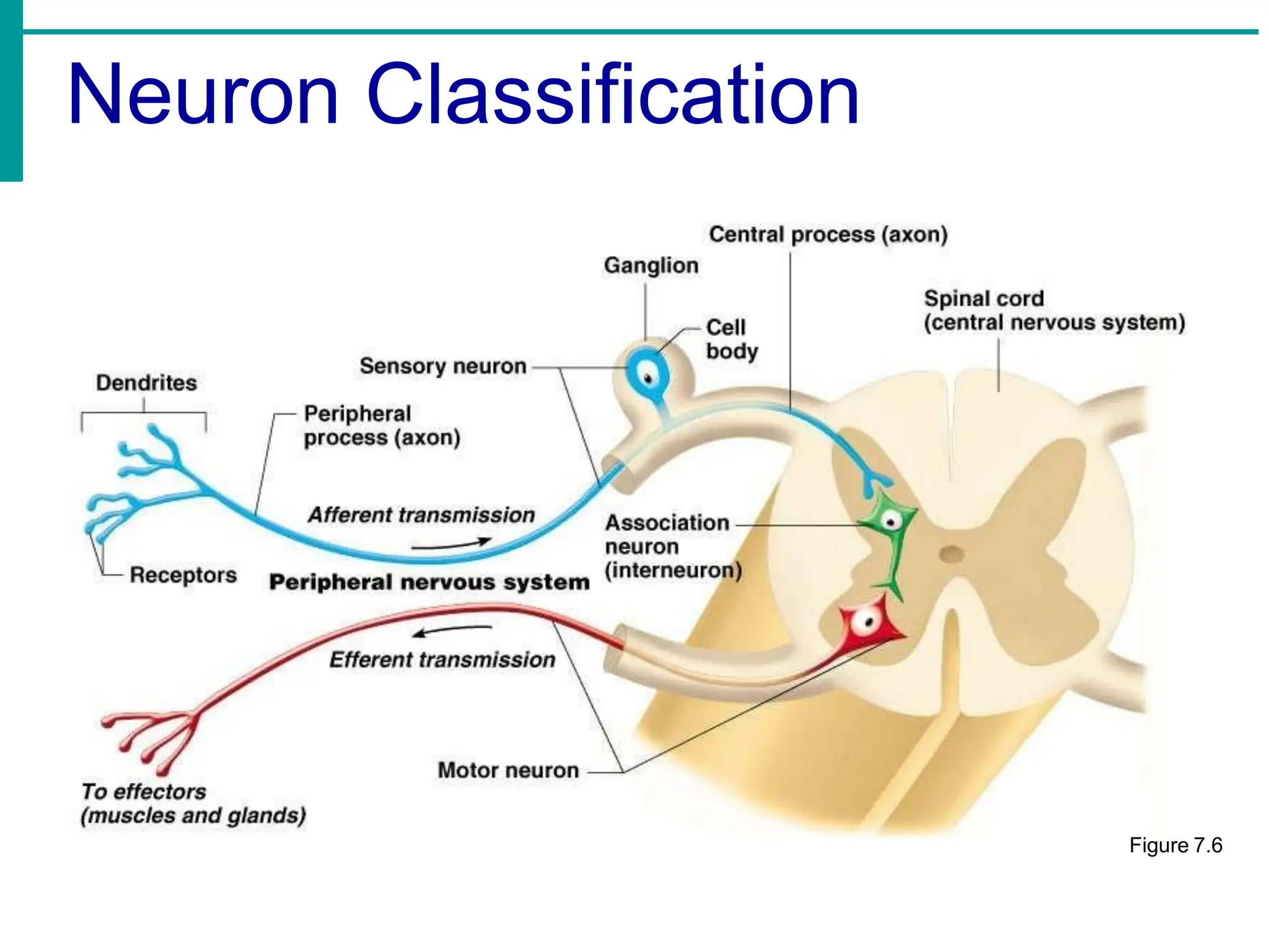
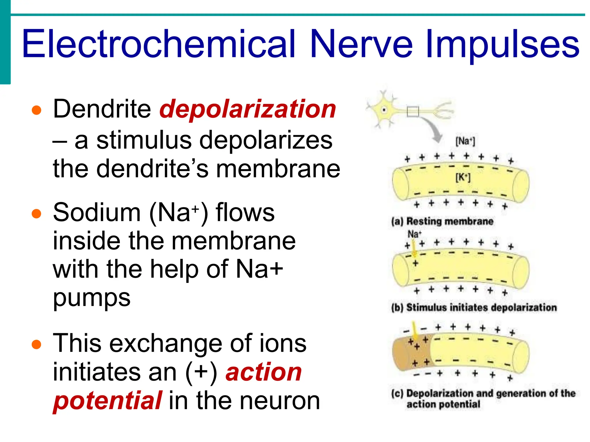
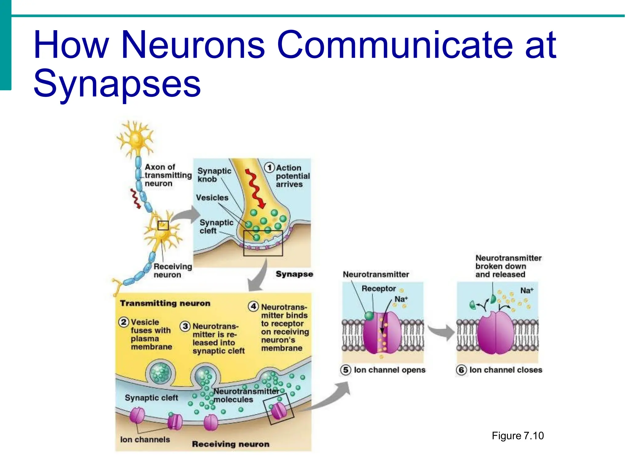
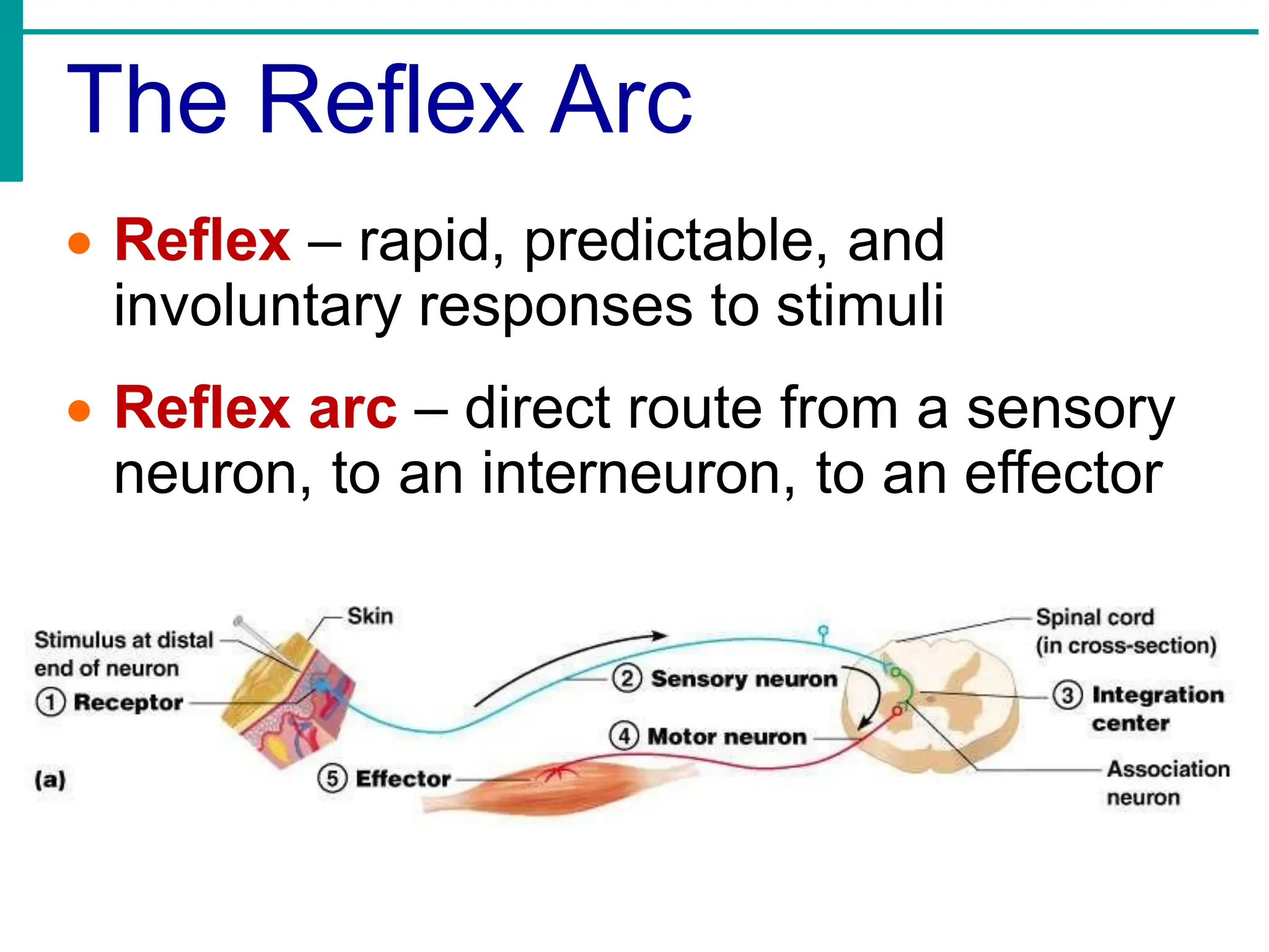
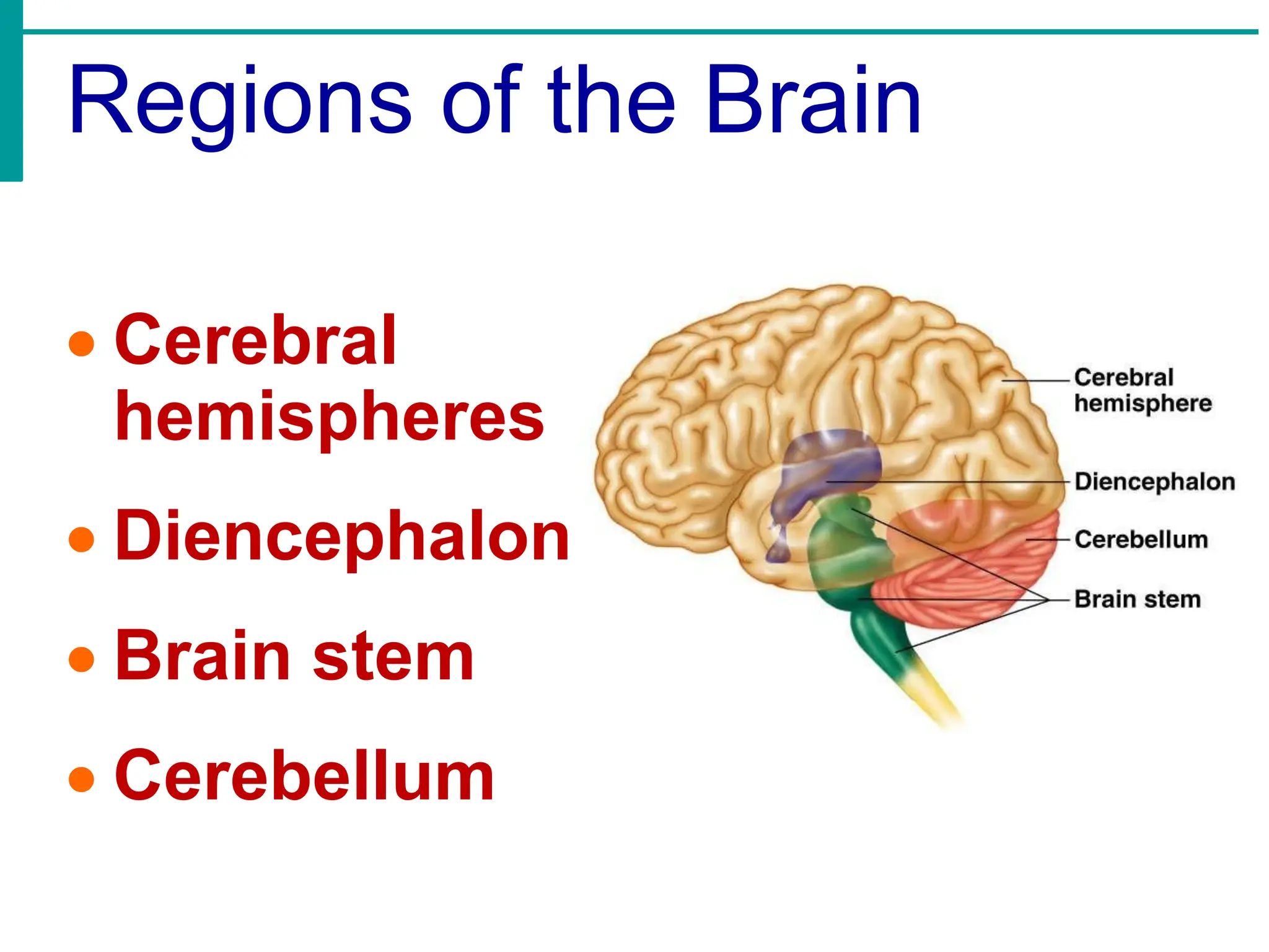
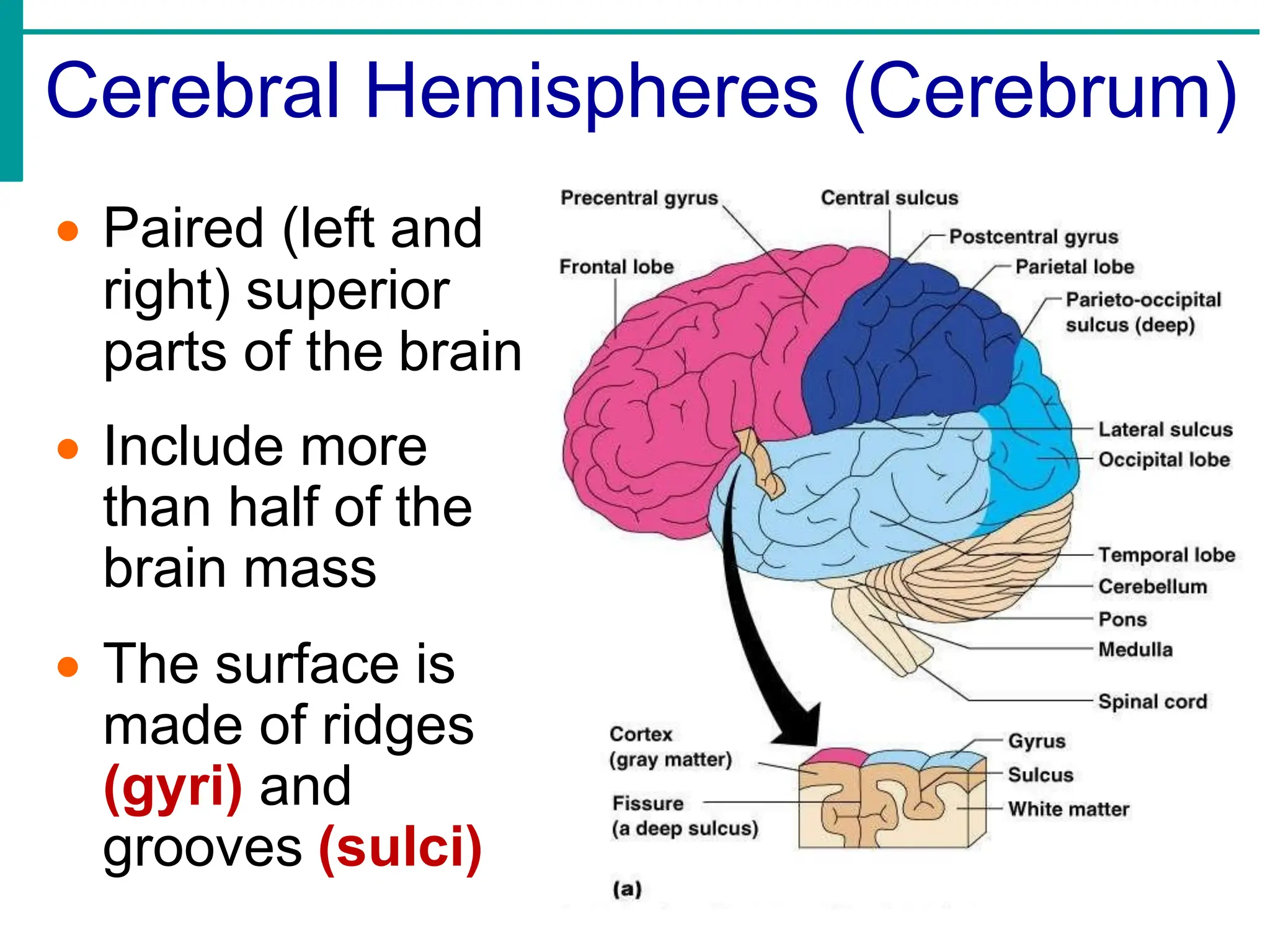
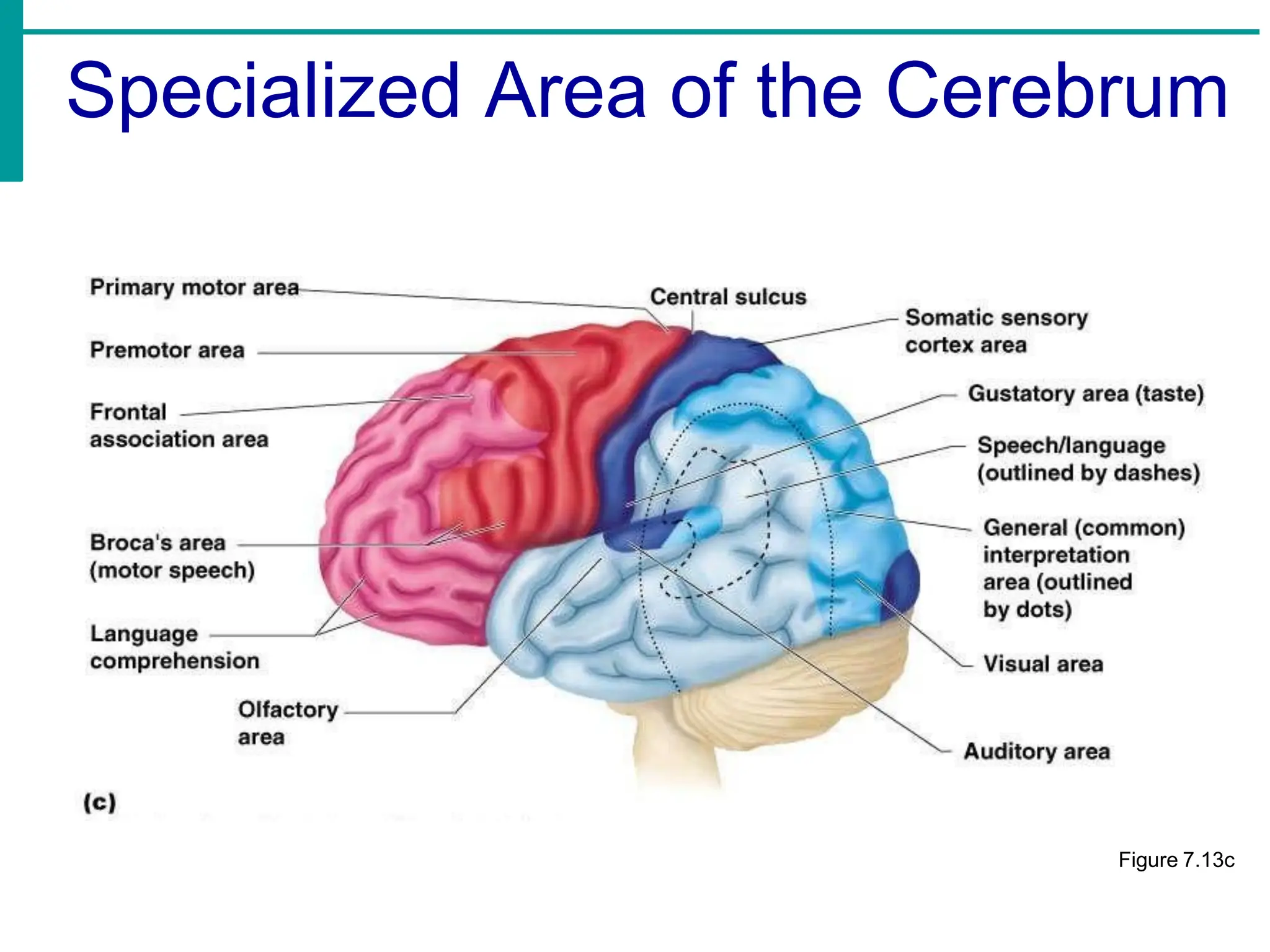
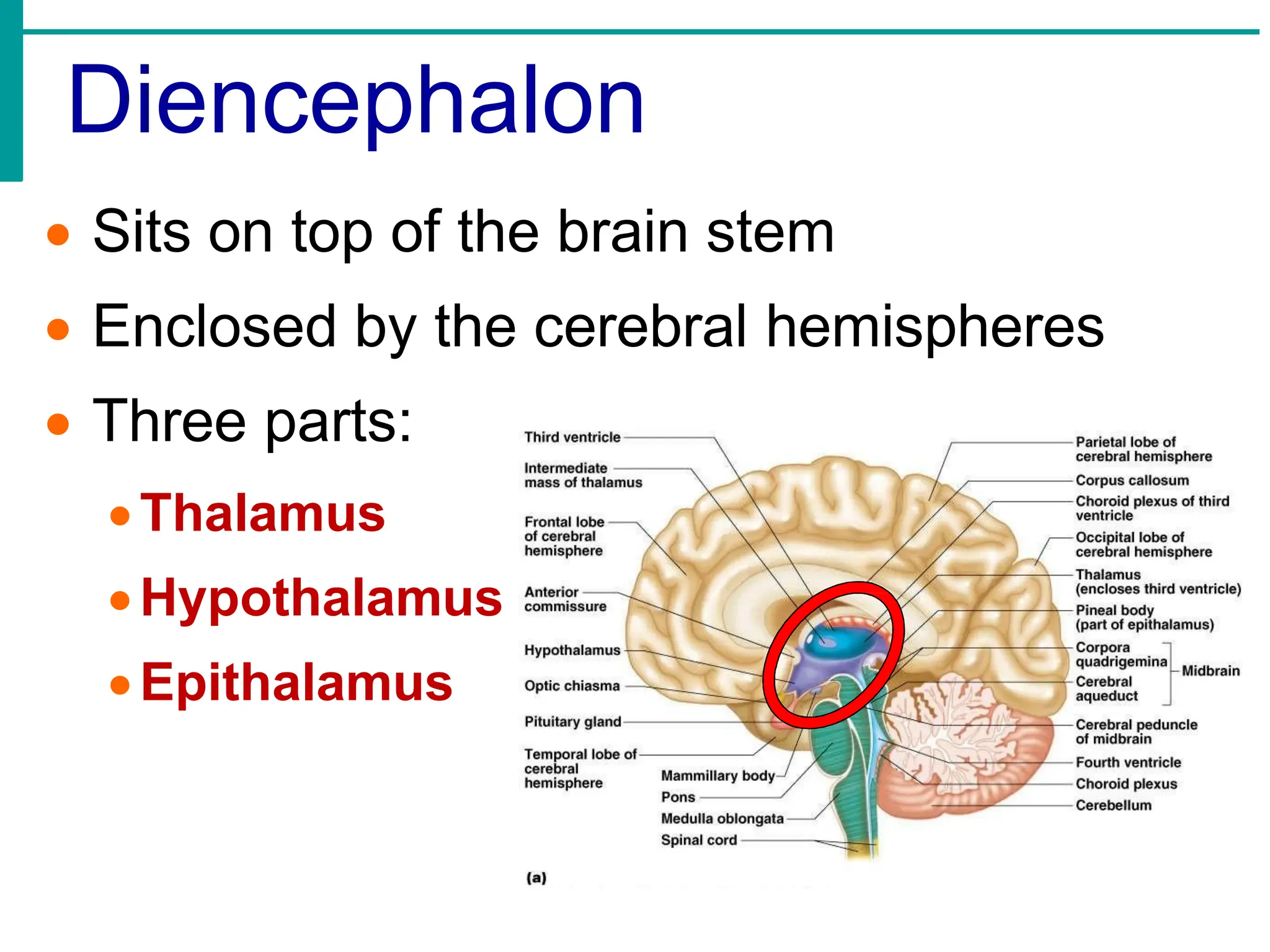
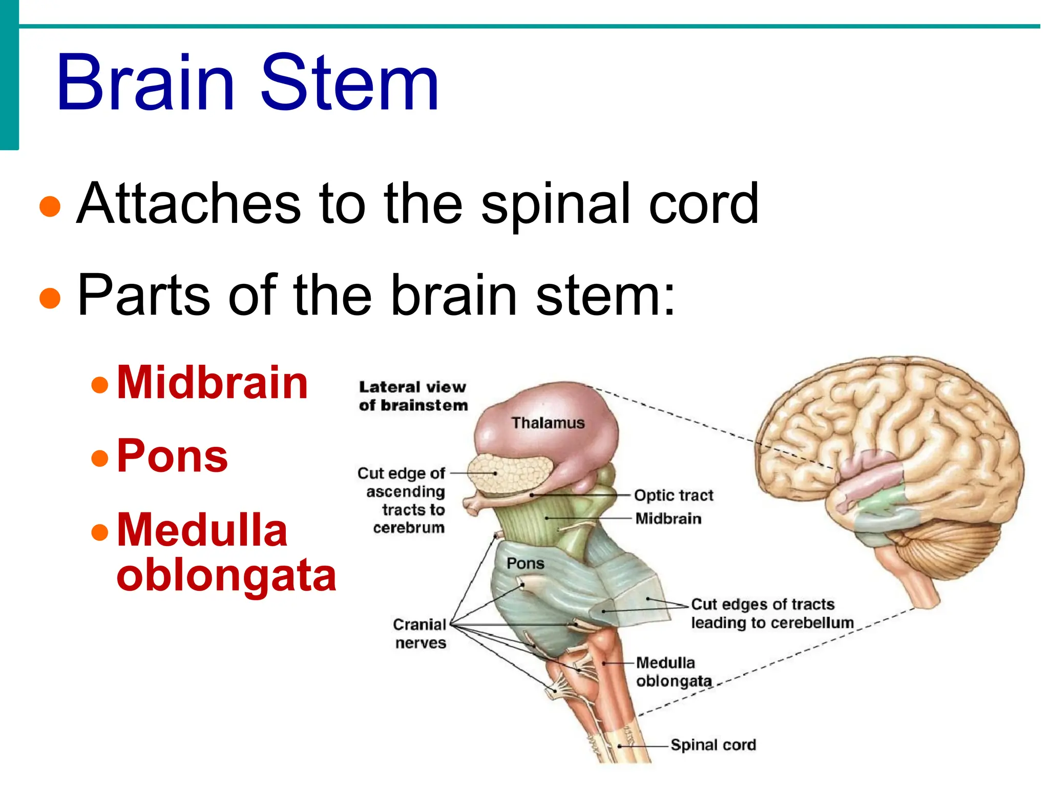
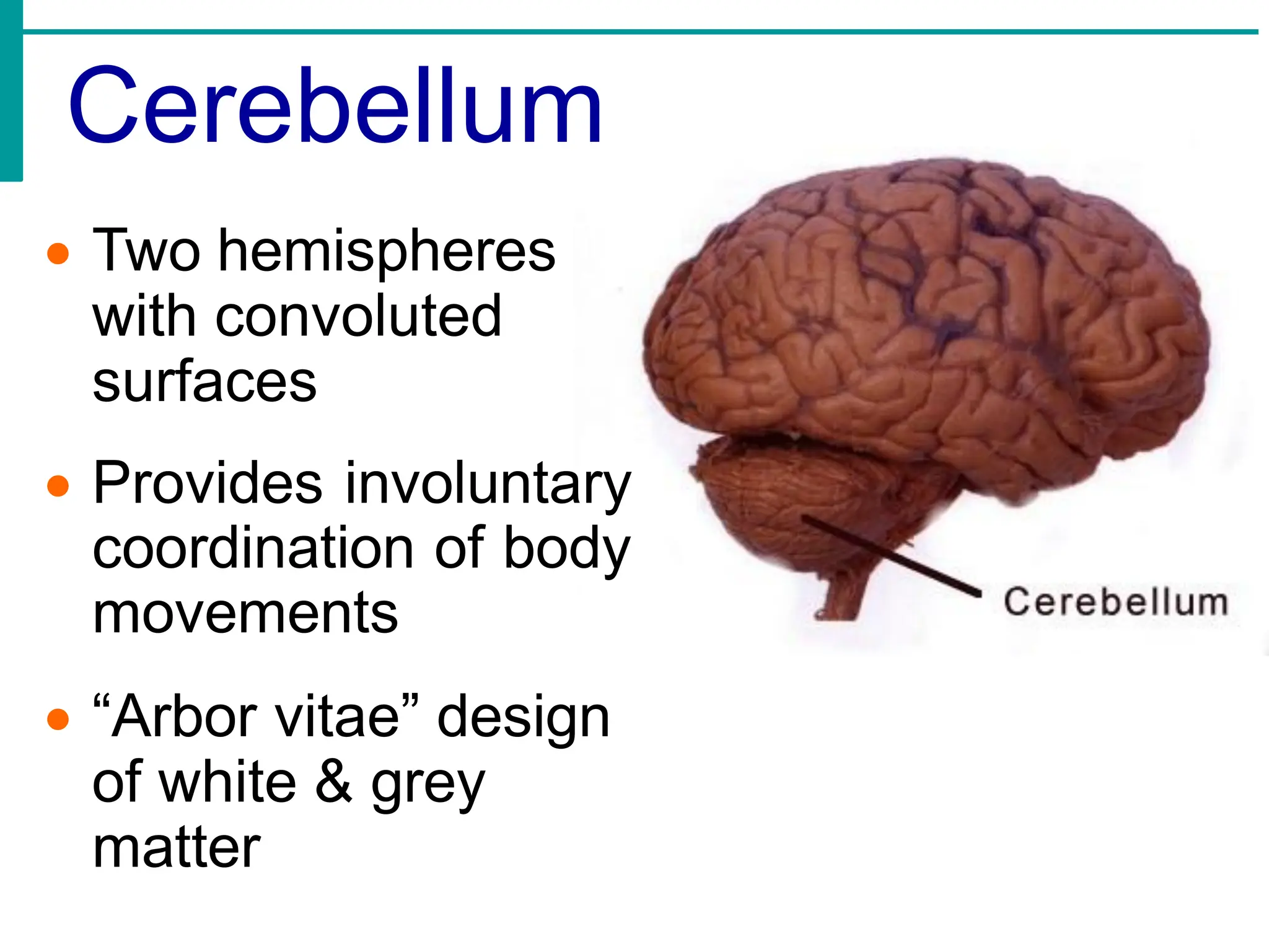
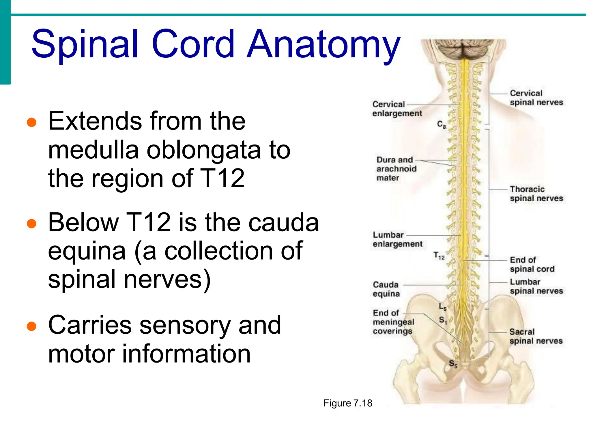
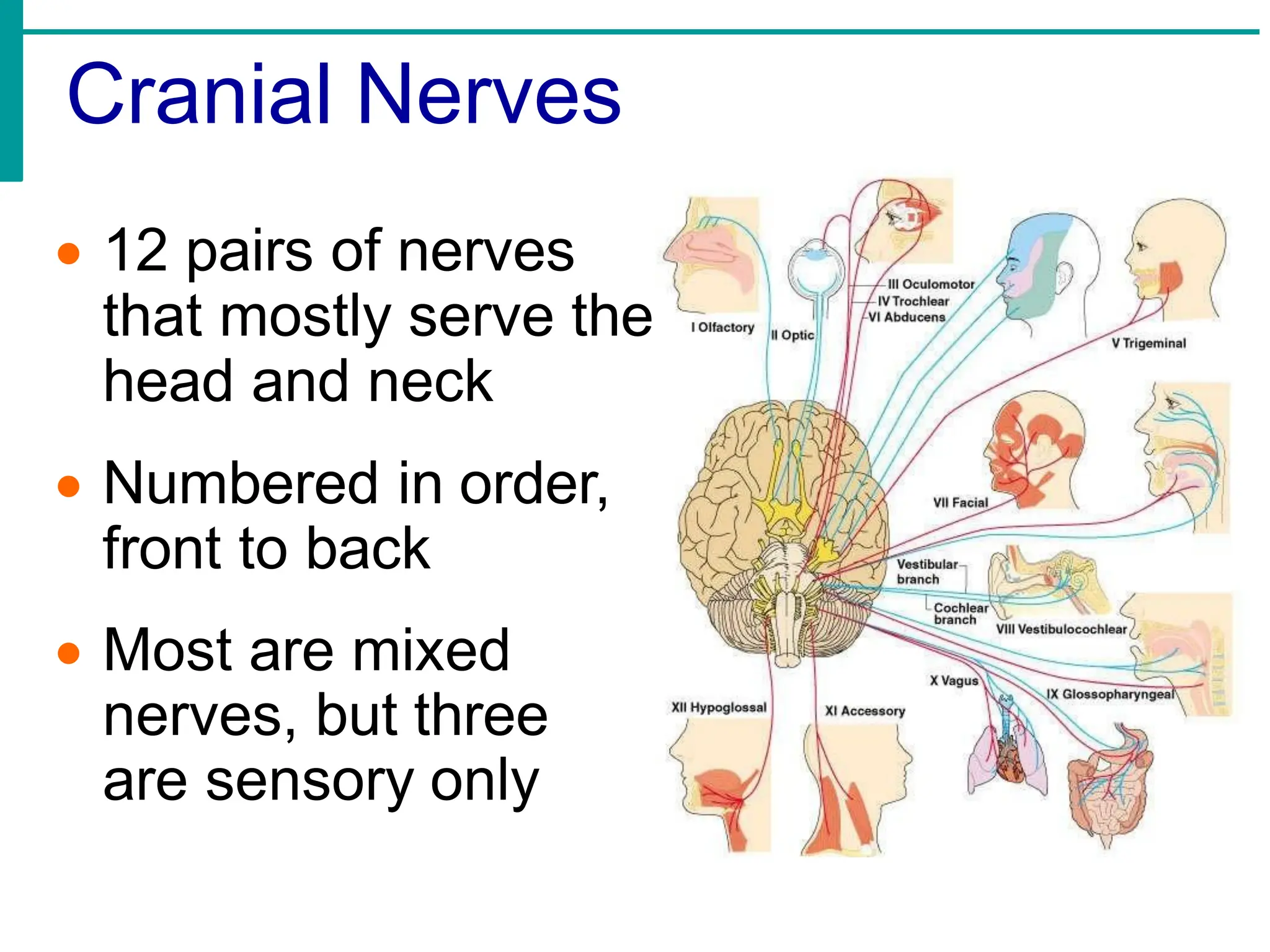
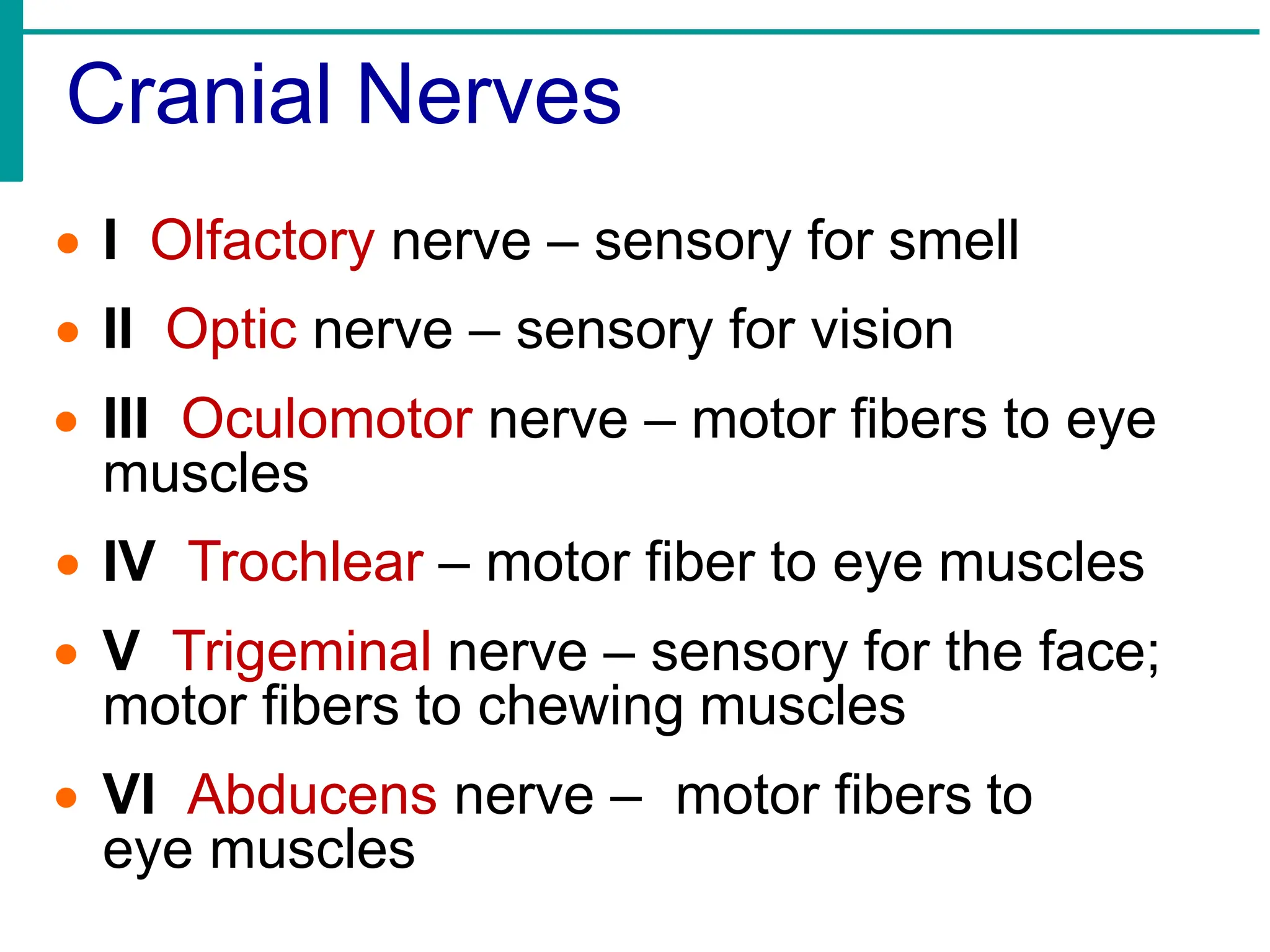
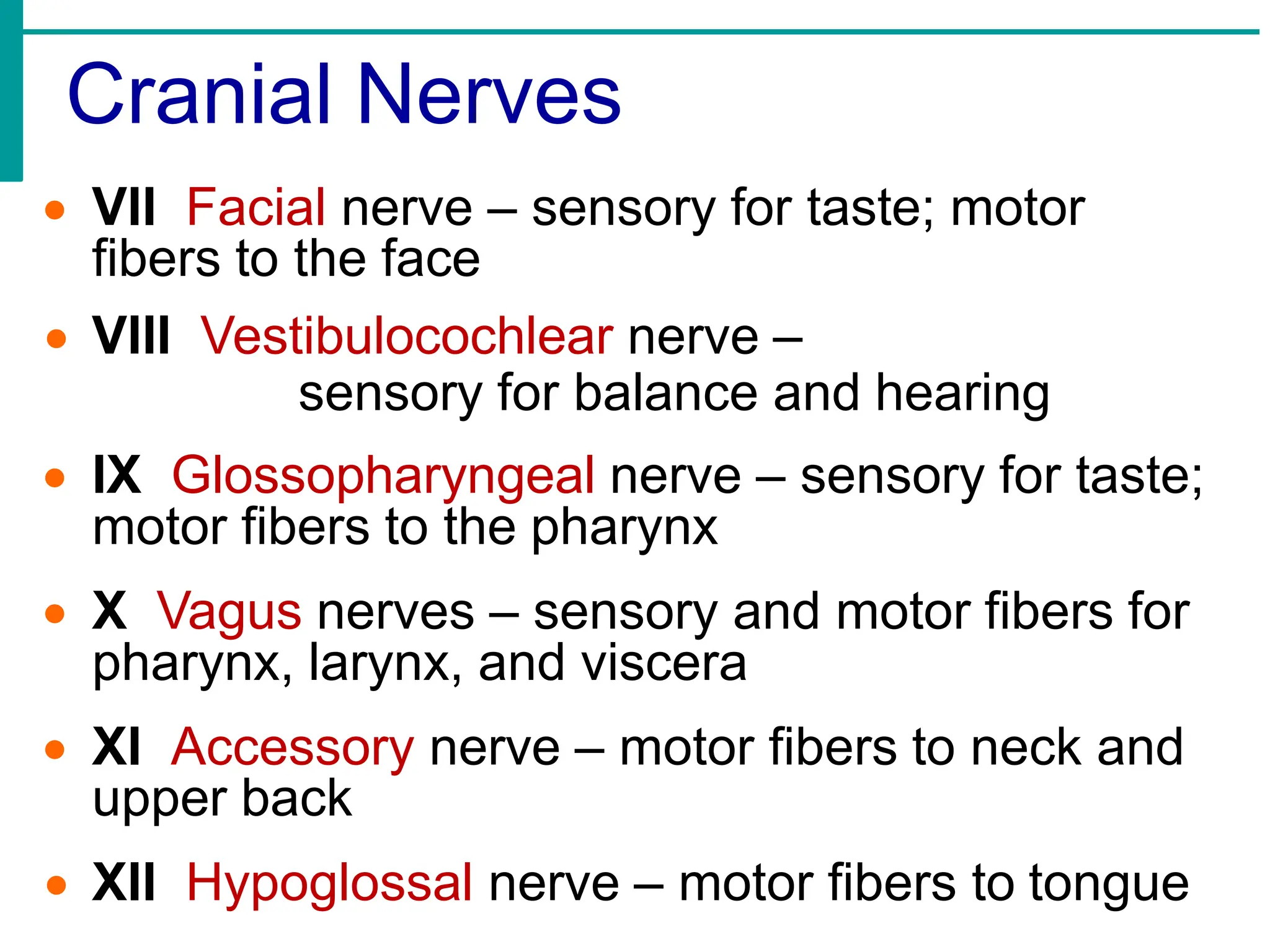
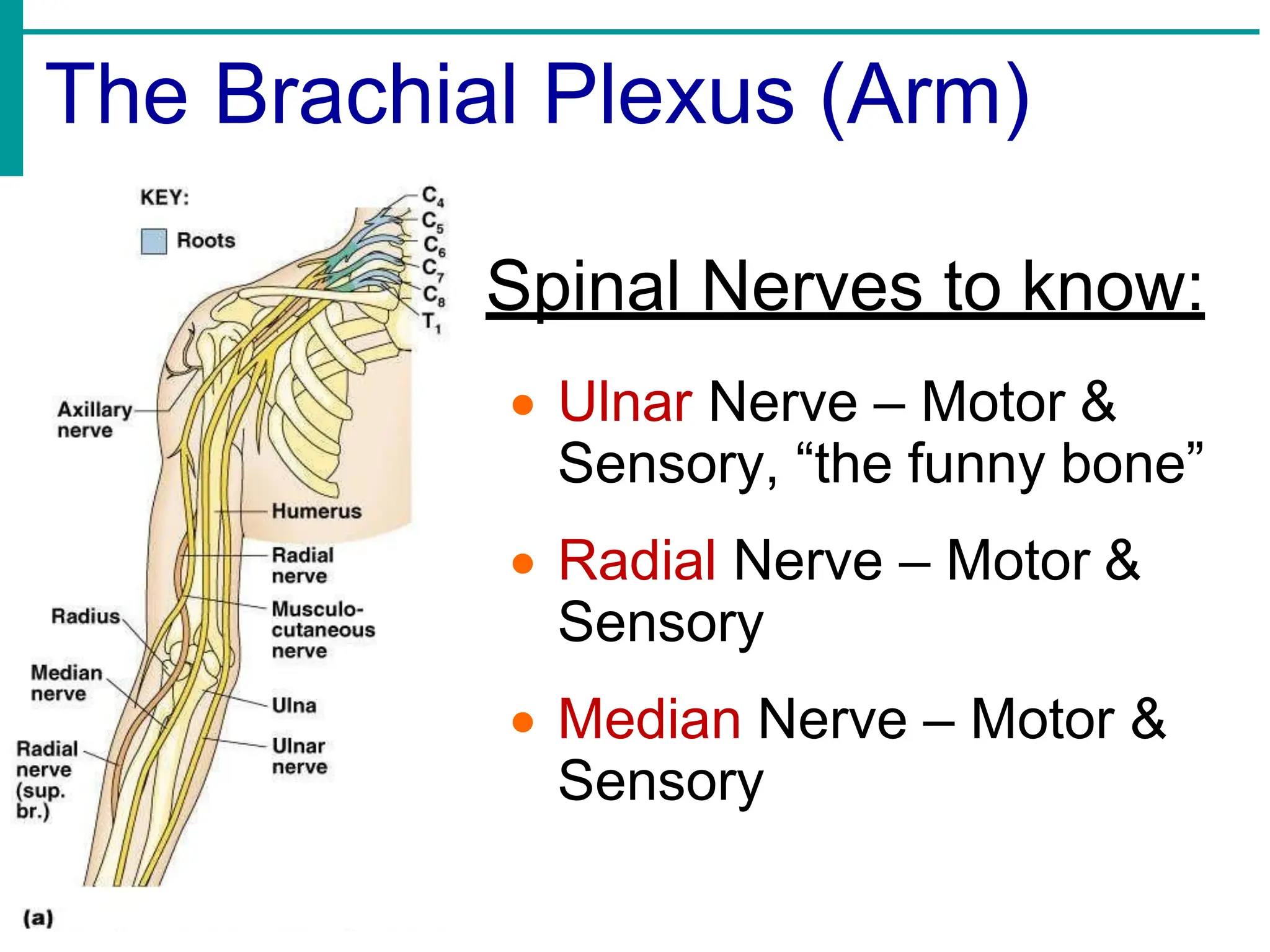
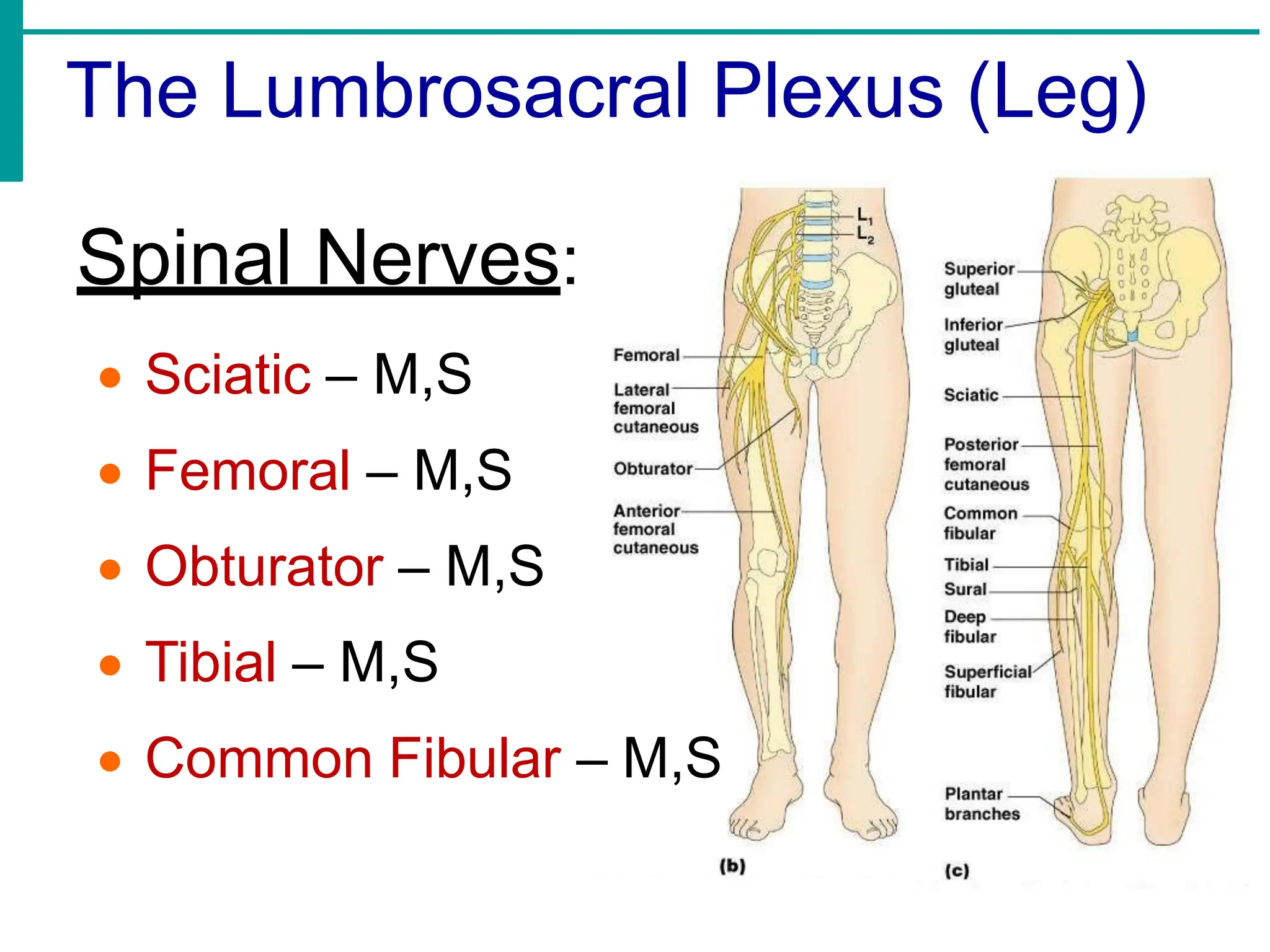
The Nervous System
Anatomy & Physiology
Department of Allied
health Sciences
Module 9
The Nervous system
Neurons: Structure, types and properties
Central nervous system: neurons, neuralgia meninges.
Ventricles of brain, CSF
Brain, spinal cord: structures, functions, peripheral nervous system.
Spinal and cranial nerves: origin distribution and functions.
Automatic nervous system
Sympathetic and para sympathetic: origin distribution and function.
Common diseases of the system.
BCCTC102
Functions of the Nervous System
• Sensory input – gathering information
• To monitor changes occurring inside
and outside the body
• Integration - To process and interpret
sensory input and decide if action is
needed
• Motor output
•A response to stimuli
•Activates muscles or glands
Classification of the Nervous System
• Central nervous
system (CNS):
•Brain
•Spinal cord
• Peripheral nervous
system (PNS):
•Nerves outside the
brain and spinal cord
The Peripheral Nervous System
• Sensory (afferent) division - Nerve
fibers that carry information to the central
nervous system
• Motor (efferent) division - Nerve fibers
that carry impulses away from the central
nervous system.
• Somatic system:
voluntary
• Autonomic system:
involuntary
Autonomic Nervous System
• The involuntary branch of the nervous
system
• Consists of only motor
nerves
• Divided into two divisions
•Sympathetic division –
“fight or flight” response
•Parasympathetic division –
“housekeeping”
Nervous System Histology
Neurons = nerve cells
•Cells specialized to transmit
electrochemical messages
•Major regions of neurons
•Cell body – nucleus and
metabolic center of the
cell
•Processes – fibers that
extend from the cell body
Neuron Anatomy
• Dendrites – conduct impulses
toward the cell body
• Cell body (soma): contains
organelles & Nissl substance
(specialized rough ER)
• Axons – conduct impulses
away from the cell body
• Schwann cells – produce myelin
sheaths in jelly-roll like
fashion
• Nodes of Ranvier – gaps in
myelin sheath along the axon
Classification of Neurons
• Sensory (afferent) neurons
•Carry impulses from the sensory receptors
•Cutaneous sense organs
•Receptors – detect stretch or tension
• Interneurons (association): “connector”
• Motor (efferent) neurons
•Carry impulses from the central nervous
system
Neuron Classification
Figure 7.6
Electrochemical Nerve Impulses
• Dendrite depolarization
– a stimulus depolarizes
the dendrite’s membrane
• Sodium (Na+) flows
inside the membrane
with the help of Na+
pumps
• This exchange of ions
initiates an (+) action
potential in the neuron
The Action Potential
• If the action potential (nerve impulse)
starts, it is propagated to the end of the
axon
• Potassium (K+) ions rush out of the
neuron after sodium ions rush in, which
repolarizes the membrane
• The sodium-potassium pump restores
the original configuration - which is
resting potential (-)
*** This action requires ATP ***
Continuation of the Nerve
Impulse between Neurons
• Impulses are able to cross the synapse
to another neuron
•Neurotransmitter is released from a
neuron’s axon terminal
•The dendrite of the next neuron has
receptors that are stimulated by the
neurotransmitter
•An action potential is started in the dendrite
How Neurons Communicate at
Synapses
Figure 7.10
The Reflex Arc
• Reflex – rapid, predictable, and
involuntary responses to stimuli
• Reflex arc – direct route from a sensory
neuron, to an interneuron, to an effector
Regions of the Brain
• Cerebral
hemispheres
• Diencephalon
• Brain stem
• Cerebellum
Cerebral Hemispheres (Cerebrum)
• Paired (left and
right) superior
parts of the brain
• Include more
than half of the
brain mass
• The surface is
made of ridges
(gyri) and
grooves (sulci)
Specialized Area of the Cerebrum
Figure 7.13c
Specialized Areas of the Cerebrum
Figure 7.13c
Diencephalon
• Sits on top of the brain stem
• Enclosed by the cerebral hemispheres
• Three parts:
•Thalamus
•Hypothalamus
•Epithalamus
Brain Stem
• Attaches to the spinal cord
• Parts of the brain stem:
•Midbrain
•Pons
•Medulla
oblongata
Cerebellum
• Two hemispheres
with convoluted
surfaces
• Provides involuntary
coordination of body
movements
• “Arbor vitae” design
of white & grey
matter
• Extends from the
medulla oblongata to
the region of T12
• Below T12 is the cauda
equina (a collection of
spinal nerves)
• Carries sensory and
motor information
Figure 7.18
Spinal Cord Anatomy
Cranial Nerves
• 12 pairs of nerves
that mostly serve the
head and neck
• Numbered in order,
front to back
• Most are mixed
nerves, but three
are sensory only
Cranial Nerves
• I Olfactory nerve – sensory for smell
• II Optic nerve – sensory for vision
• III Oculomotor nerve – motor fibers to eye
muscles
• IV Trochlear – motor fiber to eye muscles
• V Trigeminal nerve – sensory for the face;
motor fibers to chewing muscles
• VI Abducens nerve – motor fibers to
eye muscles
Cranial Nerves
• VII Facial nerve – sensory for taste; motor
fibers to the face
• VIII Vestibulocochlear nerve –
sensory for balance and hearing
• IX Glossopharyngeal nerve – sensory for taste;
motor fibers to the pharynx
• X Vagus nerves – sensory and motor fibers for
pharynx, larynx, and viscera
• XI Accessory nerve – motor fibers to neck and
upper back
• XII Hypoglossal nerve – motor fibers to tongue
Spinal Nerves to know:
• Ulnar Nerve – Motor &
Sensory, “the funny bone”
• Radial Nerve – Motor &
Sensory
• Median Nerve – Motor &
Sensory
The Brachial Plexus (Arm)
The Lumbrosacral Plexus (Leg)
Spinal Nerves:
• Sciatic – M,S
• Femoral – M,S
• Obturator – M,S
• Tibial – M,S
• Common Fibular – M,S
“You just
nailed me in
the head!”
- Travis
Bogumill (21)
Eau Claire, WI
1998

BCCTC102 Module 9 -The nervous system_PDF.pdf

  • 1.
    The Nervous System Anatomy& Physiology Department of Allied health Sciences Module 9 The Nervous system Neurons: Structure, types and properties Central nervous system: neurons, neuralgia meninges. Ventricles of brain, CSF Brain, spinal cord: structures, functions, peripheral nervous system. Spinal and cranial nerves: origin distribution and functions. Automatic nervous system Sympathetic and para sympathetic: origin distribution and function. Common diseases of the system. BCCTC102
  • 2.
    Functions of theNervous System • Sensory input – gathering information • To monitor changes occurring inside and outside the body • Integration - To process and interpret sensory input and decide if action is needed • Motor output •A response to stimuli •Activates muscles or glands
  • 3.
    Classification of theNervous System • Central nervous system (CNS): •Brain •Spinal cord • Peripheral nervous system (PNS): •Nerves outside the brain and spinal cord
  • 4.
    The Peripheral NervousSystem • Sensory (afferent) division - Nerve fibers that carry information to the central nervous system • Motor (efferent) division - Nerve fibers that carry impulses away from the central nervous system. • Somatic system: voluntary • Autonomic system: involuntary
  • 5.
    Autonomic Nervous System •The involuntary branch of the nervous system • Consists of only motor nerves • Divided into two divisions •Sympathetic division – “fight or flight” response •Parasympathetic division – “housekeeping”
  • 6.
    Nervous System Histology Neurons= nerve cells •Cells specialized to transmit electrochemical messages •Major regions of neurons •Cell body – nucleus and metabolic center of the cell •Processes – fibers that extend from the cell body
  • 7.
    Neuron Anatomy • Dendrites– conduct impulses toward the cell body • Cell body (soma): contains organelles & Nissl substance (specialized rough ER) • Axons – conduct impulses away from the cell body • Schwann cells – produce myelin sheaths in jelly-roll like fashion • Nodes of Ranvier – gaps in myelin sheath along the axon
  • 8.
    Classification of Neurons •Sensory (afferent) neurons •Carry impulses from the sensory receptors •Cutaneous sense organs •Receptors – detect stretch or tension • Interneurons (association): “connector” • Motor (efferent) neurons •Carry impulses from the central nervous system
  • 9.
  • 10.
    Electrochemical Nerve Impulses •Dendrite depolarization – a stimulus depolarizes the dendrite’s membrane • Sodium (Na+) flows inside the membrane with the help of Na+ pumps • This exchange of ions initiates an (+) action potential in the neuron
  • 11.
    The Action Potential •If the action potential (nerve impulse) starts, it is propagated to the end of the axon • Potassium (K+) ions rush out of the neuron after sodium ions rush in, which repolarizes the membrane • The sodium-potassium pump restores the original configuration - which is resting potential (-) *** This action requires ATP ***
  • 12.
    Continuation of theNerve Impulse between Neurons • Impulses are able to cross the synapse to another neuron •Neurotransmitter is released from a neuron’s axon terminal •The dendrite of the next neuron has receptors that are stimulated by the neurotransmitter •An action potential is started in the dendrite
  • 13.
    How Neurons Communicateat Synapses Figure 7.10
  • 14.
    The Reflex Arc •Reflex – rapid, predictable, and involuntary responses to stimuli • Reflex arc – direct route from a sensory neuron, to an interneuron, to an effector
  • 15.
    Regions of theBrain • Cerebral hemispheres • Diencephalon • Brain stem • Cerebellum
  • 16.
    Cerebral Hemispheres (Cerebrum) •Paired (left and right) superior parts of the brain • Include more than half of the brain mass • The surface is made of ridges (gyri) and grooves (sulci)
  • 17.
    Specialized Area ofthe Cerebrum Figure 7.13c
  • 18.
    Specialized Areas ofthe Cerebrum Figure 7.13c
  • 19.
    Diencephalon • Sits ontop of the brain stem • Enclosed by the cerebral hemispheres • Three parts: •Thalamus •Hypothalamus •Epithalamus
  • 20.
    Brain Stem • Attachesto the spinal cord • Parts of the brain stem: •Midbrain •Pons •Medulla oblongata
  • 21.
    Cerebellum • Two hemispheres withconvoluted surfaces • Provides involuntary coordination of body movements • “Arbor vitae” design of white & grey matter
  • 22.
    • Extends fromthe medulla oblongata to the region of T12 • Below T12 is the cauda equina (a collection of spinal nerves) • Carries sensory and motor information Figure 7.18 Spinal Cord Anatomy
  • 23.
    Cranial Nerves • 12pairs of nerves that mostly serve the head and neck • Numbered in order, front to back • Most are mixed nerves, but three are sensory only
  • 24.
    Cranial Nerves • IOlfactory nerve – sensory for smell • II Optic nerve – sensory for vision • III Oculomotor nerve – motor fibers to eye muscles • IV Trochlear – motor fiber to eye muscles • V Trigeminal nerve – sensory for the face; motor fibers to chewing muscles • VI Abducens nerve – motor fibers to eye muscles
  • 25.
    Cranial Nerves • VIIFacial nerve – sensory for taste; motor fibers to the face • VIII Vestibulocochlear nerve – sensory for balance and hearing • IX Glossopharyngeal nerve – sensory for taste; motor fibers to the pharynx • X Vagus nerves – sensory and motor fibers for pharynx, larynx, and viscera • XI Accessory nerve – motor fibers to neck and upper back • XII Hypoglossal nerve – motor fibers to tongue
  • 26.
    Spinal Nerves toknow: • Ulnar Nerve – Motor & Sensory, “the funny bone” • Radial Nerve – Motor & Sensory • Median Nerve – Motor & Sensory The Brachial Plexus (Arm)
  • 27.
    The Lumbrosacral Plexus(Leg) Spinal Nerves: • Sciatic – M,S • Femoral – M,S • Obturator – M,S • Tibial – M,S • Common Fibular – M,S
  • 28.
    “You just nailed mein the head!” - Travis Bogumill (21) Eau Claire, WI 1998