Downloaded 854 times

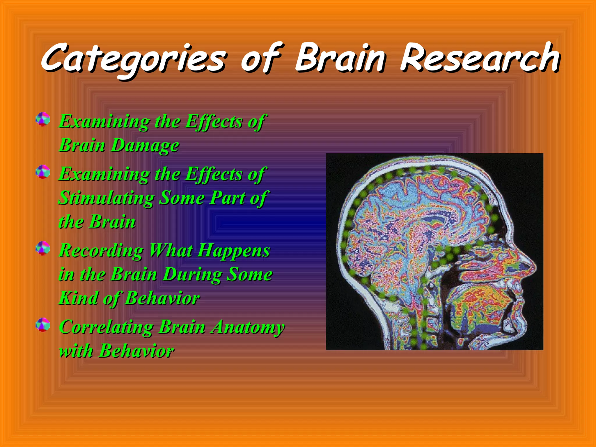
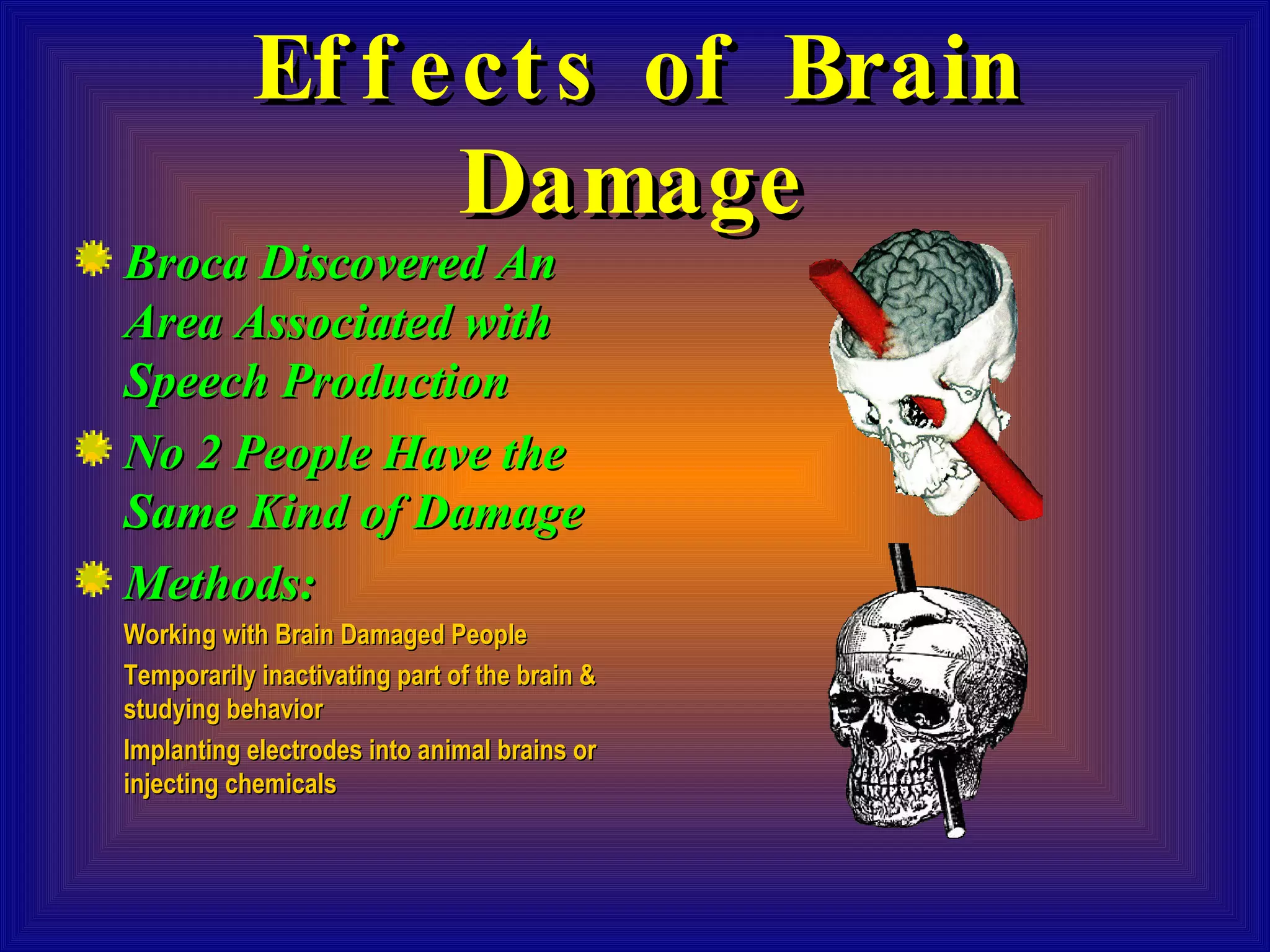
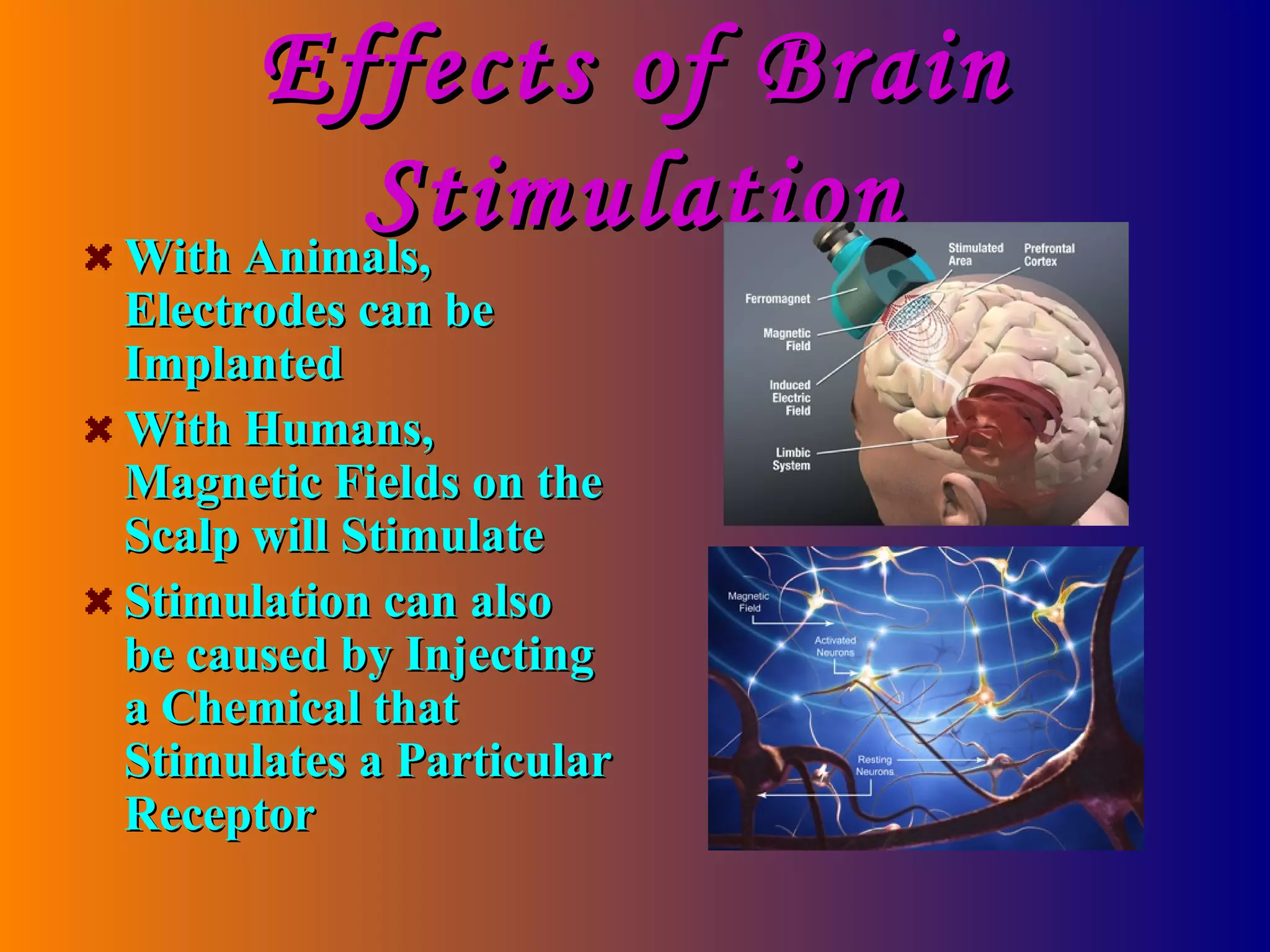
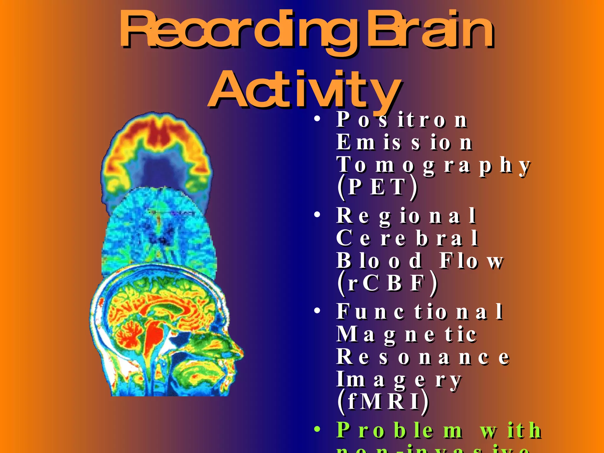
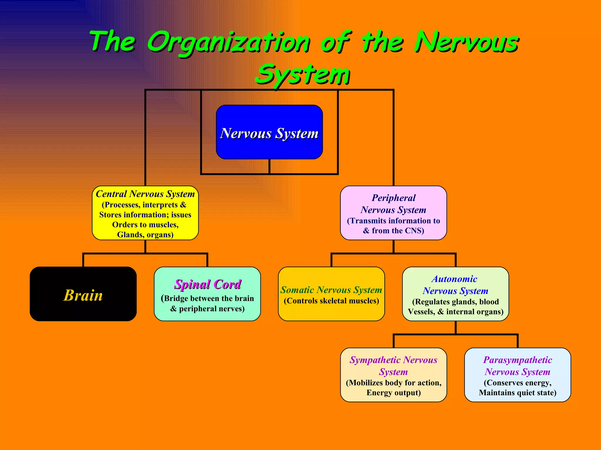

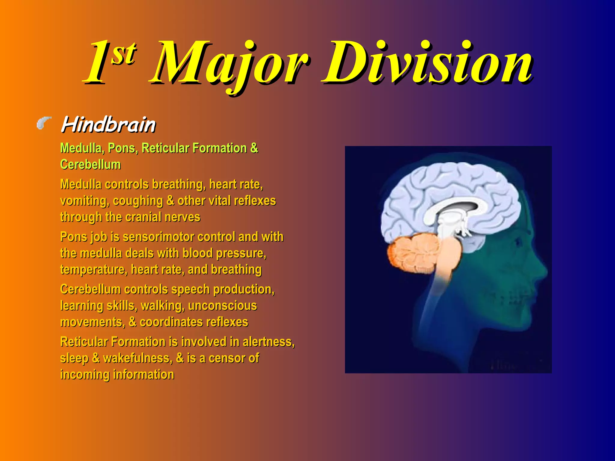
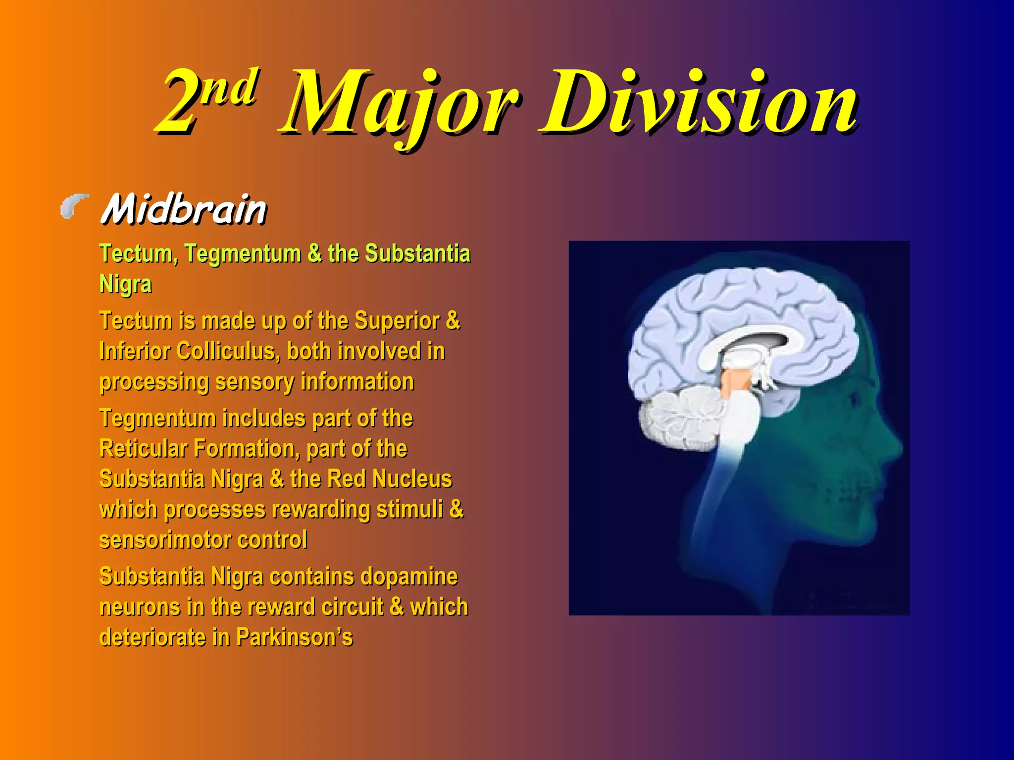
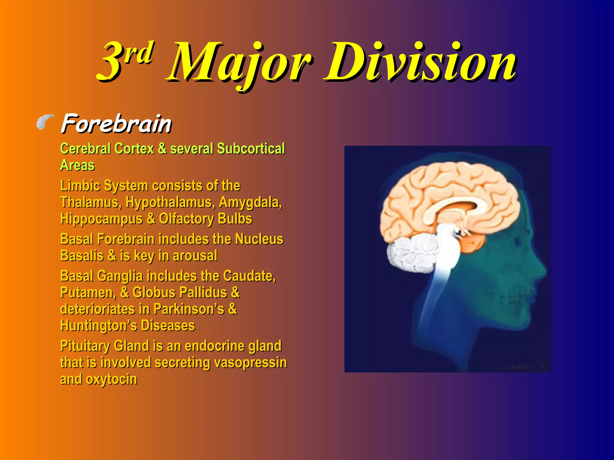

The document summarizes different categories of brain research and provides details about the anatomy and organization of the nervous system. It discusses how brain research involves examining the effects of brain damage and stimulation, as well as recording brain activity during behaviors. It then describes the central and peripheral nervous systems before explaining the detailed anatomy of different brain regions and lobes.


![Chapter 2 Psych 1 Online Stud 1199299912883466 2[1]](https://cdn.slidesharecdn.com/ss_thumbnails/chapter-2-psych-1-online-stud-1199299912883466-21-090621223402-phpapp02-thumbnail.jpg?width=640&height=640&fit=bounds)


