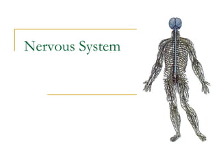
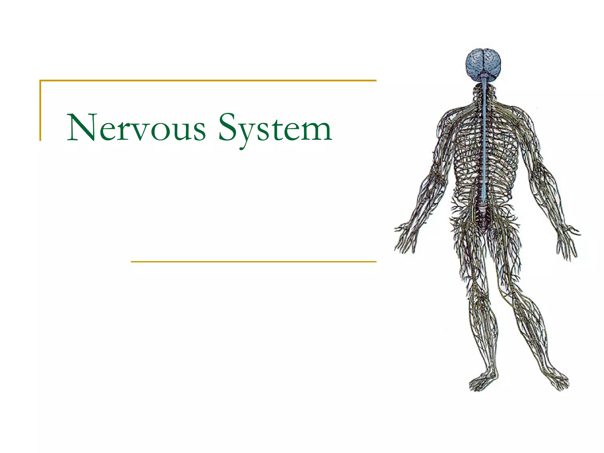
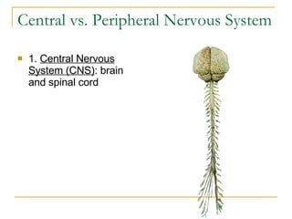
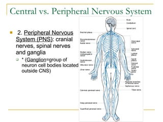
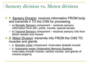
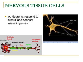
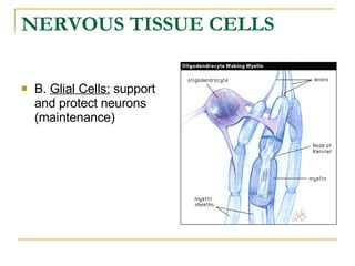
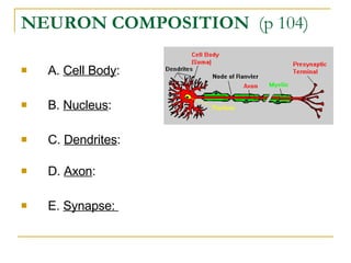
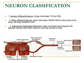
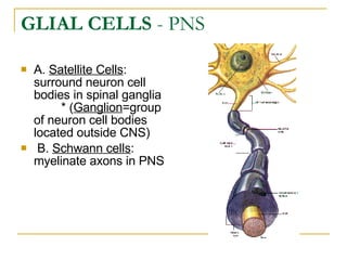
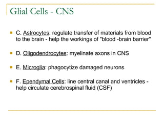
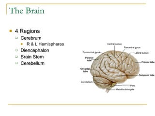
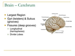
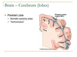
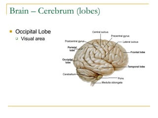
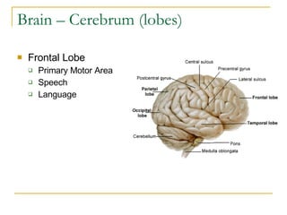
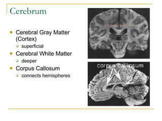
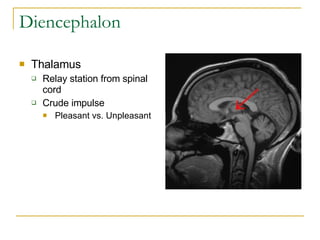
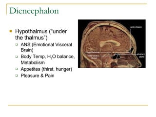
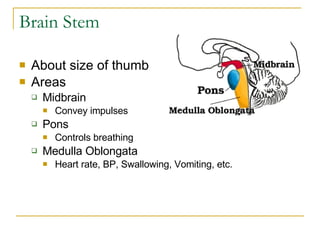
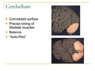
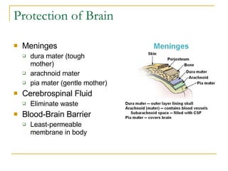
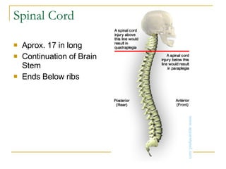
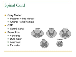
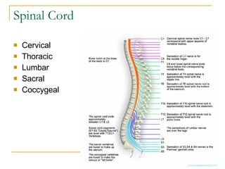
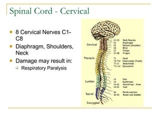
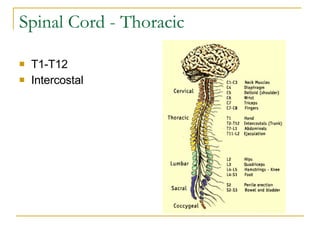
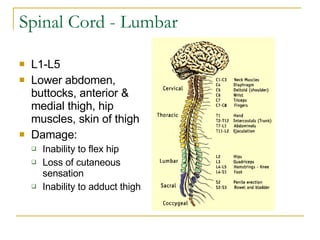
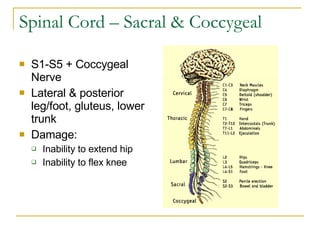
Nervous System
Central vs. Peripheral Nervous System 1.  Central Nervous System (CNS) : brain and spinal cord   
Central vs. Peripheral Nervous System   2.  Peripheral Nervous System (PNS) : cranial nerves, spinal nerves and ganglia * ( Ganglion =group of neuron cell bodies located outside CNS)
Sensory division vs. Motor division  1.  Sensory Division : receives information FROM body and transmits it TO the CNS for processing a)  Somatic Sensory  component – receives sensory information from skin, joints, muscle, special senses  b)  Visceral Sensory  component – receives sensory info from blood vessels and viscera 2.  Motor Division : transmits info FROM the CNS TO muscles and glands  a.  Somatic motor  component: innervates skeletal muscle  b.  Autonomic motor (Autonomic Nervous System) : innervates smooth muscle, cardiac muscle, and glands of viscera (organs)
NERVOUS TISSUE CELLS A.  Neurons : respond to stimuli and conduct nerve impulses
NERVOUS TISSUE CELLS   B.  Glial Cells:  support and protect neurons (maintenance)
NEURON COMPOSITION   (p 104) A.  Cell Body :       B.  Nucleus :       C.  Dendrites :                D.  Axon :  E.  Synapse:        
NEURON CLASSIFICATION   1.  Sensory (Afferent) Neuron : brings information TO the CNS        2.  Motor (Efferent) Neuron : takes information FROM CNS to other parts of the body, cell body located in CNS        3.  Interneuron (Association Neurons) : helps coordinate and integrate info between sensory and motor neurons, cell body located in CNS
GLIAL CELLS  - PNS A.  Satellite Cells : surround neuron cell bodies in spinal ganglia          * ( Ganglion =group of neuron cell bodies located outside CNS)    B.  Schwann cells : myelinate axons in PNS   
Glial Cells - CNS C.  Astrocytes : regulate transfer of materials from blood to the brain - help the workings of "blood -brain barrier"     D.  Oligodendrocytes : myelinate axons in CNS     E.  Microglia : phagocytize damaged neurons     F.  Ependymal Cells : line central canal and ventricles - help circulate cerebrospinal fluid (CSF)   
MYELINATION OF AXONS   wrapping an axon with myelin
The Brain 4 Regions Cerebrum R & L Hemispheres Diencephalon Brain Stem Cerebellum
Brain – Cerebrum Largest Region Gyri (twisters) & Sulcus (grooves) Fissures (deep grooves) Longitudinal (hemispheres) Divide Lobes
Brain – Cerebrum (lobes) Parietal Lobe Somatic sensory area “ homunculus”
Brain – Cerebrum (lobes) Occipital Lobe Visual area
Brain – Cerebrum (lobes) Temporal Lobe Olfactory Deep inside
Brain – Cerebrum (lobes) Frontal Lobe Primary Motor Area Speech  Language
Cerebrum Cerebral Gray Matter (Cortex) superficial  Cerebral White Matter deeper  Corpus Callosum connects hemispheres
Diencephalon (interbrain) Sits atop brain stem Enclosed by cerebrum Structures  Thalamus Hypothalamus Epithalamus
Diencephalon Thalamus Relay station from spinal cord Crude impulse Pleasant vs. Unpleasant
Diencephalon Hypothalmus (“under the thalmus”) ANS (Emotional Visceral Brain) Body Temp, H 2 O balance, Metabolism Appetites (thirst, hunger) Pleasure & Pain
Diencephalon Epithalamus Pineal  body (endocrine system) Choroid plexus (cerebrospinal fluid)
Brain Stem About size of thumb Areas Midbrain Convey impulses Pons Controls breathing Medulla Oblongata Heart rate, BP, Swallowing, Vomiting, etc.
Cerebellum Convoluted surface Precise timing of Skeletal muscles Balance “ Auto-Pilot”
Protection of Brain Meninges dura mater (tough mother) arachnoid mater pia mater (gentle mother) Cerebrospinal Fluid Eliminate waste Blood-Brain Barrier Least-permeable membrane in body
Spinal Cord Aprox. 17 in long Continuation of Brain Stem Ends Below ribs
Spinal Cord Gray Matter Posterior Horns (dorsal) Anterior Horns (ventral) CSF Central Canal Protection Vertebrae Dura mater Arachnoid Pia mater
Spinal Cord Cervical Thoracic Lumbar Sacral Coccygeal
Spinal Cord - Cervical 8 Cervical Nerves C1-C8 Diaphragm, Shoulders, Neck Damage may result in: Respiratory Paralysis
Spinal Cord - Thoracic T1-T12 Intercostal
Spinal Cord - Lumbar L1-L5 Lower abdomen, buttocks, anterior & medial thigh, hip muscles, skin of thigh Damage: Inability to flex hip  Loss of cutaneous sensation Inability to adduct thigh
Spinal Cord – Sacral & Coccygeal S1-S5 + Coccygeal Nerve Lateral & posterior leg/foot, gluteus, lower trunk Damage: Inability to extend hip Inability to flex knee

Nervous System

  • 1.
  • 2.
    Central vs. PeripheralNervous System 1. Central Nervous System (CNS) : brain and spinal cord  
  • 3.
    Central vs. PeripheralNervous System   2. Peripheral Nervous System (PNS) : cranial nerves, spinal nerves and ganglia * ( Ganglion =group of neuron cell bodies located outside CNS)
  • 4.
    Sensory division vs.Motor division 1. Sensory Division : receives information FROM body and transmits it TO the CNS for processing a) Somatic Sensory component – receives sensory information from skin, joints, muscle, special senses b) Visceral Sensory component – receives sensory info from blood vessels and viscera 2. Motor Division : transmits info FROM the CNS TO muscles and glands a. Somatic motor component: innervates skeletal muscle b. Autonomic motor (Autonomic Nervous System) : innervates smooth muscle, cardiac muscle, and glands of viscera (organs)
  • 5.
    NERVOUS TISSUE CELLSA. Neurons : respond to stimuli and conduct nerve impulses
  • 6.
    NERVOUS TISSUE CELLS B. Glial Cells: support and protect neurons (maintenance)
  • 7.
    NEURON COMPOSITION (p 104) A. Cell Body :     B. Nucleus :     C. Dendrites :             D. Axon : E. Synapse:        
  • 8.
    NEURON CLASSIFICATION 1. Sensory (Afferent) Neuron : brings information TO the CNS      2. Motor (Efferent) Neuron : takes information FROM CNS to other parts of the body, cell body located in CNS       3. Interneuron (Association Neurons) : helps coordinate and integrate info between sensory and motor neurons, cell body located in CNS
  • 9.
    GLIAL CELLS - PNS A. Satellite Cells : surround neuron cell bodies in spinal ganglia         * ( Ganglion =group of neuron cell bodies located outside CNS)   B. Schwann cells : myelinate axons in PNS  
  • 10.
    Glial Cells -CNS C. Astrocytes : regulate transfer of materials from blood to the brain - help the workings of "blood -brain barrier"   D. Oligodendrocytes : myelinate axons in CNS   E. Microglia : phagocytize damaged neurons   F. Ependymal Cells : line central canal and ventricles - help circulate cerebrospinal fluid (CSF)  
  • 11.
    MYELINATION OF AXONS wrapping an axon with myelin
  • 12.
    The Brain 4Regions Cerebrum R & L Hemispheres Diencephalon Brain Stem Cerebellum
  • 13.
    Brain – CerebrumLargest Region Gyri (twisters) & Sulcus (grooves) Fissures (deep grooves) Longitudinal (hemispheres) Divide Lobes
  • 14.
    Brain – Cerebrum(lobes) Parietal Lobe Somatic sensory area “ homunculus”
  • 15.
    Brain – Cerebrum(lobes) Occipital Lobe Visual area
  • 16.
    Brain – Cerebrum(lobes) Temporal Lobe Olfactory Deep inside
  • 17.
    Brain – Cerebrum(lobes) Frontal Lobe Primary Motor Area Speech Language
  • 18.
    Cerebrum Cerebral GrayMatter (Cortex) superficial Cerebral White Matter deeper Corpus Callosum connects hemispheres
  • 19.
    Diencephalon (interbrain) Sitsatop brain stem Enclosed by cerebrum Structures Thalamus Hypothalamus Epithalamus
  • 20.
    Diencephalon Thalamus Relaystation from spinal cord Crude impulse Pleasant vs. Unpleasant
  • 21.
    Diencephalon Hypothalmus (“underthe thalmus”) ANS (Emotional Visceral Brain) Body Temp, H 2 O balance, Metabolism Appetites (thirst, hunger) Pleasure & Pain
  • 22.
    Diencephalon Epithalamus Pineal body (endocrine system) Choroid plexus (cerebrospinal fluid)
  • 23.
    Brain Stem Aboutsize of thumb Areas Midbrain Convey impulses Pons Controls breathing Medulla Oblongata Heart rate, BP, Swallowing, Vomiting, etc.
  • 24.
    Cerebellum Convoluted surfacePrecise timing of Skeletal muscles Balance “ Auto-Pilot”
  • 25.
    Protection of BrainMeninges dura mater (tough mother) arachnoid mater pia mater (gentle mother) Cerebrospinal Fluid Eliminate waste Blood-Brain Barrier Least-permeable membrane in body
  • 26.
    Spinal Cord Aprox.17 in long Continuation of Brain Stem Ends Below ribs
  • 27.
    Spinal Cord GrayMatter Posterior Horns (dorsal) Anterior Horns (ventral) CSF Central Canal Protection Vertebrae Dura mater Arachnoid Pia mater
  • 28.
    Spinal Cord CervicalThoracic Lumbar Sacral Coccygeal
  • 29.
    Spinal Cord -Cervical 8 Cervical Nerves C1-C8 Diaphragm, Shoulders, Neck Damage may result in: Respiratory Paralysis
  • 30.
    Spinal Cord -Thoracic T1-T12 Intercostal
  • 31.
    Spinal Cord -Lumbar L1-L5 Lower abdomen, buttocks, anterior & medial thigh, hip muscles, skin of thigh Damage: Inability to flex hip Loss of cutaneous sensation Inability to adduct thigh
  • 32.
    Spinal Cord –Sacral & Coccygeal S1-S5 + Coccygeal Nerve Lateral & posterior leg/foot, gluteus, lower trunk Damage: Inability to extend hip Inability to flex knee