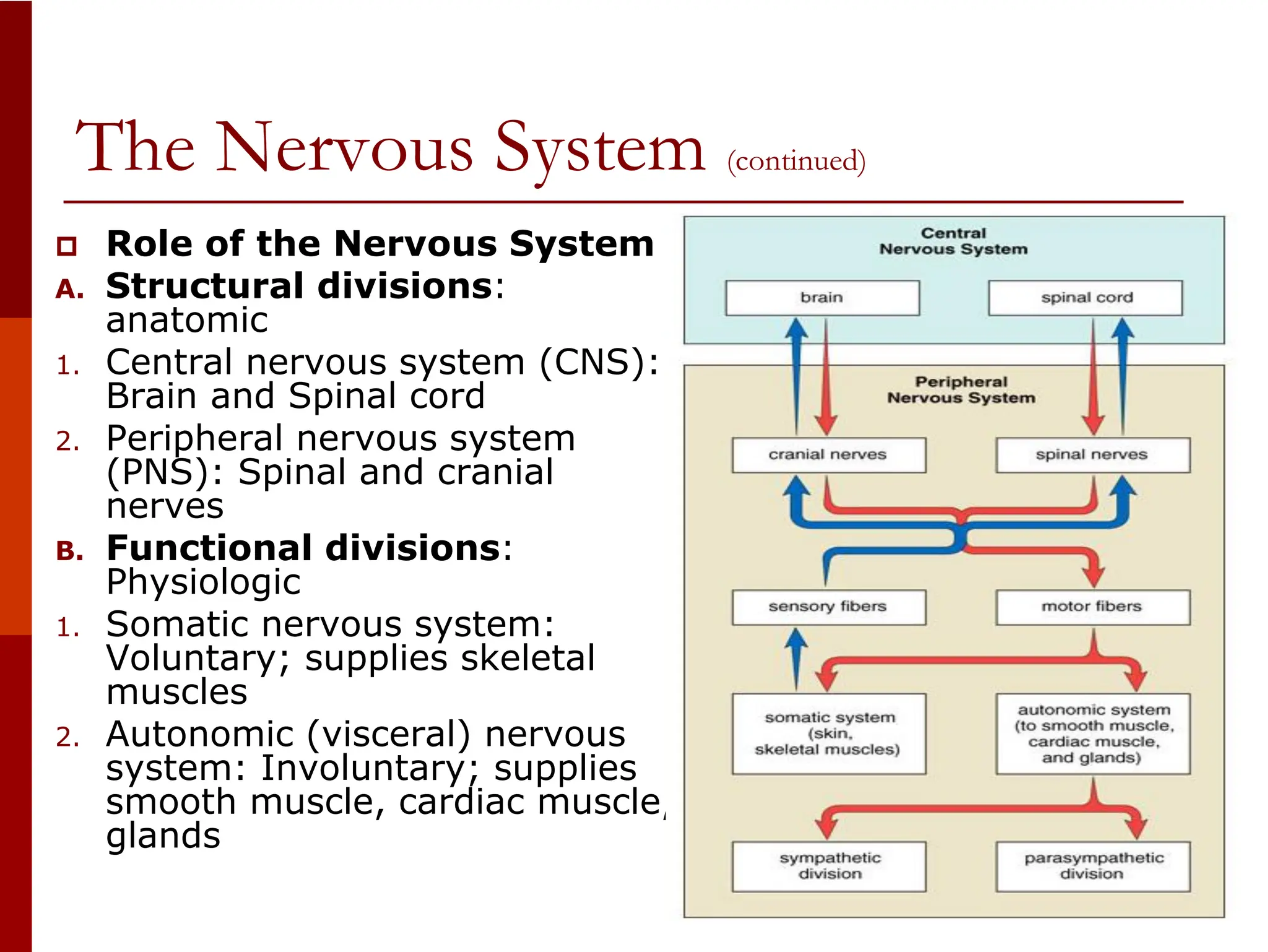
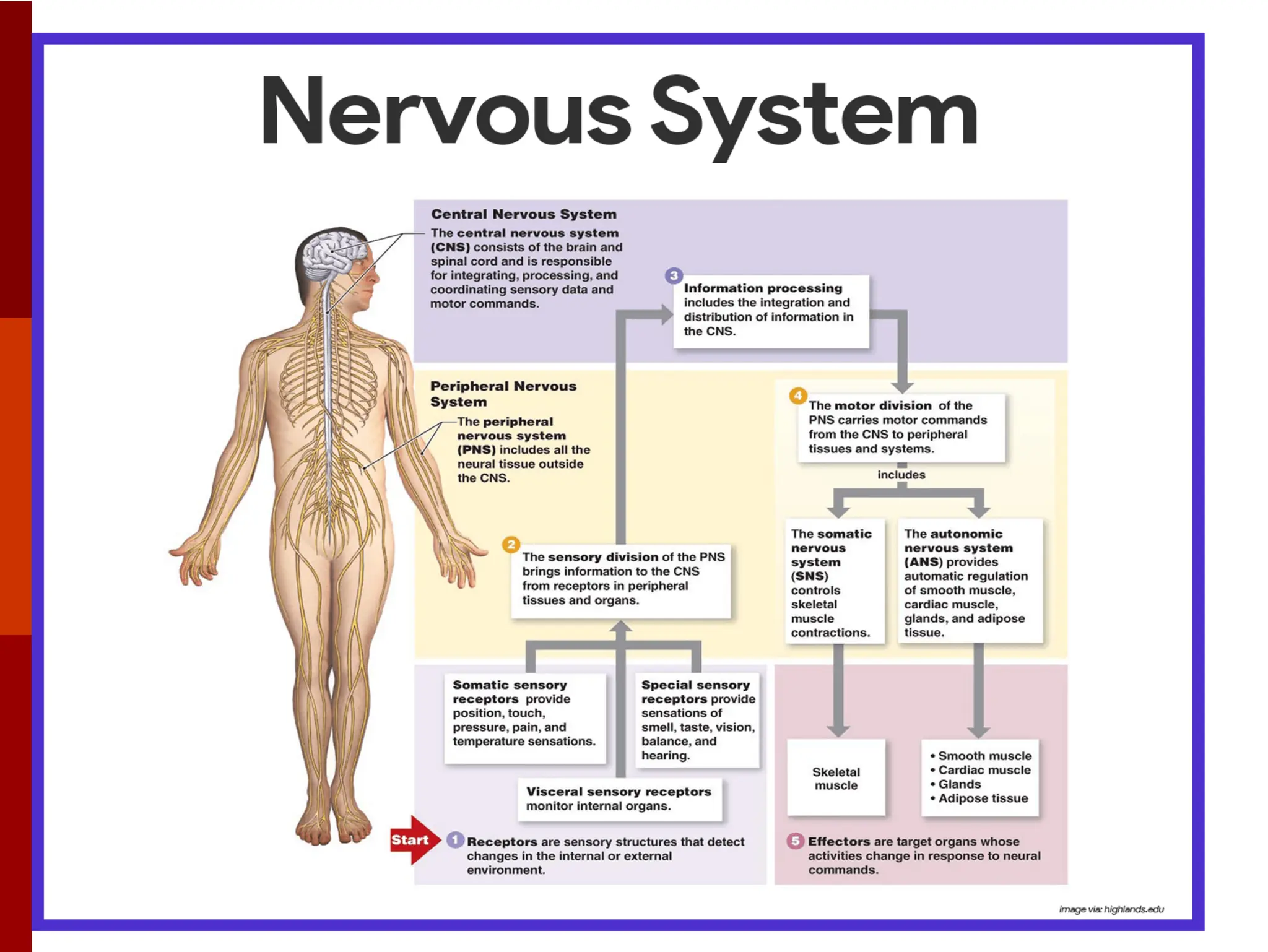
2. The Nervous System
Nervous system:
Specialized for processing
information and sending
messages from one part of
the body to another.
Two major divisions:
• Central nervous system
(CNS): Consists of the
brain and the spinal cord.
• Peripheral nervous
system (PNS): Consists of
nerves.
4. The Nervous System (continued)
Role of the Nervous System
A. Structural divisions:
anatomic
1. Central nervous system (CNS):
Brain and Spinal cord
2. Peripheral nervous system
(PNS): Spinal and cranial
nerves
B. Functional divisions:
Physiologic
1. Somatic nervous system:
Voluntary; supplies skeletal
muscles
2. Autonomic (visceral) nervous
system: Involuntary; supplies
smooth muscle, cardiac muscle,
glands
6. Nervous Tissue
Nervous tissue
Nerve tissue contains two types of cells:
• Neurons: Nerve cells that transmit impulses.
• Neuroglia: Support and nourish the neurons.
7. Neurons
Neurons and their functions
A. Structure of a neuron
1. Cell body
2. Cell fibers
• Dendrite: Carries impulses to cell
body
• Axon: Carries impulses away from
cell body
3. Myelin sheath
• Covers and protects some axon
• Speed conduction
• Made by Schwann cells in PNS;
other cells in CNS
• Neurilemma: Outer layer of
Schwann cell; aids axon repair
• White matter: Myelinated tissue;
gray matter (unmyelinated tissue)
9. Neurons (continued)
B. Types of neurons
1. Sensory (afferent): Carry
impulses towards CNS
2. Motor (efferent): Carry
impulses away from CNS
3. Interneurons: In CNS
C. Nerves and Tracts:
Bundles of neuron fibers
1. Nerve: In peripheral
nervous system Held
together by connective
tissue
• Endoneurium: Around a
single fiber
• Perineurium: Around each
fascicle
• Epineurium: Around whole
nerve
11. Neurons (continued)
Types of nerves
• Sensory (afferent): Contains only fibers
that carry impulses toward the CNS (from a
receptor)
• Motor (efferent) nerve: Contains only fibers
that carry impulses away from the CNS (to
an effector)
• Mixed nerve: Contains both sensory and
motor fibers
2. Tract: In central nervous system (CNS)
12. Neuroglia
Neuroglia
• Nonconducting cells
• Protects and support nervous
tissue
• Smaller cells that surround and
wrap the more delicate neurons.
• Support and nourish neurons.
a. Microglial cell: Engulf and
destroy microbes and cellular
debris in the CNS.
b. Oligodendrocyte: Give support
to neurons in CNS.
c. Astrocyte: Maintain the proper
balance of K+ for generation of
nerve impulses by CNS neurrons.
d. Ependymal cells: Probably
assist in the circulation of
cerebrospinal fluid (CSF) in the
ventricles of the brain and the
central canal of the spinal cord.
13. The Spinal cord
Spinal cord
• In vertebral column
• Ends between first and
second lumbar vertebrae
A. Structure of the spinal
cord
• H-shaped area of gray
matter
• White matter around gray
matter
a. Ascending tracts: Carry
impulses towards brain
b. Descending tracts: Carry
impulses away from brain
17. The Spinal cord (continued)
B. Reflex arc: Pathway through the nervous system
1. Components
• Receptor: Detect stimulus
• Sensory neuron: Receptor to CNS
• Central neuron: In CNS
• Motor neuron: CNS to effector
• Effector: Muscle or gland that responds
18. The Spinal cord (continued)
2. Reflex activities: Simple reflex is rapid, automatic response
using few neurons
• Examples: Stretch reflex, eye blink, withdrawal reflex
• Spinal reflex: Coordinated in spinal cord
21. The Spinal cord (continued)
C. Medical procedures involving in spinal cord
• Lumbar puncture: It is sometimes necessary to
remove a small amount of cerebrospinal fluid
(CSF). The sample that is removed can then be
studied in the laboratory for evidence of disease or
injury.
• Administration of drugs: Anesthetics or
medications are sometimes injected into the space
below the cord. Injection of anesthetic into the
epidural space in the lumbar region of the spine
(an “epidural”) is often used during labor and
childbirth. The spinal route also can be used to
administer pain medication.
22. The Spinal cord (continued)
• Different degrees of loss are named using
the root-plegia, meaning “paralysis,” for
example:
Monoplegia: Paralysis of one limb
Diplegia: Paralysis of both upper or both
lower limbs
Paraplegia: Paralysis of both lower limbs
Hemiplegia: Paralysis of one side of the
body
Tetraplegia or quadriplegia: Paralysis of
all four limbs
27. Somatic Nervous System
Somatic Nervous
System (SNS)
• Conveys information
from the CNS to
skeletal muscles.
• Skeletal muscles,
under conscious
control or voluntary.
28. Autonomic Nervous System
Autonomic Nervous
System
(Visceral nervous
system)
• Involuntary
• Control glands, smooth
muscle, heart (cardiac)
muscle
• Two motor neurons
(preganglionic and
postganglionic)
29. Autonomic Nervous System (continued)
A. Divisions of the Autonomic Nervous System (ANS)
1. Sympathetic Nervous System
• The sympathetic motor neurons originate in the spinal cord with cell bodies in the
thoracic and lumbar regions, the thoracolumbar area.
• This system is described as adrenergic, which means “activated by adrenaline.”
• Synapses in sympathetic chains and three collateral ganglia (celiac, superior
mesenteric, inferior mesenteric)
30. Autonomic Nervous System (continued)
2. Parasympathetic system
• The parasympathetic motor pathways begin in the craniosacral areas.
• This system is described as cholinergic, which means “activated by
acetylcholine.”
• Synapses in terminal ganglia in or near effector organs
31.
32. Autonomic Nervous System (continued)
B. Functions of the Autonomic Nervous System
1. Sympathetic: Stimulates fight-or-flight (stress) response
2. Parasympathetic: Returns body to normal
3. Usually have opposite effects on an organ
• Increase in the rate and force of heart contractions (increase
in blood pressure).
• Contraction of small arteries in the skin and the internal
organs.
• Dilation of blood vessels to skeletal muscle, bringing more
blood to these tissues.
• Dilation of the bronchial tubes to allow more oxygen to enter.
• Stimulation of the central portion of the adrenal gland.
• Increase in basal metabolic rate (BMR). BMR is the resting
metabolic rate of a normal fasting subject under homeostatic
conditions.
• Dilation of the pupil and decrease in focusing ability (for near
objects).
33. SNS and ANS
Comparison of Somatic Motor and Autonomic
Motor Pathways
34. Brain Protective Structures
Protective Structures of the Brain and Spinal Cord
A. Meninges: Three layers
• Dura mater: Tough outermost layer
• Arachnoid: Weblike middle layer
• Pia mater: Vascular innermost layer
36. Brain Protective Structures
B. Cerebrospinal fluid
(CSF)
1. Circulates around and
within brain and spinal
cord
2. Cushions and protects
3. Ventricles: Four spaces
within brain where CSF
is produced
Choroid plexus:
Vascular network in
ventricle that produces
CSF by filtration of the
blood and by cellular
secretion.
44. Divisions of the Brain
Divisions of the Brain
A. Cerebral hemispheres
1. Lobes: Frontal, parietal, temporal, occipital and insula
Insula: It is a small fifth lobe deep within each hemisphere that cannot
be seen from the surface.
48. Divisions of the Brain
B. Diencephalon: Area
between cerebral
hemispheres and brain
stem
1. Thalamus: Directs
sensory impulses to cortex
2. Hypothalamus: Maintains
homeostasis, controls
pituitary gland
Limbic system:
1. Contains parts of cerebrum
and diencephalon
2. Controls emotion and
behaviors
49. Divisions of the Brain (continued)
C. Brain Stem
• Midbrain: involved in eye
and ear reflexes
• Pons: Connecting link for
other divisions
• Medulla oblongata:
1. Connects with spinal cord
2. Contains vital centers for
respiration, heart rate,
vasomotor activity
(regulates the contraction
of smooth muscle in the
blood vessel walls and thus
controls blood flow and
blood pressure)
D. The Cerebellum:
Regulates coordination,
balance, muscle tone
50. Cranial Nerves
Cranial nerves: 12 pairs
attached to brain
A. Name and functions of the
cranial nerves
1. Functions
• Carry special and general
sensory impulses
• Carry somatic (skeletal
muscles) and visceral
(internal organs) motor
impulses
• Sensory (I, II, VIII)
• Motor (III, IV, VI, XI, XII)
• Mixed (V, VII, IX, X)
B. Disorders involving the
cranial nerves
1. Bell palsy: Facial paralysis
(cranial nerve VII)
2. Trigeminal neuralgia: Pain in
Vth cranial nerve