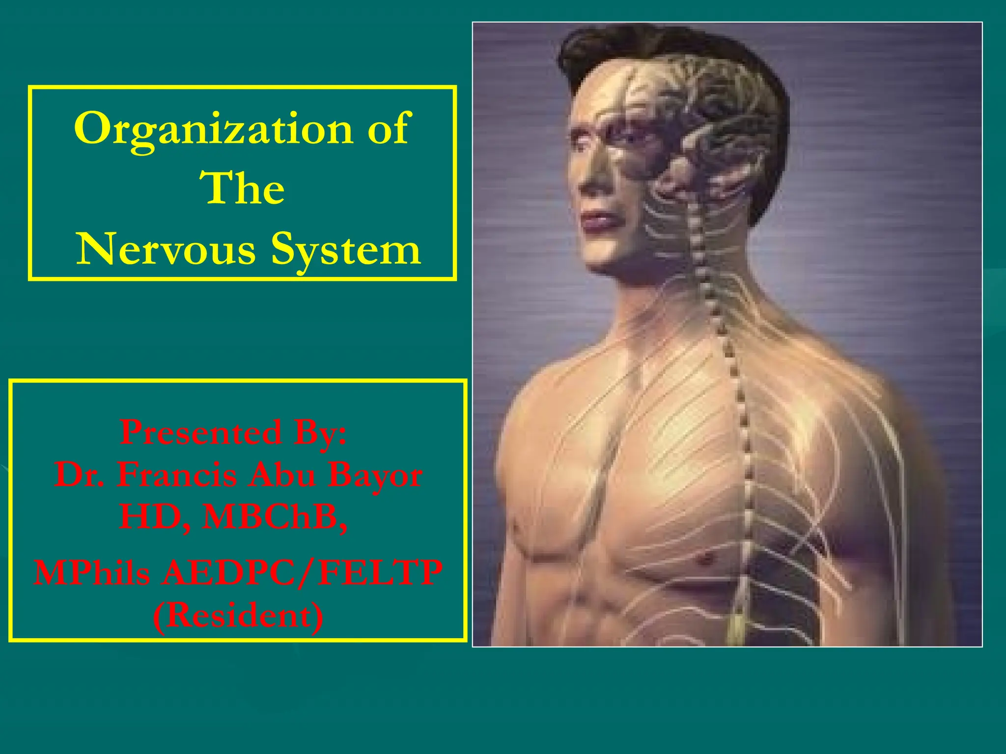
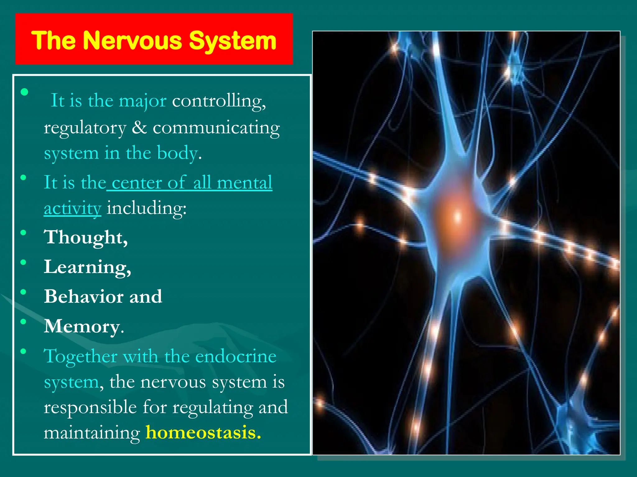
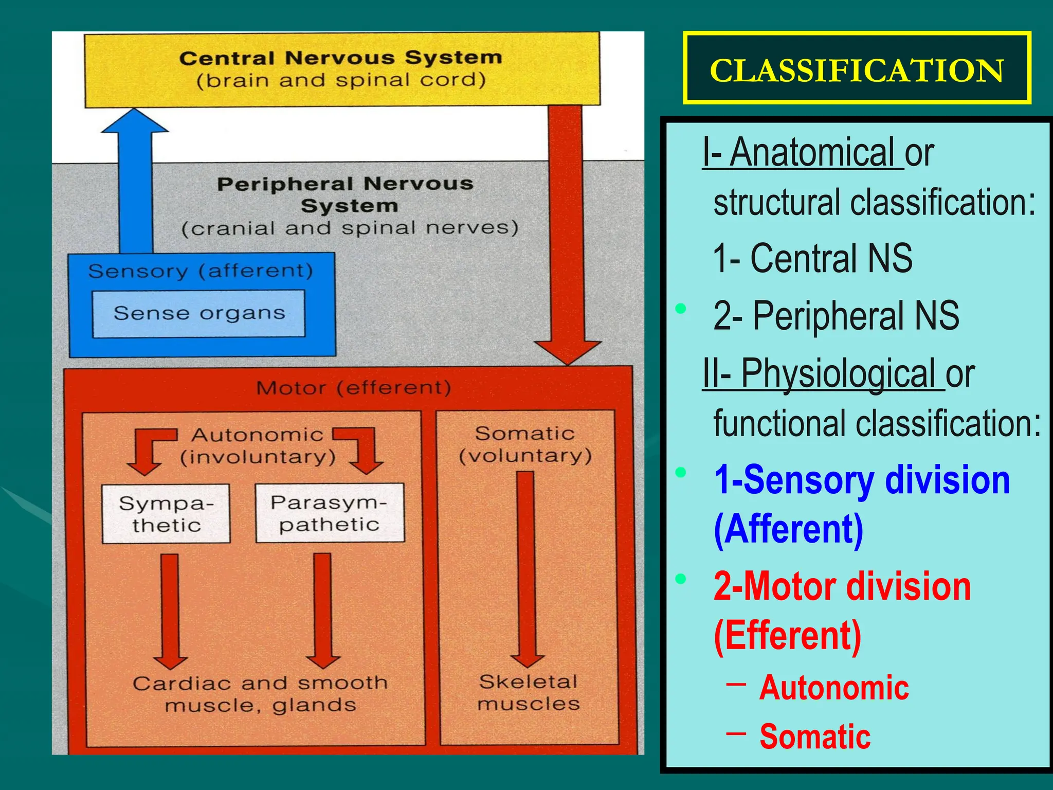
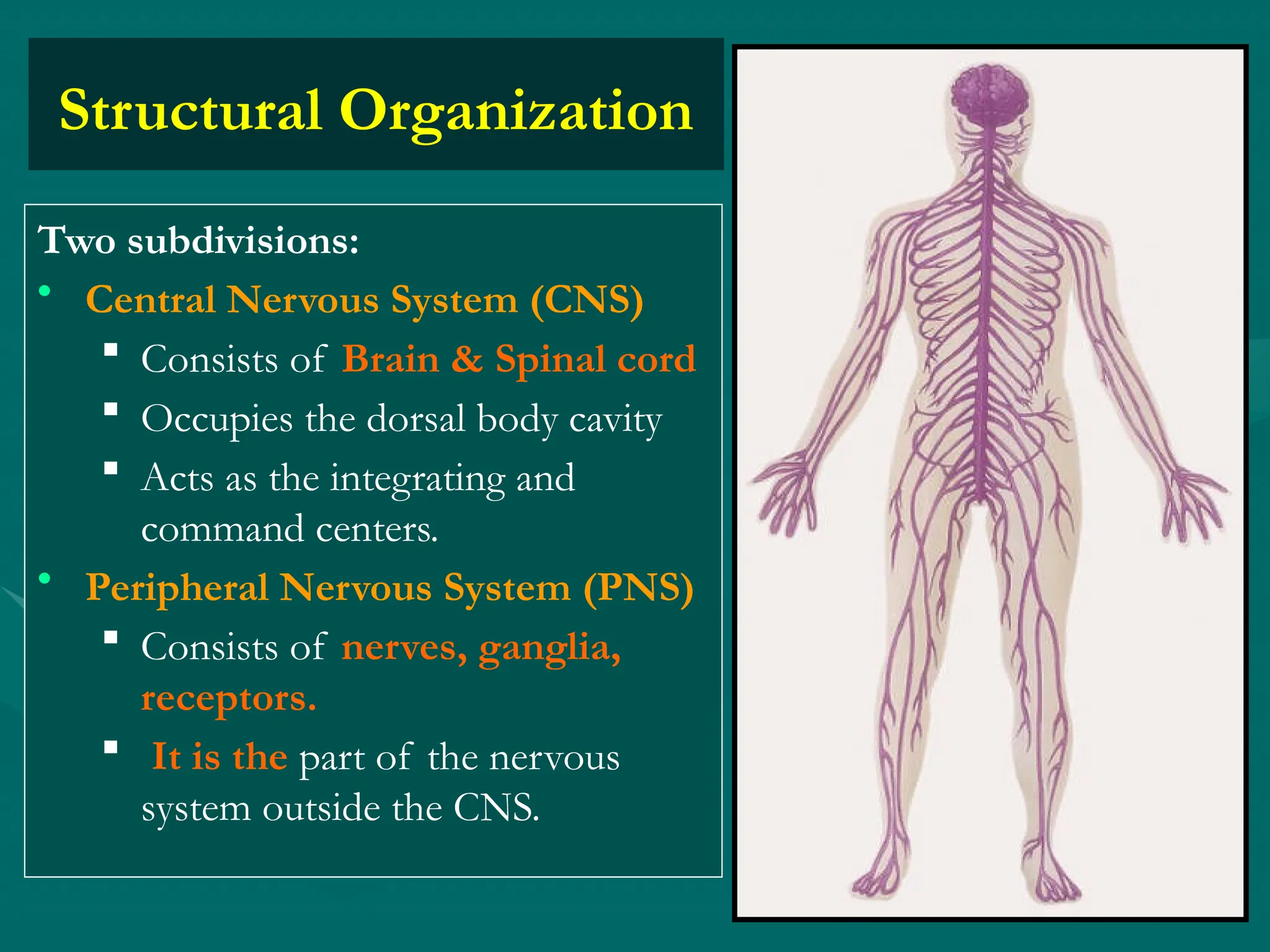
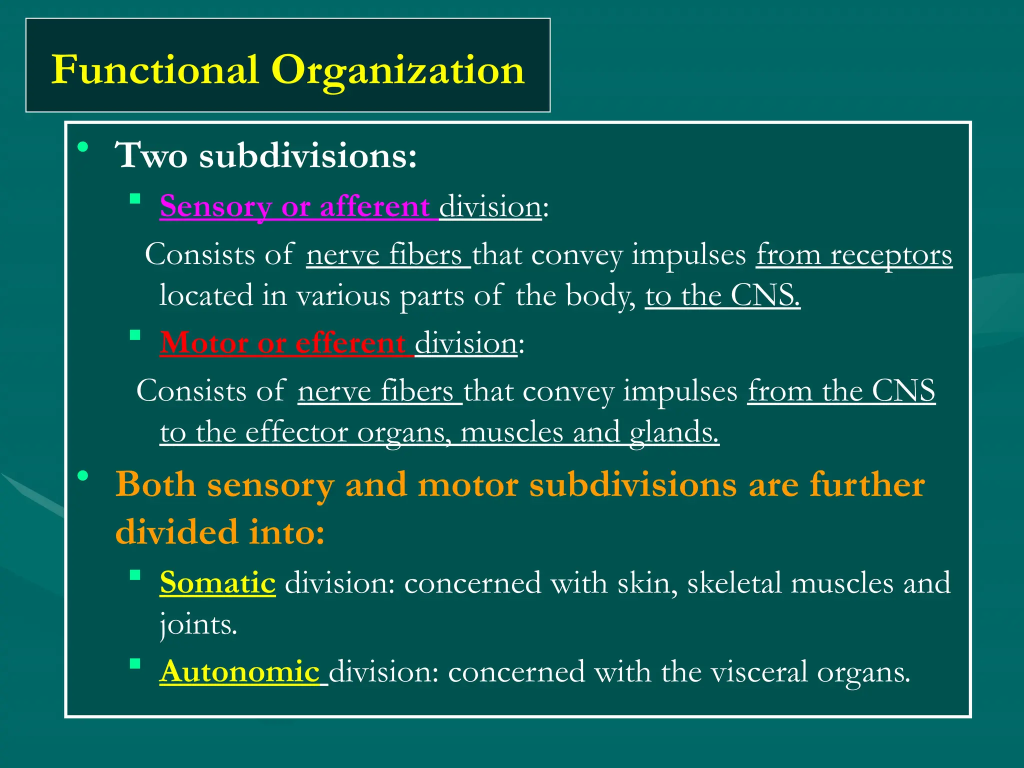
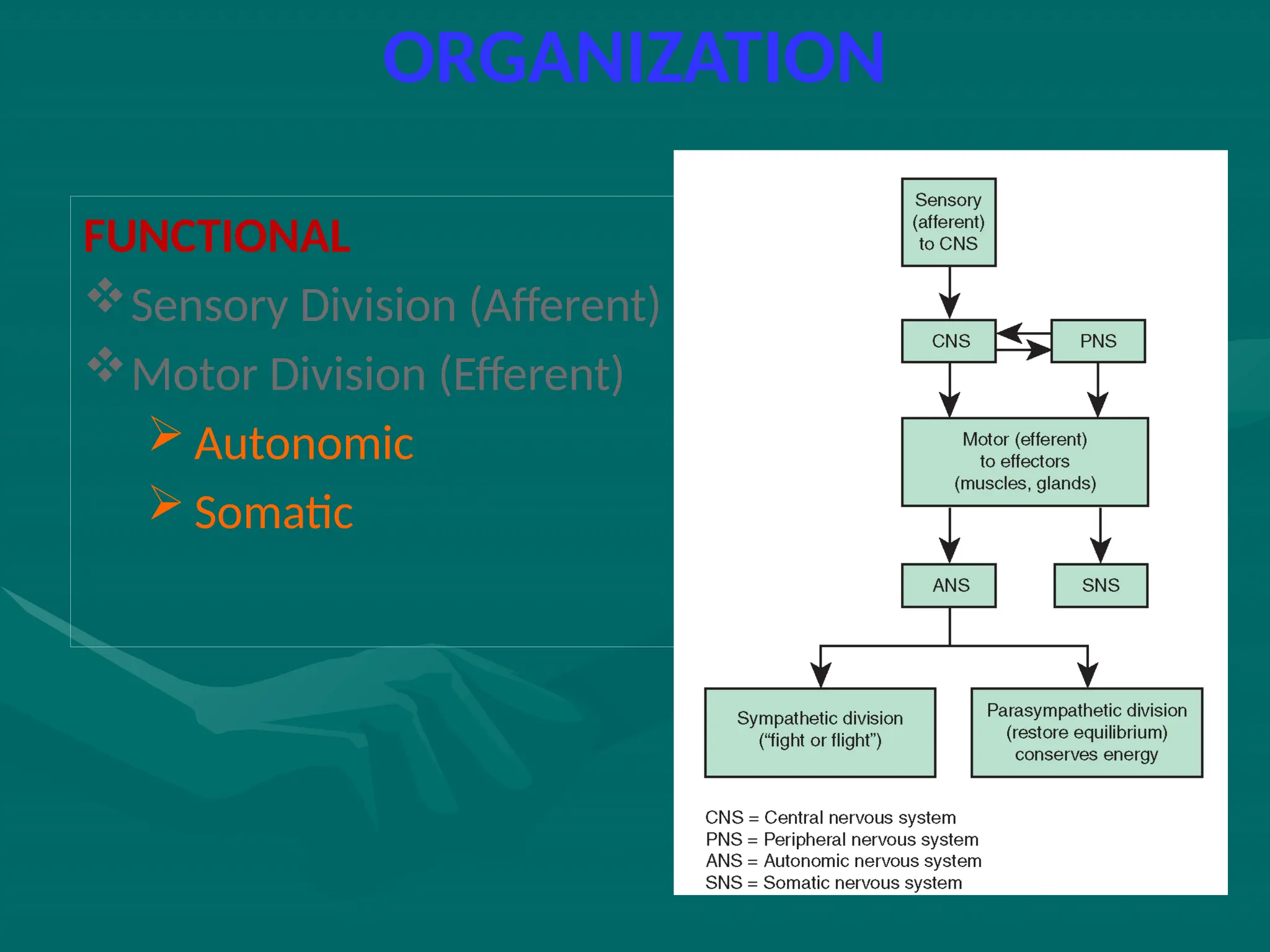
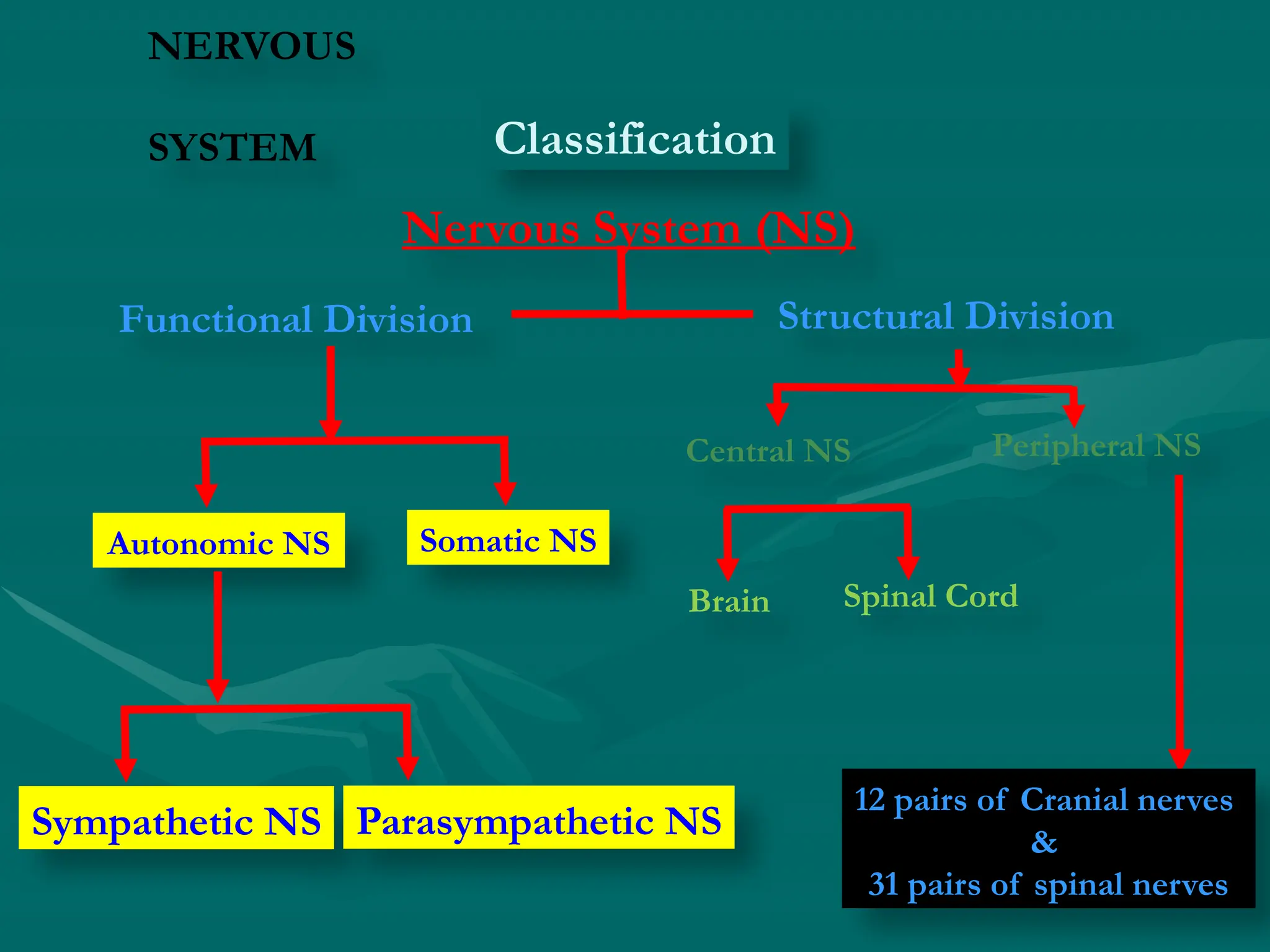
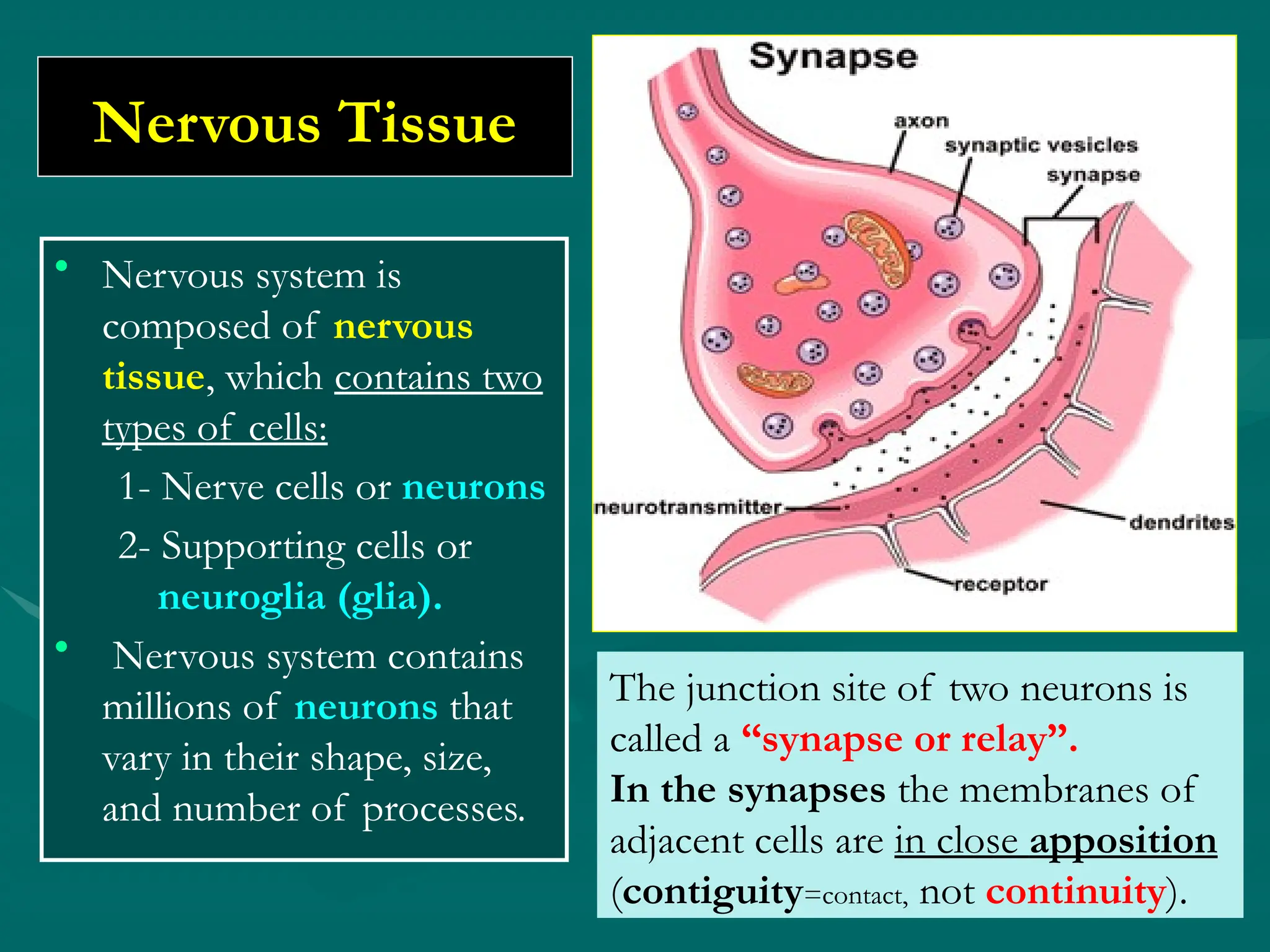
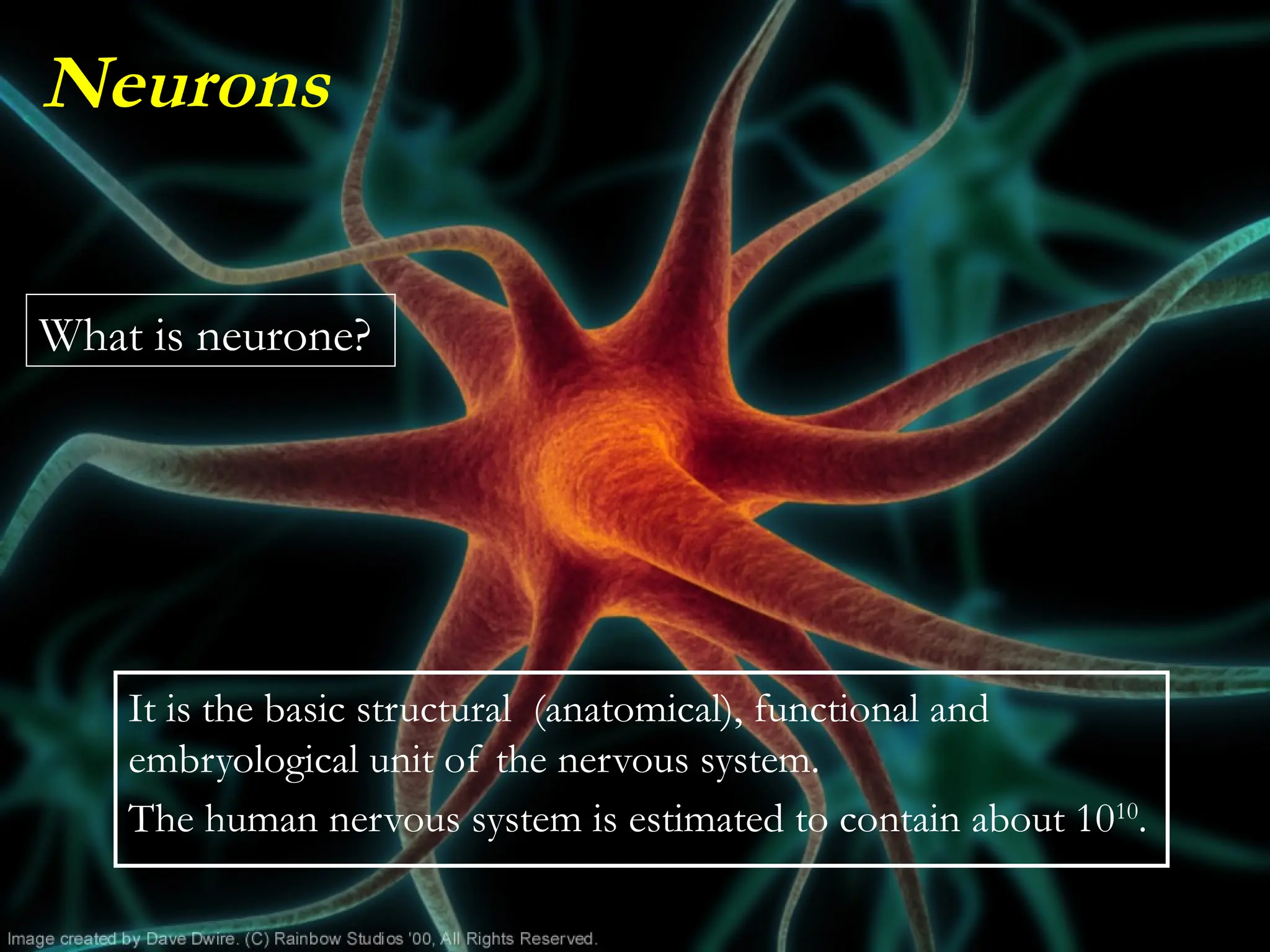
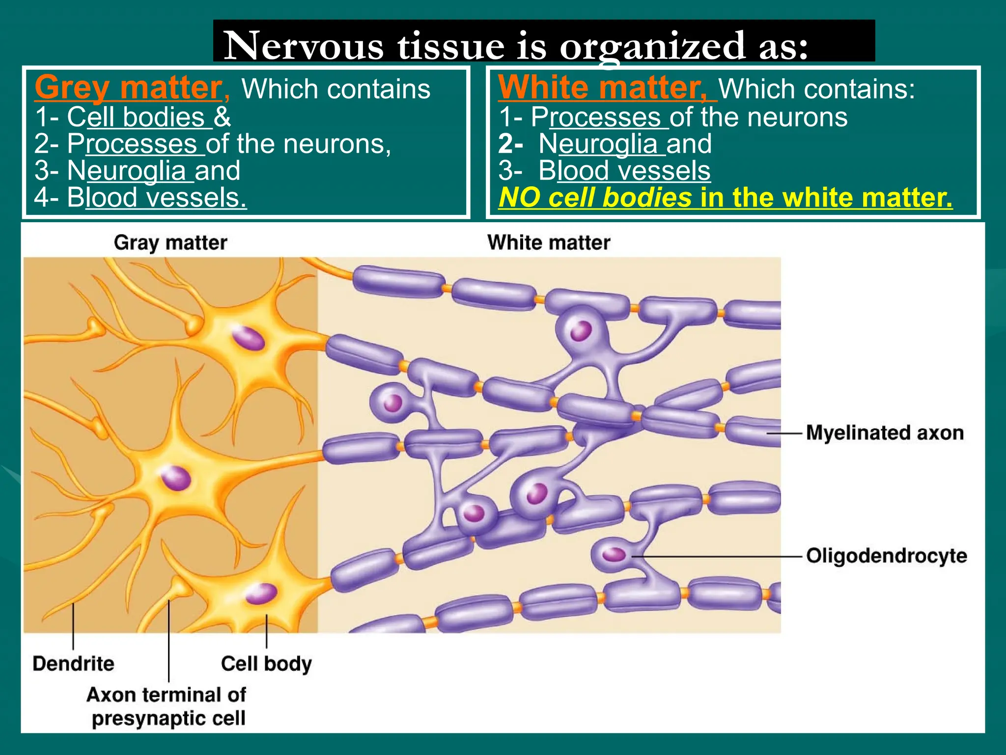
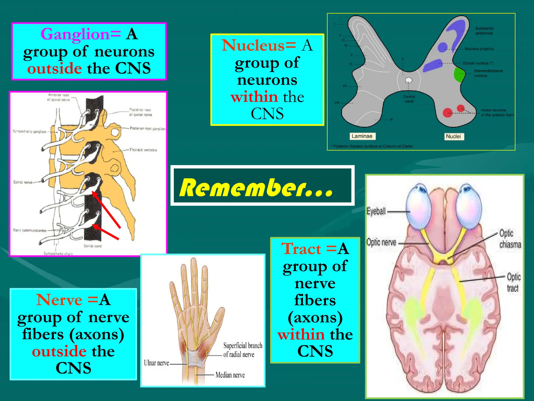
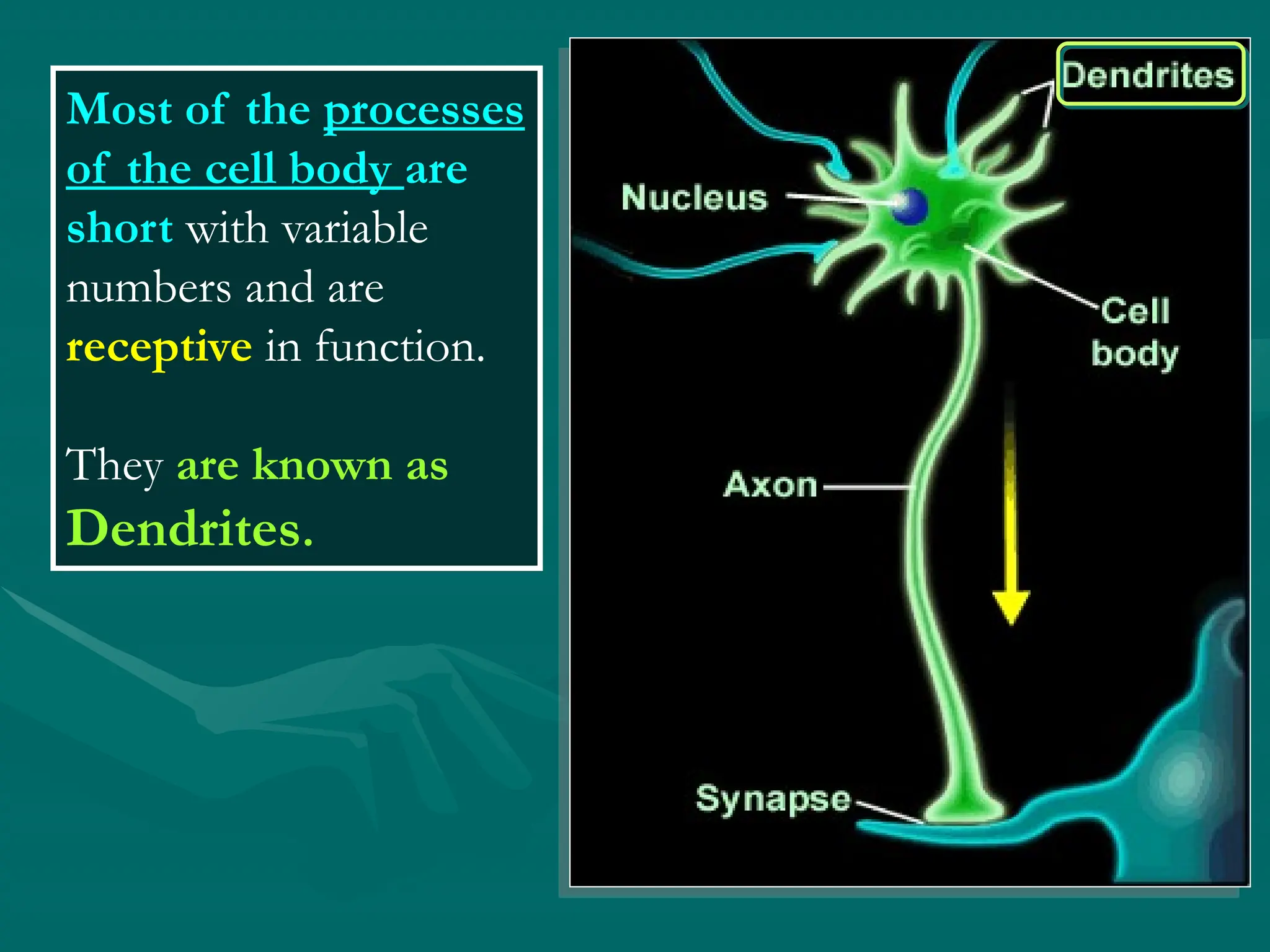
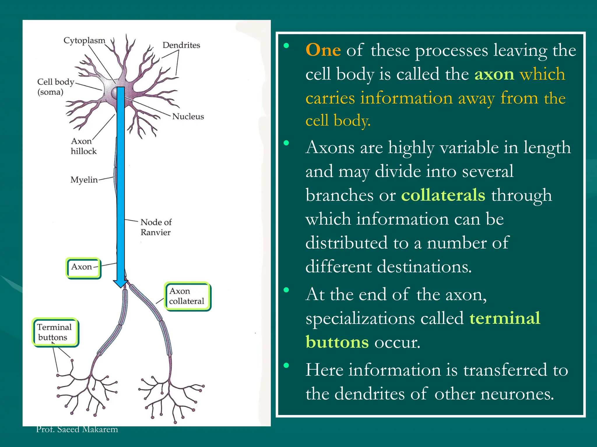
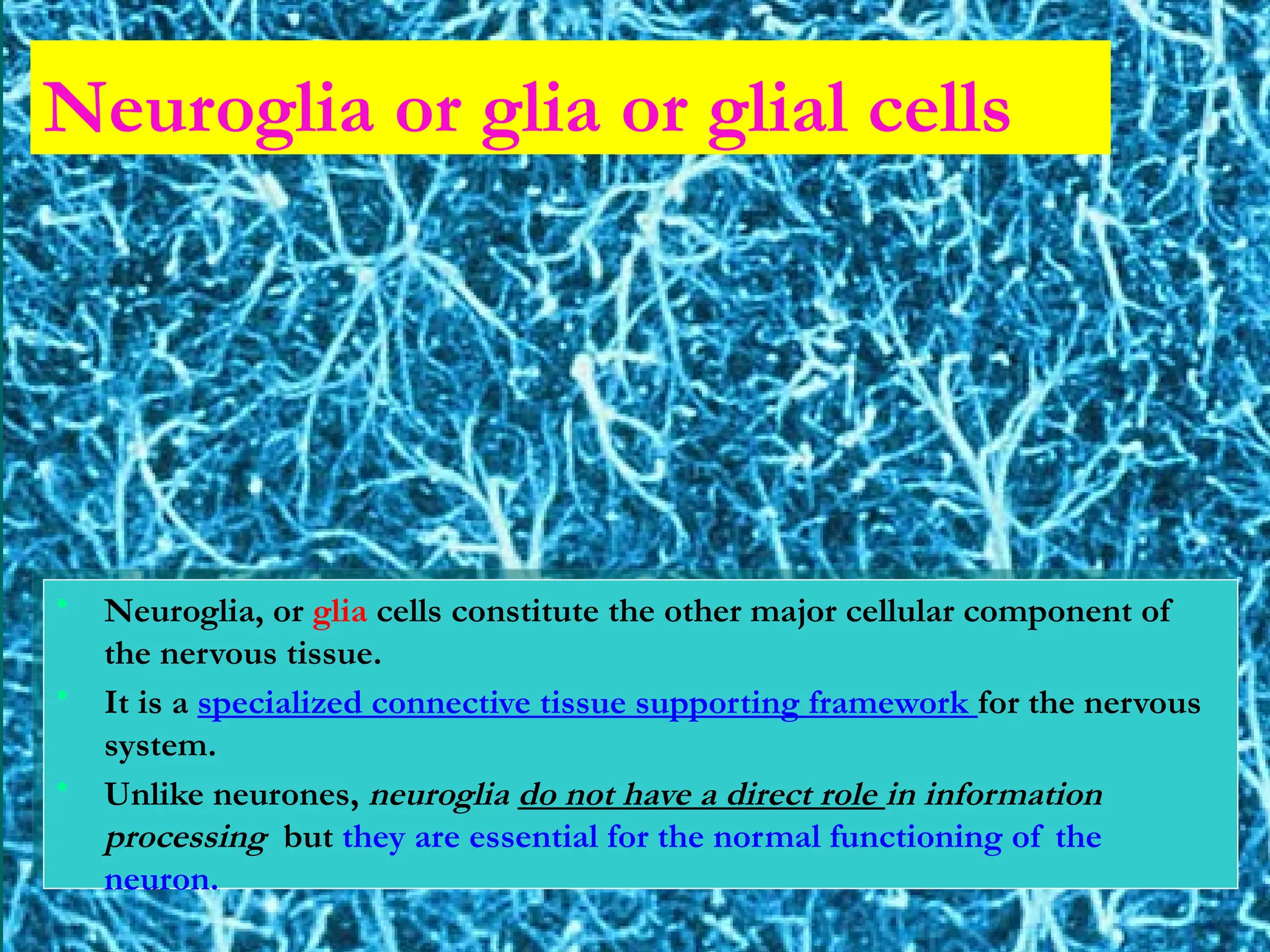
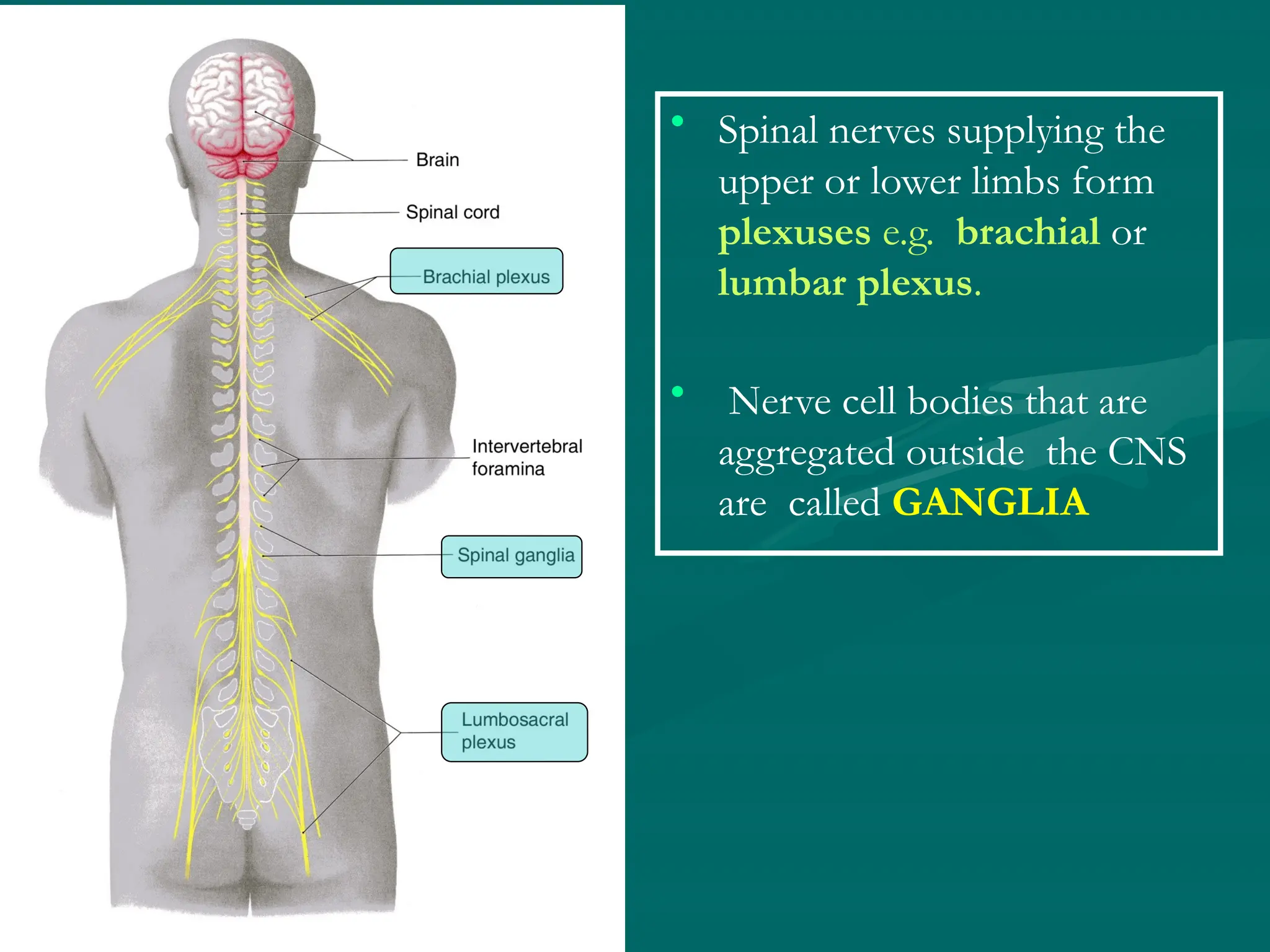
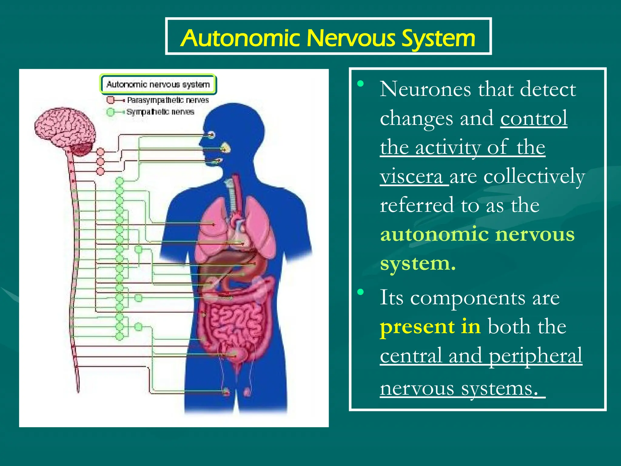
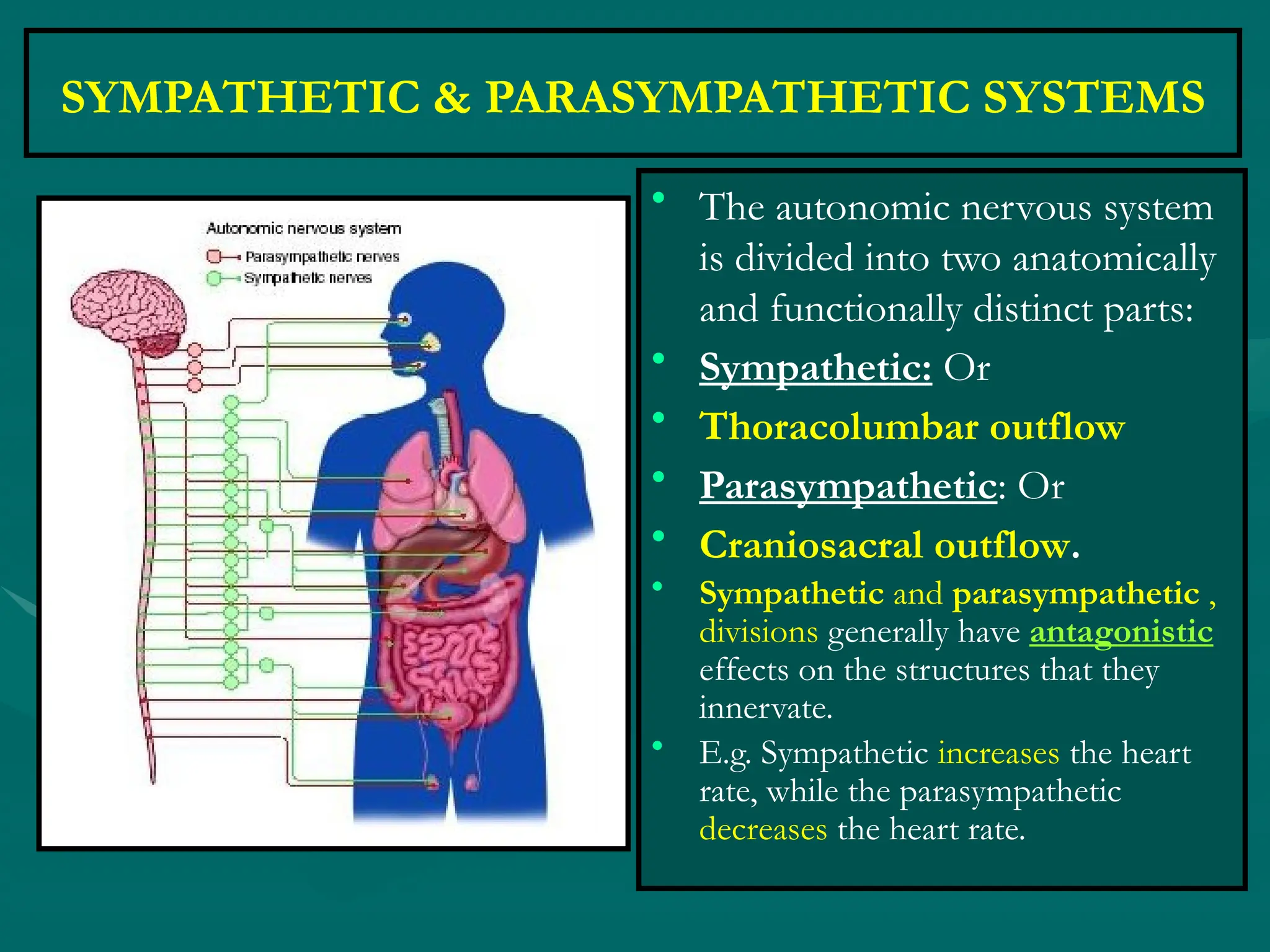
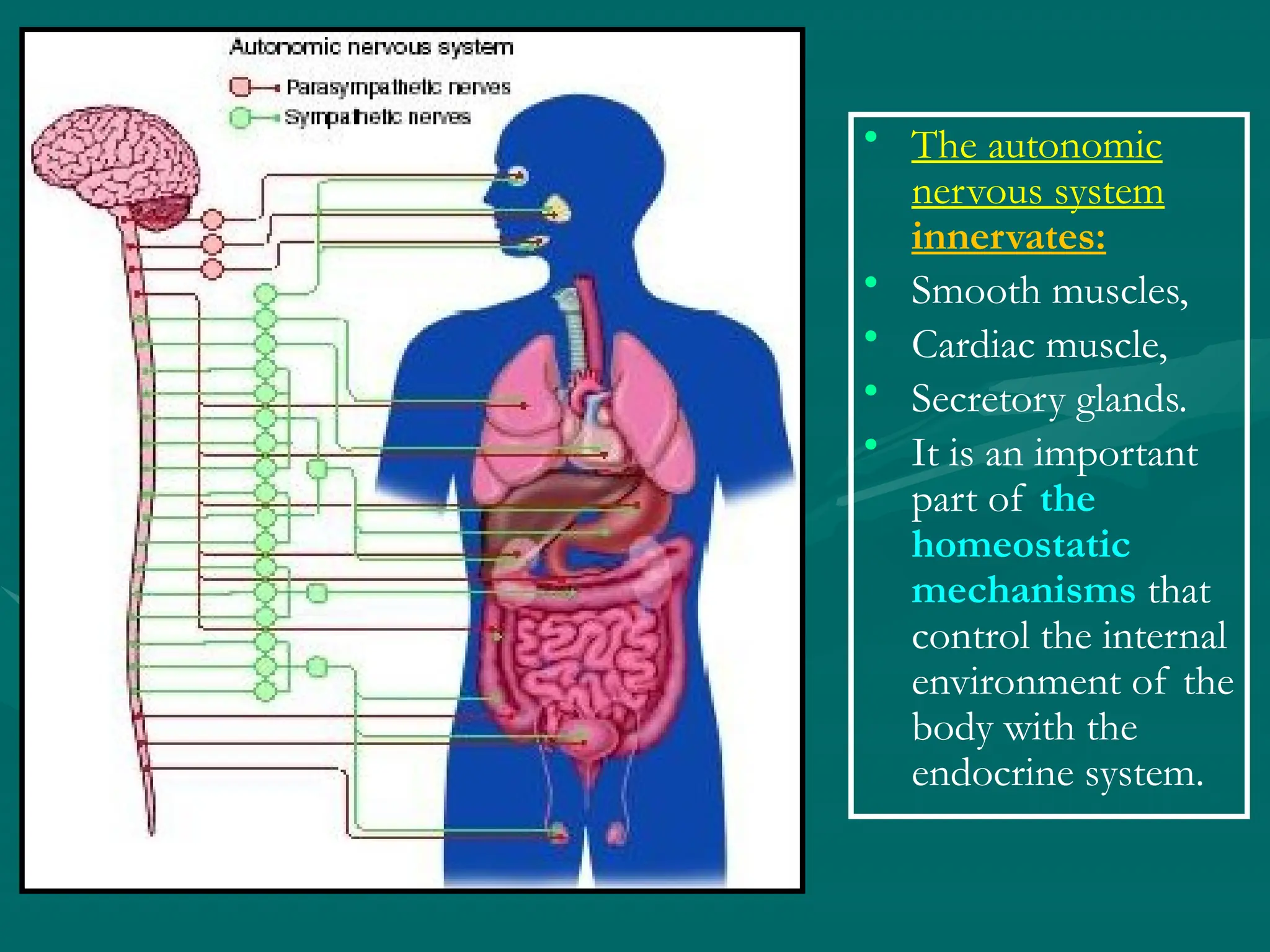
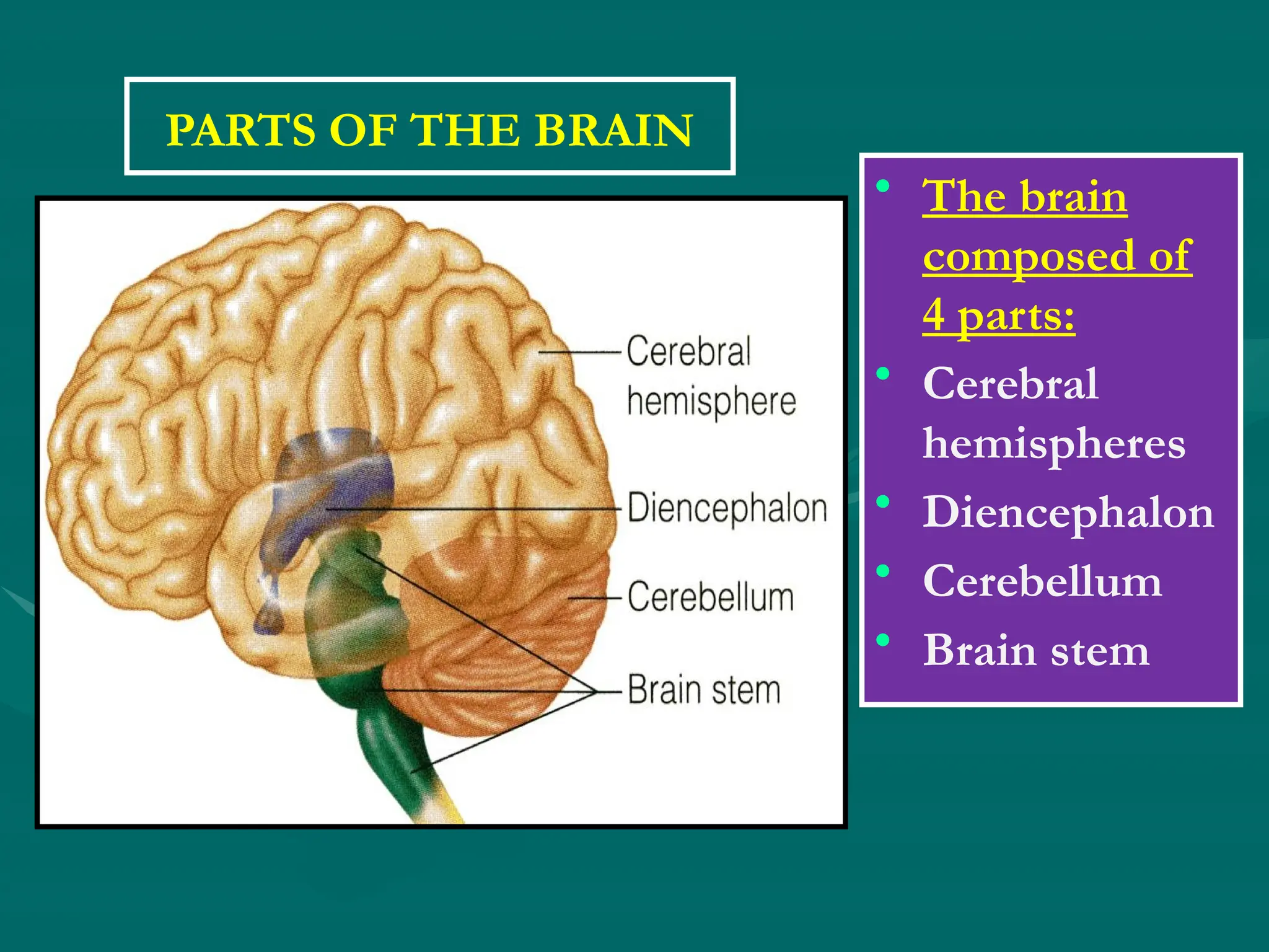
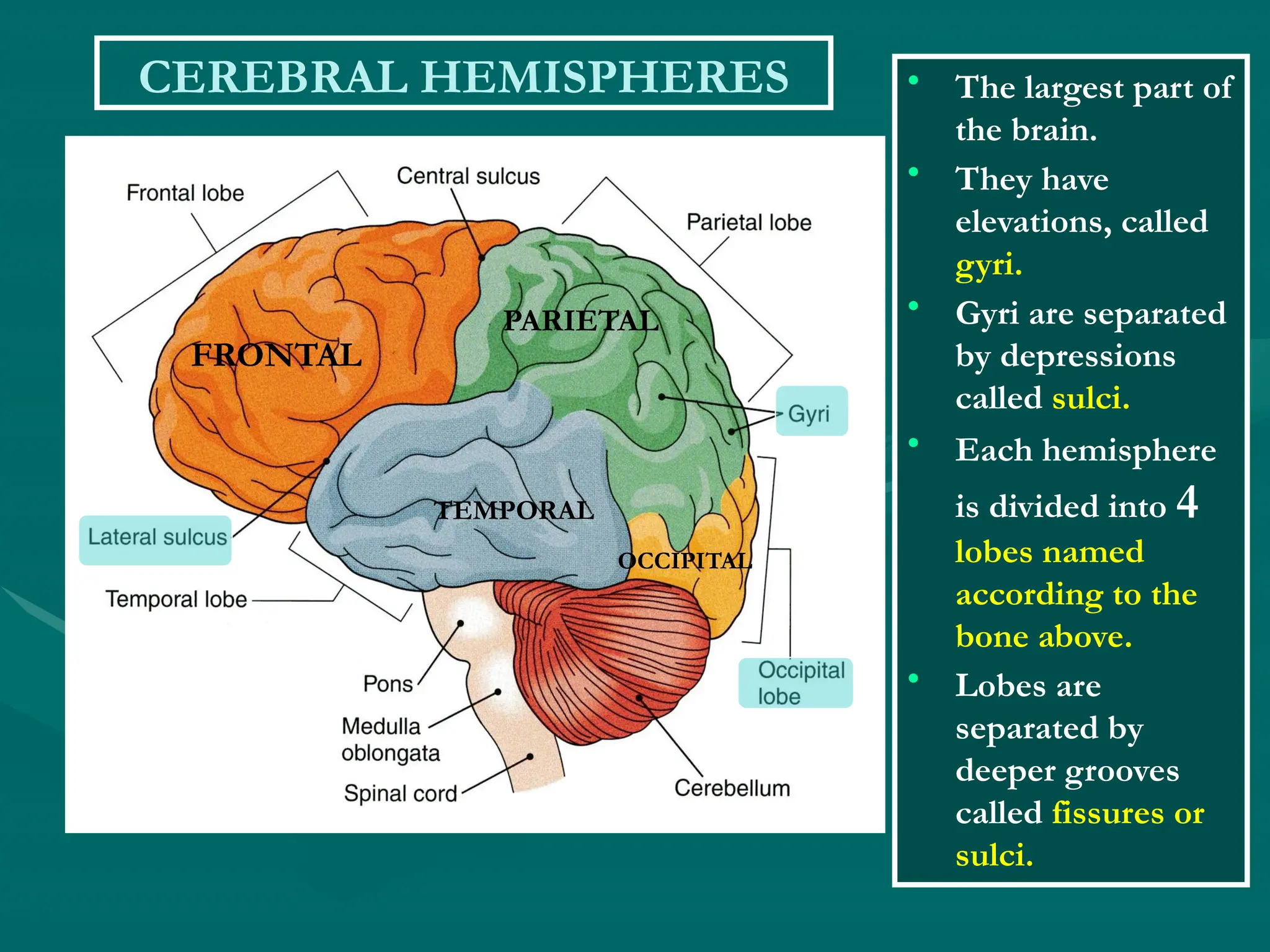
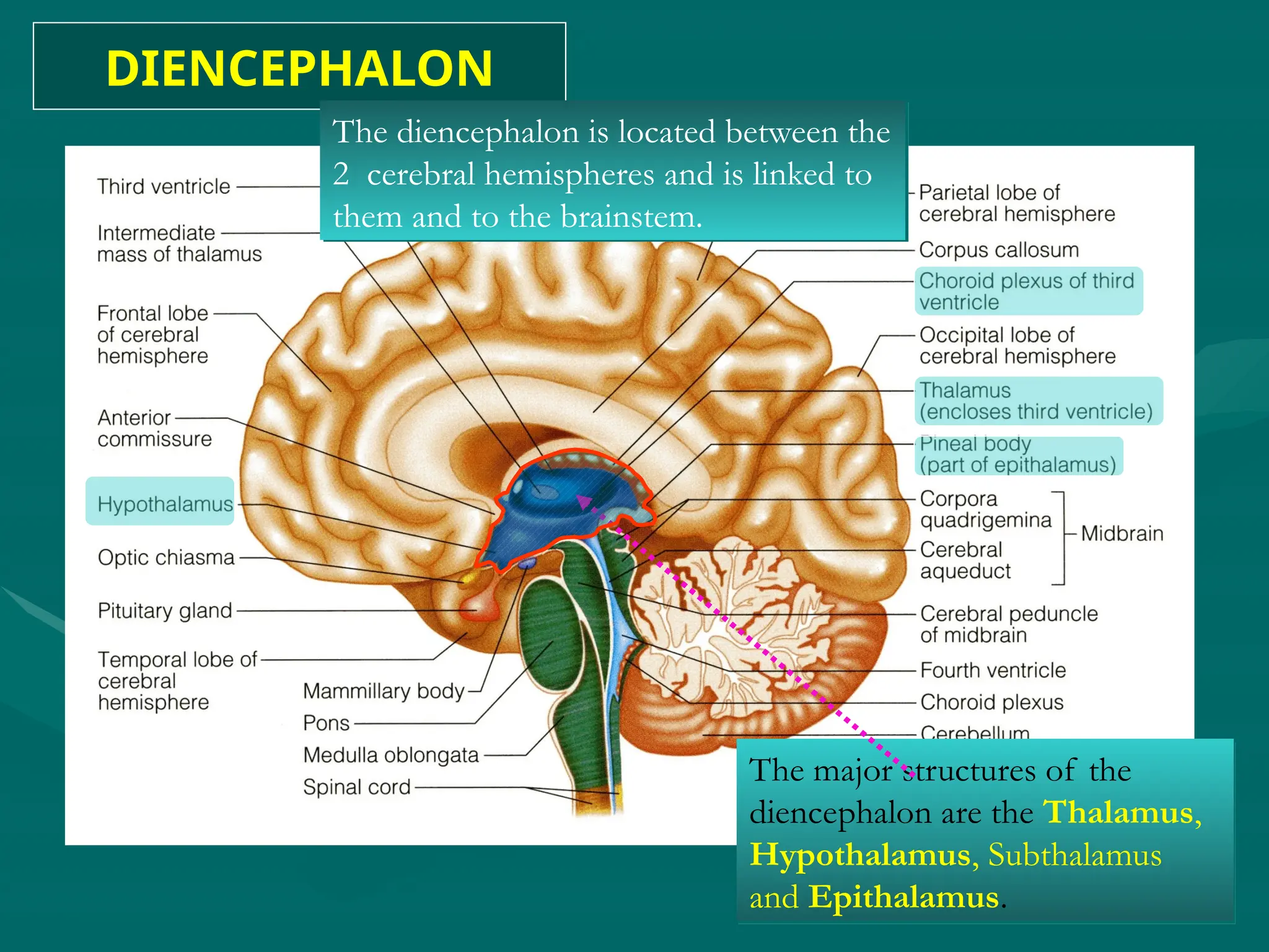
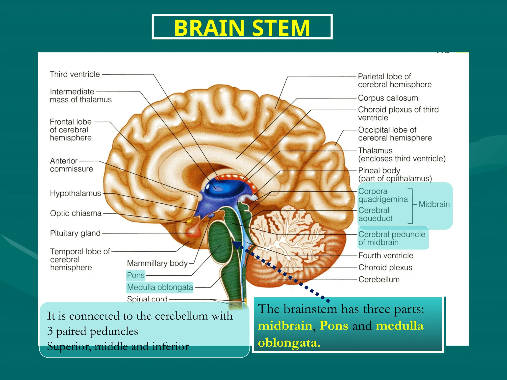
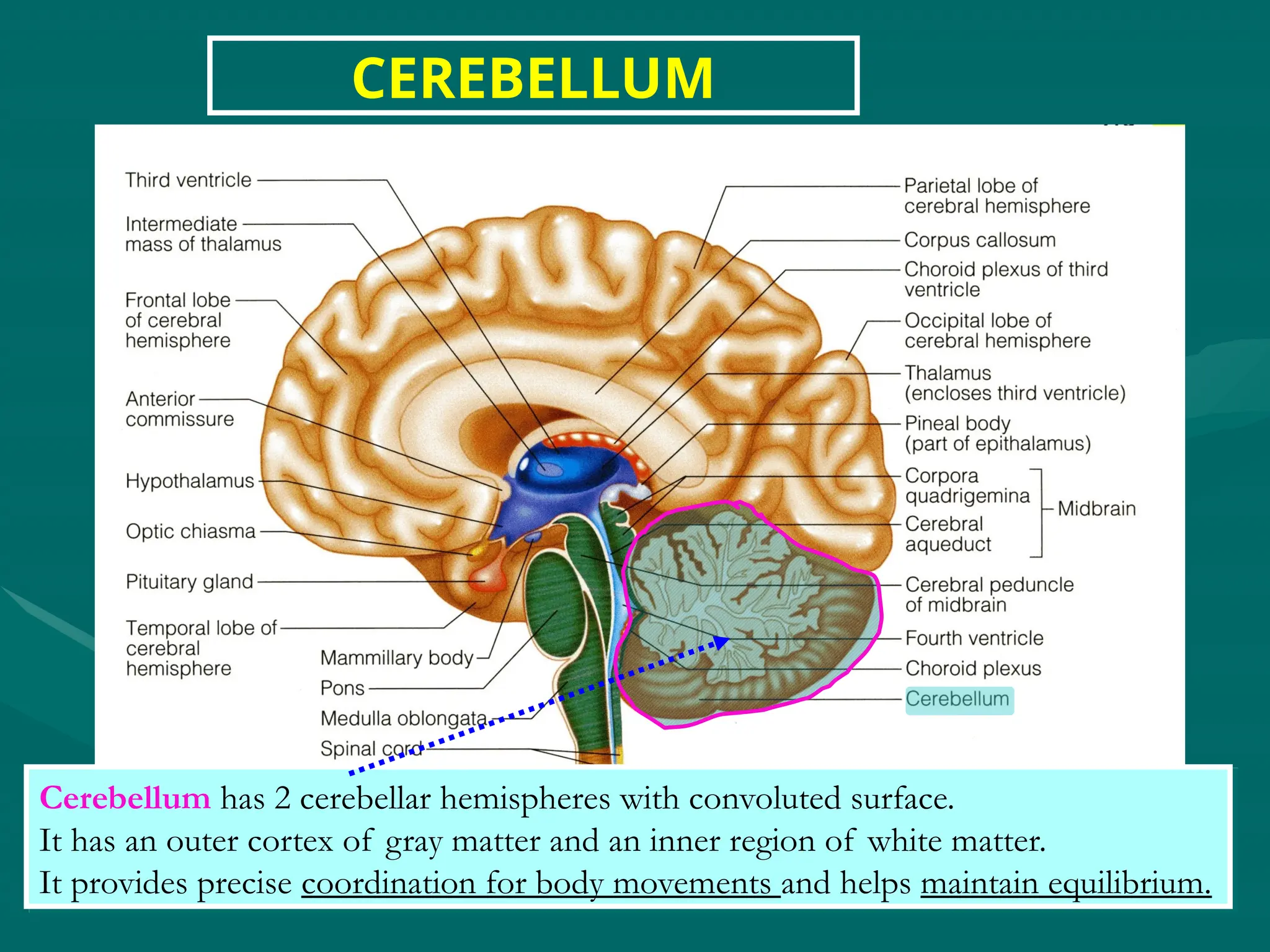
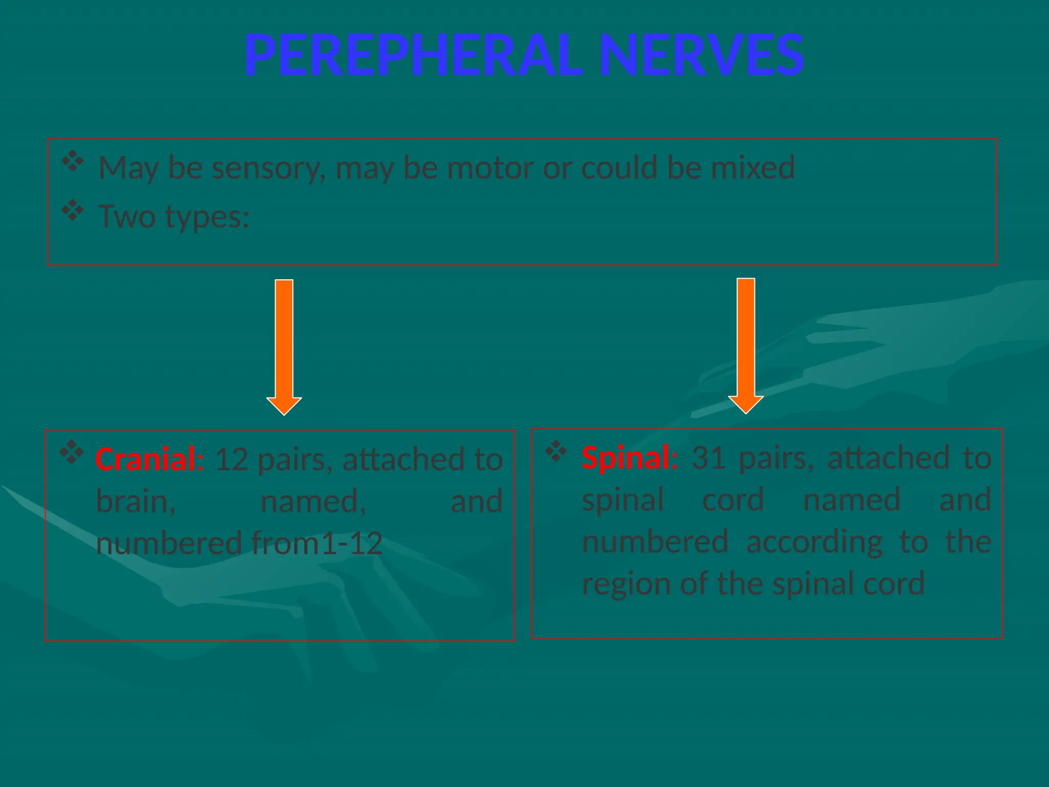
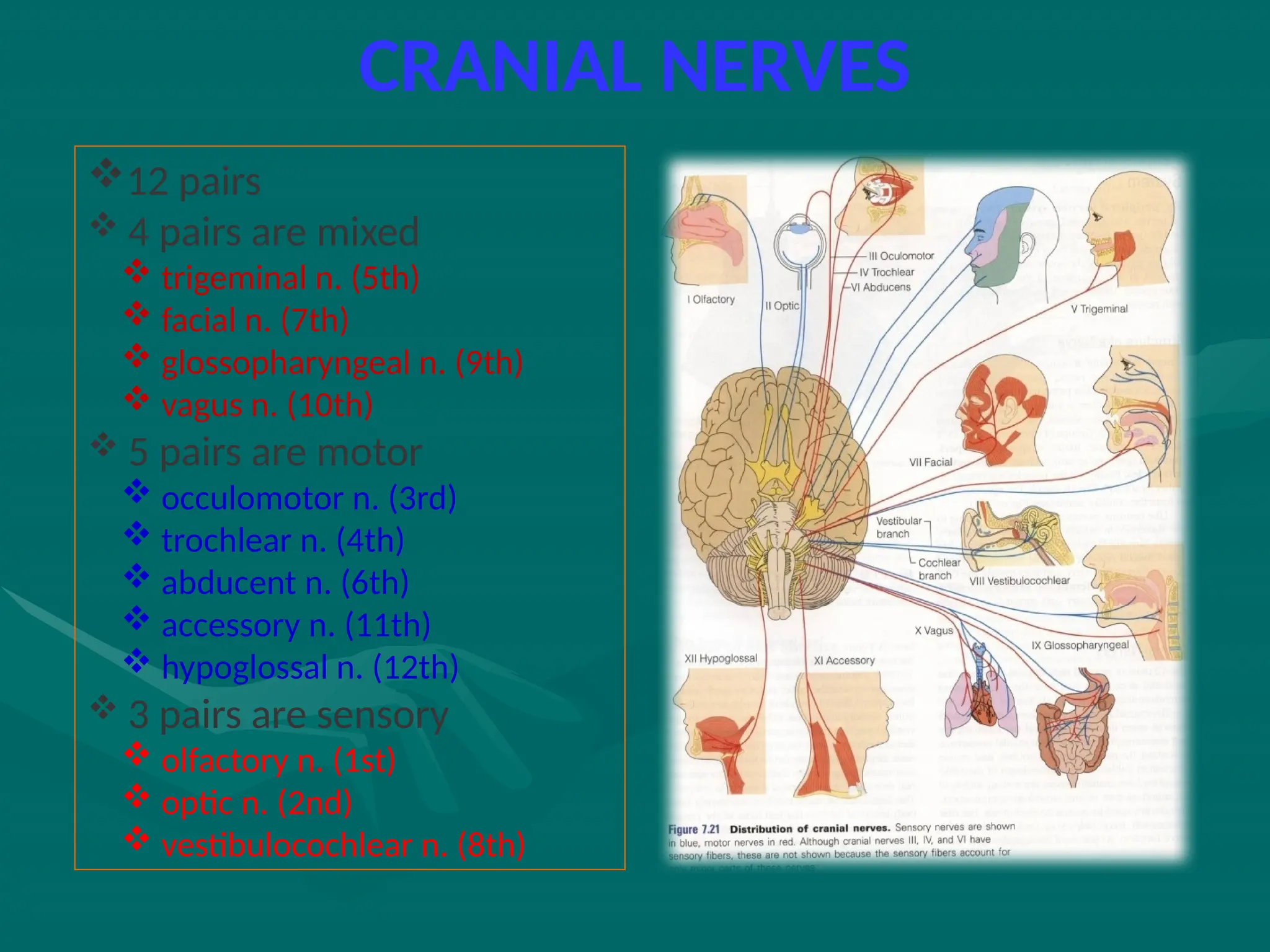
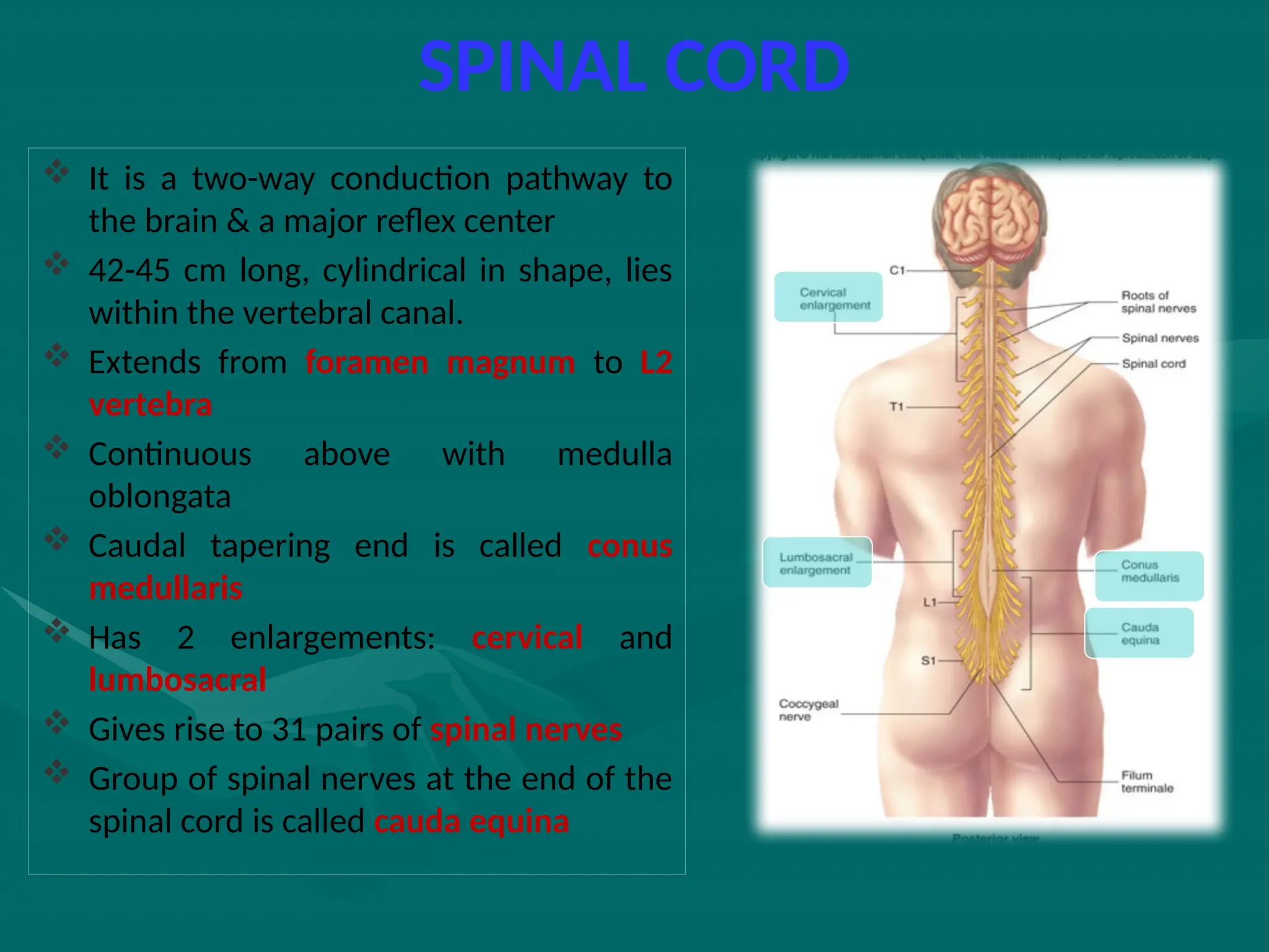
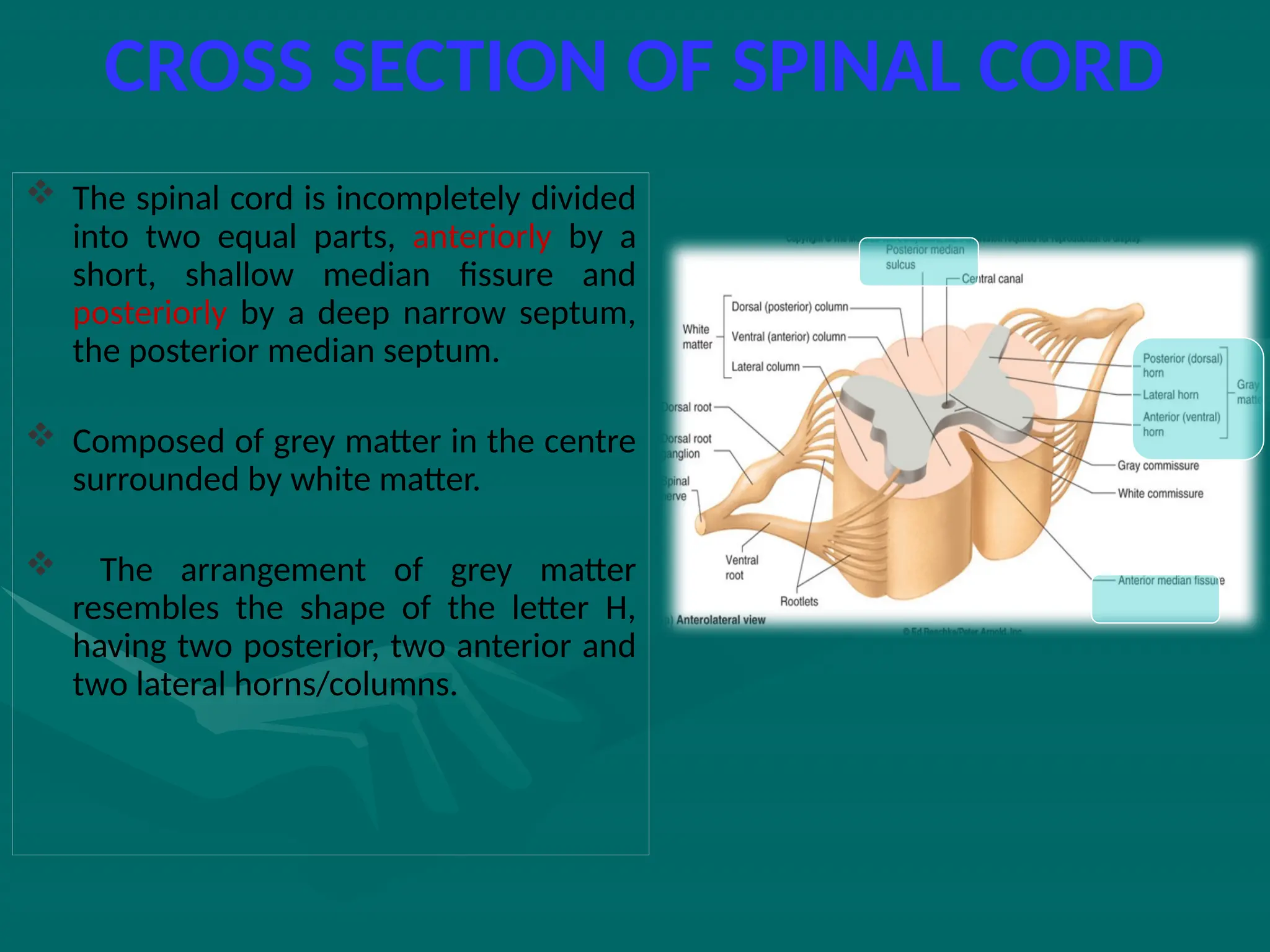
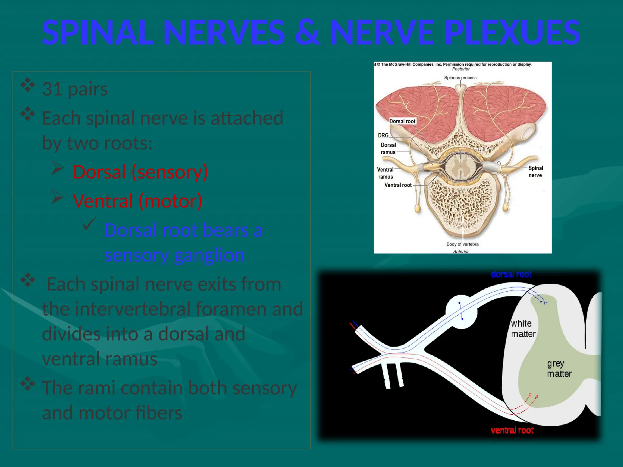
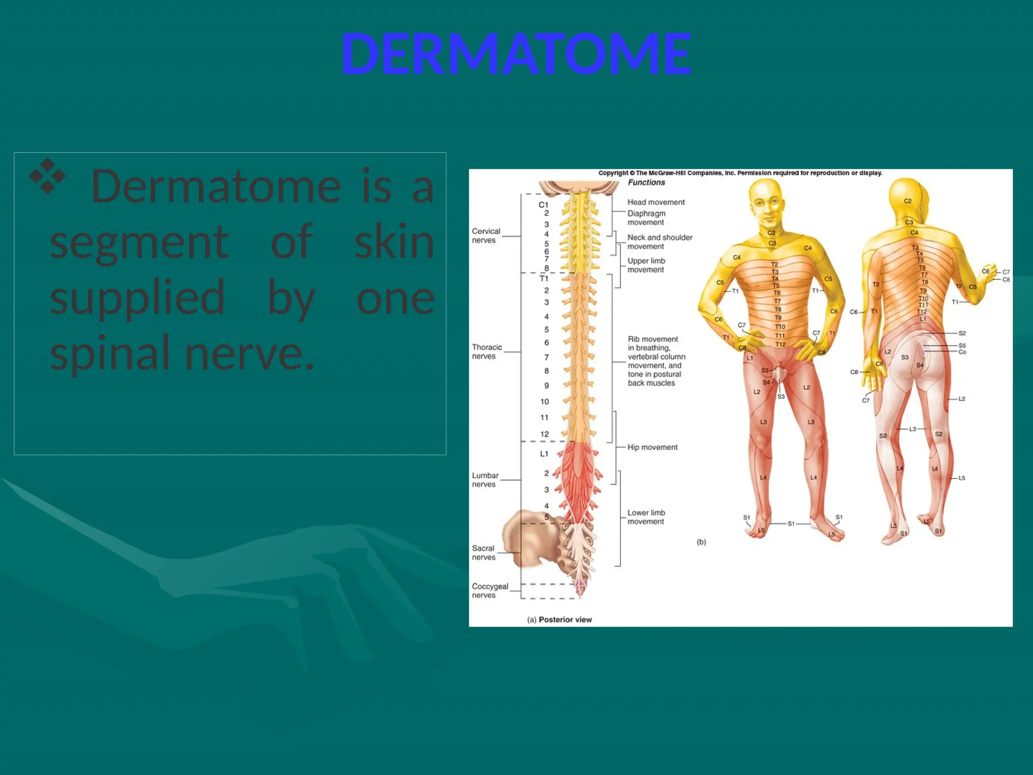
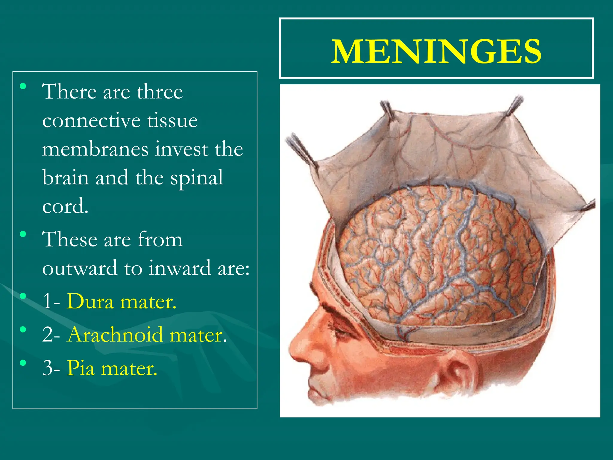
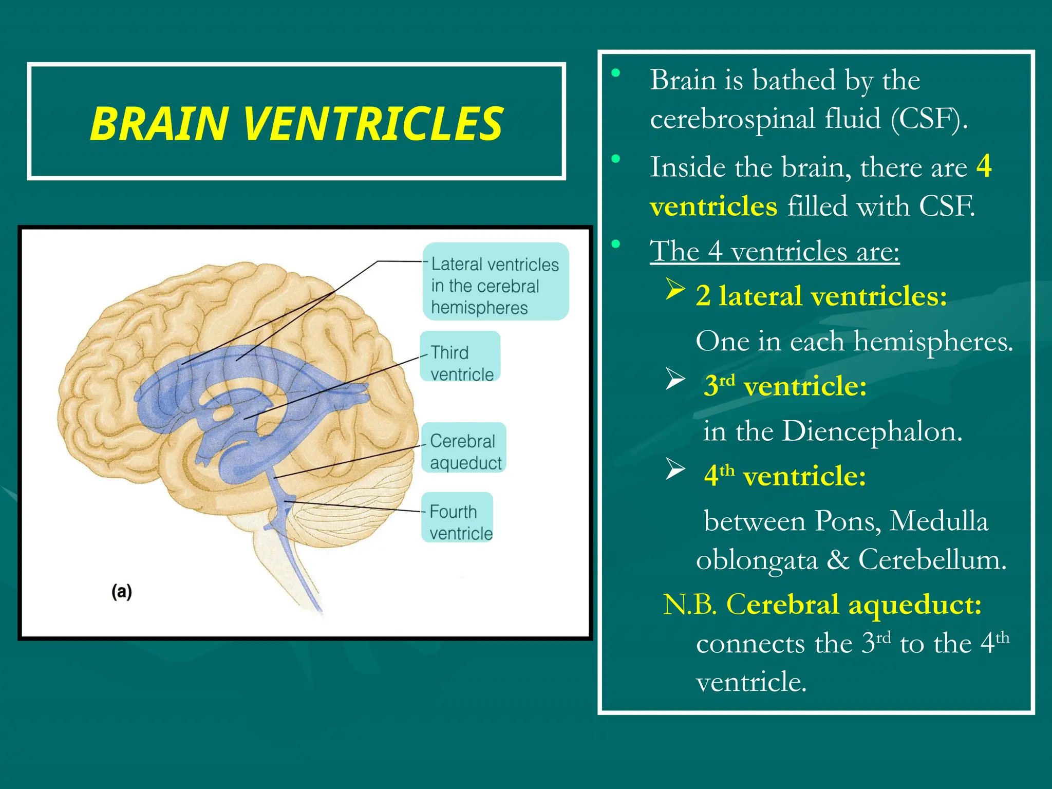
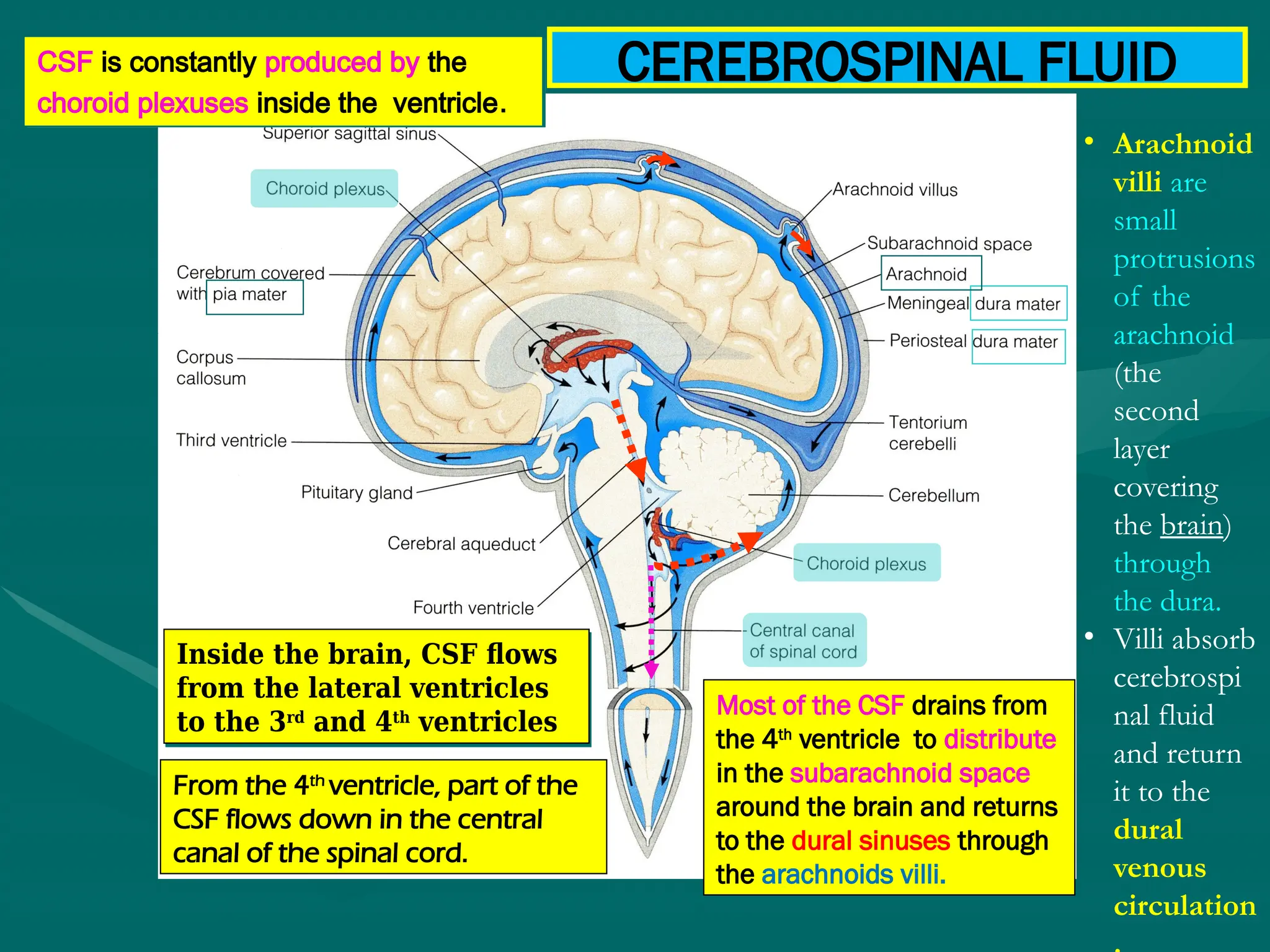
Organization of
The
Nervous System
Presented By:
Dr. Francis Abu Bayor
HD, MBChB,
MPhils AEDPC/FELTP
(Resident)
Objectives
At the end of the lecture, the students should be able to:
• List the parts of the nervous system.
• To gain a three-dimensional appreciation of the parts of the
brain and their relative positions to one another.
• To understand the basic organization of the main structures
that form the nervous system
• List the function of the nervous system.
• Describe the Structural & Functional Organizations.
• Define the terms:
– Nervous tissue, nucleus
– grey matter, ganglion
– white matter,. Tract
– nerve
• List the parts of the brain.
• List the structures protecting the central nervous system.
The Nervous System
• It is the major controlling,
regulatory & communicating
system in the body.
• It is the center of all mental
activity including:
• Thought,
• Learning,
• Behavior and
• Memory.
• Together with the endocrine
system, the nervous system is
responsible for regulating and
maintaining homeostasis.
How does the nervous
system work ?
The nervous system has
three functions:
 Collection of sensory input:
Identifies changes occurring
inside or outside the body by
using sensory receptors. These
changes are called stimuli.
 Integration:
Processes, analyzes and
interprets these changes and
makes decisions.
 Motor output, or response
by activating muscles or glands
(effectors).
INTRODUCTION
CLASSIFICATION
I- Anatomical or
structural classification:
1- Central NS
• 2- Peripheral NS
II- Physiological or
functional classification:
• 1-Sensory division
(Afferent)
• 2-Motor division
(Efferent)
– Autonomic
– Somatic
Structural Organization
Two subdivisions:
• Central Nervous System (CNS)
 Consists of Brain & Spinal cord
 Occupies the dorsal body cavity
 Acts as the integrating and
command centers.
• Peripheral Nervous System (PNS)
 Consists of nerves, ganglia,
receptors.
 It is the part of the nervous
system outside the CNS.
Functional Organization
• Two subdivisions:
 Sensory or afferent division:
Consists of nerve fibers that convey impulses from receptors
located in various parts of the body, to the CNS.
 Motor or efferent division:
Consists of nerve fibers that convey impulses from the CNS
to the effector organs, muscles and glands.
• Both sensory and motor subdivisions are further
divided into:
 Somatic division: concerned with skin, skeletal muscles and
joints.
 Autonomic division: concerned with the visceral organs.
ORGANIZATION
FUNCTIONAL
Sensory Division (Afferent)
Motor Division (Efferent)
 Autonomic
 Somatic
Nervous System (NS)
Peripheral NS
Central NS
Autonomic NS Somatic NS
Sympathetic NS Parasympathetic NS
Brain Spinal Cord
Functional Division
12 pairs of Cranial nerves
&
31 pairs of spinal nerves
Structural Division
Classification
NERVOUS
SYSTEM
Nervous Tissue
• Nervous system is
composed of nervous
tissue, which contains two
types of cells:
1- Nerve cells or neurons
2- Supporting cells or
neuroglia (glia).
• Nervous system contains
millions of neurons that
vary in their shape, size,
and number of processes.
The junction site of two neurons is
called a “synapse or relay”.
In the synapses the membranes of
adjacent cells are in close apposition
(contiguity=contact, not continuity).
Prof. Saeed Makarem
It is the basic structural (anatomical), functional and
embryological unit of the nervous system.
The human nervous system is estimated to contain about 1010
.
Neurons
What is neurone?
Nervous tissue is organized as:
Grey matter, Which contains
1- Cell bodies &
2- Processes of the neurons,
3- Neuroglia and
4- Blood vessels.
White matter, Which contains:
1- Processes of the neurons
2- Neuroglia and
3- Blood vessels
NO cell bodies in the white matter.
Nucleus= A
group of
neurons
within the
CNS
Ganglion= A
group of neurons
outside the CNS
Tract =A
group of
nerve
fibers
(axons)
within the
CNS
Nerve =A
group of nerve
fibers (axons)
outside the
CNS
Remember…
Most of the processes
of the cell body are
short with variable
numbers and are
receptive in function.
They are known as
Dendrites.
Prof. Saeed Makarem
• One of these processes leaving the
cell body is called the axon which
carries information away from the
cell body.
• Axons are highly variable in length
and may divide into several
branches or collaterals through
which information can be
distributed to a number of
different destinations.
• At the end of the axon,
specializations called terminal
buttons occur.
• Here information is transferred to
the dendrites of other neurones.
• Neuroglia, or glia cells constitute the other major cellular component of
the nervous tissue.
• It is a specialized connective tissue supporting framework for the nervous
system.
• Unlike neurones, neuroglia do not have a direct role in information
processing but they are essential for the normal functioning of the
neuron.
Neuroglia or glia or glial cells
• Spinal nerves supplying the
upper or lower limbs form
plexuses e.g. brachial or
lumbar plexus.
• Nerve cell bodies that are
aggregated outside the CNS
are called GANGLIA
• Neurones that detect
changes and control
the activity of the
viscera are collectively
referred to as the
autonomic nervous
system.
• Its components are
present in both the
central and peripheral
nervous systems.
Autonomic Nervous System
SYMPATHETIC & PARASYMPATHETIC SYSTEMS
• The autonomic nervous system
is divided into two anatomically
and functionally distinct parts:
• Sympathetic: Or
• Thoracolumbar outflow
• Parasympathetic: Or
• Craniosacral outflow.
• Sympathetic and parasympathetic ,
divisions generally have antagonistic
effects on the structures that they
innervate.
• E.g. Sympathetic increases the heart
rate, while the parasympathetic
decreases the heart rate.
• The autonomic
nervous system
innervates:
• Smooth muscles,
• Cardiac muscle,
• Secretory glands.
• It is an important
part of the
homeostatic
mechanisms that
control the internal
environment of the
body with the
endocrine system.
PARTS OF THE BRAIN
• The brain
composed of
4 parts:
• Cerebral
hemispheres
• Diencephalon
• Cerebellum
• Brain stem
CEREBRAL HEMISPHERES • The largest part of
the brain.
• They have
elevations, called
gyri.
• Gyri are separated
by depressions
called sulci.
• Each hemisphere
is divided into 4
lobes named
according to the
bone above.
• Lobes are
separated by
deeper grooves
called fissures or
sulci.
FRONTAL
PARIETAL
TEMPORAL
OCCIPITAL
TISSUE OF THE CEREBRAL
HEMISPHERES
• The outer layer is the
gray matter or
cortex
• Deeper is located the
white matter,
composed of bundles
of nerve fibers,
carrying impulses to
and from the cortex
• Basal nuclei are gray
matter that are
located deep within
the white matter
• They help the motor
cortex in regulation
of voluntary
motor activities.
Basal nuclei
DIENCEPHALON
The diencephalon is located between the
2 cerebral hemispheres and is linked to
them and to the brainstem.
The major structures of the
diencephalon are the Thalamus,
Hypothalamus, Subthalamus
and Epithalamus.
BRAIN STEM
The brainstem has three parts:
midbrain, Pons and medulla
oblongata.
It is connected to the cerebellum with
3 paired peduncles
Superior, middle and inferior
CEREBELLUM
Cerebellum has 2 cerebellar hemispheres with convoluted surface.
It has an outer cortex of gray matter and an inner region of white matter.
It provides precise coordination for body movements and helps maintain equilibrium.
PEREPHERAL NERVES
 Cranial: 12 pairs, attached to
brain, named, and
numbered from1-12
 May be sensory, may be motor or could be mixed
 Two types:
 Spinal: 31 pairs, attached to
spinal cord named and
numbered according to the
region of the spinal cord
CRANIAL NERVES
12 pairs
 4 pairs are mixed
 trigeminal n. (5th)
 facial n. (7th)
 glossopharyngeal n. (9th)
 vagus n. (10th)
 5 pairs are motor
 occulomotor n. (3rd)
 trochlear n. (4th)
 abducent n. (6th)
 accessory n. (11th)
 hypoglossal n. (12th)
 3 pairs are sensory
 olfactory n. (1st)
 optic n. (2nd)
 vestibulocochlear n. (8th)
SPINAL CORD
 It is a two-way conduction pathway to
the brain & a major reflex center
 42-45 cm long, cylindrical in shape, lies
within the vertebral canal.
 Extends from foramen magnum to L2
vertebra
 Continuous above with medulla
oblongata
 Caudal tapering end is called conus
medullaris
 Has 2 enlargements: cervical and
lumbosacral
 Gives rise to 31 pairs of spinal nerves
 Group of spinal nerves at the end of the
spinal cord is called cauda equina
CROSS SECTION OF SPINAL CORD
 The spinal cord is incompletely divided
into two equal parts, anteriorly by a
short, shallow median fissure and
posteriorly by a deep narrow septum,
the posterior median septum.
 Composed of grey matter in the centre
surrounded by white matter.
 The arrangement of grey matter
resembles the shape of the letter H,
having two posterior, two anterior and
two lateral horns/columns.
SPINAL NERVES & NERVE PLEXUES
 31 pairs
 Each spinal nerve is attached
by two roots:
 Dorsal (sensory)
 Ventral (motor)
 Dorsal root bears a
sensory ganglion
 Each spinal nerve exits from
the intervertebral foramen and
divides into a dorsal and
ventral ramus
 The rami contain both sensory
and motor fibers
SPINAL NERVES & NERVE PLEXUES
 The dorsal rami are distributed
individually.
 Supply the skin and muscles of
the back
 the ventral rami form plexuses
(except in thoracic region where
they form the intercostal nerves)
 Supply the anterior part of the
body
SPINAL NERVES & NERVE PLEXUES
 The dorsal rami are distributed
individually.
 Supply the skin and muscles of
the back
 the ventral rami form plexuses
(except in thoracic region where
they form the intercostal nerves)
 Supply the anterior part of the
body
DERMATOME
 Dermatome is a
segment of skin
supplied by one
spinal nerve.
MENINGES
• There are three
connective tissue
membranes invest the
brain and the spinal
cord.
• These are from
outward to inward are:
• 1- Dura mater.
• 2- Arachnoid mater.
• 3- Pia mater.
BRAIN VENTRICLES
• Brain is bathed by the
cerebrospinal fluid (CSF).
• Inside the brain, there are 4
ventricles filled with CSF.
• The 4 ventricles are:
 2 lateral ventricles:
One in each hemispheres.
 3rd
ventricle:
in the Diencephalon.
 4th
ventricle:
between Pons, Medulla
oblongata & Cerebellum.
N.B. Cerebral aqueduct:
connects the 3rd
to the 4th
ventricle.
CEREBROSPINAL FLUID
CSF is constantly produced by the
choroid plexuses inside the ventricle.
Inside the brain, CSF flows
from the lateral ventricles
to the 3rd
and 4th
ventricles
From the 4th
ventricle, part of the
CSF flows down in the central
canal of the spinal cord.
Most of the CSF drains from
the 4th
ventricle to distribute
in the subarachnoid space
around the brain and returns
to the dural sinuses through
the arachnoids villi.
• Arachnoid
villi are
small
protrusions
of the
arachnoid
(the
second
layer
covering
the brain)
through
the dura.
• Villi absorb
cerebrospi
nal fluid
and return
it to the
dural
venous
circulation
THANK YOU
AND GOOD
LUCK

20 Organization of the nervous system.pptx

  • 1.
    Organization of The Nervous System PresentedBy: Dr. Francis Abu Bayor HD, MBChB, MPhils AEDPC/FELTP (Resident)
  • 2.
    Objectives At the endof the lecture, the students should be able to: • List the parts of the nervous system. • To gain a three-dimensional appreciation of the parts of the brain and their relative positions to one another. • To understand the basic organization of the main structures that form the nervous system • List the function of the nervous system. • Describe the Structural & Functional Organizations. • Define the terms: – Nervous tissue, nucleus – grey matter, ganglion – white matter,. Tract – nerve • List the parts of the brain. • List the structures protecting the central nervous system.
  • 3.
    The Nervous System •It is the major controlling, regulatory & communicating system in the body. • It is the center of all mental activity including: • Thought, • Learning, • Behavior and • Memory. • Together with the endocrine system, the nervous system is responsible for regulating and maintaining homeostasis.
  • 4.
    How does thenervous system work ? The nervous system has three functions:  Collection of sensory input: Identifies changes occurring inside or outside the body by using sensory receptors. These changes are called stimuli.  Integration: Processes, analyzes and interprets these changes and makes decisions.  Motor output, or response by activating muscles or glands (effectors). INTRODUCTION
  • 5.
    CLASSIFICATION I- Anatomical or structuralclassification: 1- Central NS • 2- Peripheral NS II- Physiological or functional classification: • 1-Sensory division (Afferent) • 2-Motor division (Efferent) – Autonomic – Somatic
  • 6.
    Structural Organization Two subdivisions: •Central Nervous System (CNS)  Consists of Brain & Spinal cord  Occupies the dorsal body cavity  Acts as the integrating and command centers. • Peripheral Nervous System (PNS)  Consists of nerves, ganglia, receptors.  It is the part of the nervous system outside the CNS.
  • 7.
    Functional Organization • Twosubdivisions:  Sensory or afferent division: Consists of nerve fibers that convey impulses from receptors located in various parts of the body, to the CNS.  Motor or efferent division: Consists of nerve fibers that convey impulses from the CNS to the effector organs, muscles and glands. • Both sensory and motor subdivisions are further divided into:  Somatic division: concerned with skin, skeletal muscles and joints.  Autonomic division: concerned with the visceral organs.
  • 8.
    ORGANIZATION FUNCTIONAL Sensory Division (Afferent) MotorDivision (Efferent)  Autonomic  Somatic
  • 9.
    Nervous System (NS) PeripheralNS Central NS Autonomic NS Somatic NS Sympathetic NS Parasympathetic NS Brain Spinal Cord Functional Division 12 pairs of Cranial nerves & 31 pairs of spinal nerves Structural Division Classification NERVOUS SYSTEM
  • 10.
    Nervous Tissue • Nervoussystem is composed of nervous tissue, which contains two types of cells: 1- Nerve cells or neurons 2- Supporting cells or neuroglia (glia). • Nervous system contains millions of neurons that vary in their shape, size, and number of processes. The junction site of two neurons is called a “synapse or relay”. In the synapses the membranes of adjacent cells are in close apposition (contiguity=contact, not continuity).
  • 11.
    Prof. Saeed Makarem Itis the basic structural (anatomical), functional and embryological unit of the nervous system. The human nervous system is estimated to contain about 1010 . Neurons What is neurone?
  • 12.
    Nervous tissue isorganized as: Grey matter, Which contains 1- Cell bodies & 2- Processes of the neurons, 3- Neuroglia and 4- Blood vessels. White matter, Which contains: 1- Processes of the neurons 2- Neuroglia and 3- Blood vessels NO cell bodies in the white matter.
  • 13.
    Nucleus= A group of neurons withinthe CNS Ganglion= A group of neurons outside the CNS Tract =A group of nerve fibers (axons) within the CNS Nerve =A group of nerve fibers (axons) outside the CNS Remember…
  • 14.
    Most of theprocesses of the cell body are short with variable numbers and are receptive in function. They are known as Dendrites.
  • 15.
    Prof. Saeed Makarem •One of these processes leaving the cell body is called the axon which carries information away from the cell body. • Axons are highly variable in length and may divide into several branches or collaterals through which information can be distributed to a number of different destinations. • At the end of the axon, specializations called terminal buttons occur. • Here information is transferred to the dendrites of other neurones.
  • 16.
    • Neuroglia, orglia cells constitute the other major cellular component of the nervous tissue. • It is a specialized connective tissue supporting framework for the nervous system. • Unlike neurones, neuroglia do not have a direct role in information processing but they are essential for the normal functioning of the neuron. Neuroglia or glia or glial cells
  • 17.
    • Spinal nervessupplying the upper or lower limbs form plexuses e.g. brachial or lumbar plexus. • Nerve cell bodies that are aggregated outside the CNS are called GANGLIA
  • 18.
    • Neurones thatdetect changes and control the activity of the viscera are collectively referred to as the autonomic nervous system. • Its components are present in both the central and peripheral nervous systems. Autonomic Nervous System
  • 19.
    SYMPATHETIC & PARASYMPATHETICSYSTEMS • The autonomic nervous system is divided into two anatomically and functionally distinct parts: • Sympathetic: Or • Thoracolumbar outflow • Parasympathetic: Or • Craniosacral outflow. • Sympathetic and parasympathetic , divisions generally have antagonistic effects on the structures that they innervate. • E.g. Sympathetic increases the heart rate, while the parasympathetic decreases the heart rate.
  • 20.
    • The autonomic nervoussystem innervates: • Smooth muscles, • Cardiac muscle, • Secretory glands. • It is an important part of the homeostatic mechanisms that control the internal environment of the body with the endocrine system.
  • 21.
    PARTS OF THEBRAIN • The brain composed of 4 parts: • Cerebral hemispheres • Diencephalon • Cerebellum • Brain stem
  • 22.
    CEREBRAL HEMISPHERES •The largest part of the brain. • They have elevations, called gyri. • Gyri are separated by depressions called sulci. • Each hemisphere is divided into 4 lobes named according to the bone above. • Lobes are separated by deeper grooves called fissures or sulci. FRONTAL PARIETAL TEMPORAL OCCIPITAL
  • 23.
    TISSUE OF THECEREBRAL HEMISPHERES • The outer layer is the gray matter or cortex • Deeper is located the white matter, composed of bundles of nerve fibers, carrying impulses to and from the cortex • Basal nuclei are gray matter that are located deep within the white matter • They help the motor cortex in regulation of voluntary motor activities. Basal nuclei
  • 24.
    DIENCEPHALON The diencephalon islocated between the 2 cerebral hemispheres and is linked to them and to the brainstem. The major structures of the diencephalon are the Thalamus, Hypothalamus, Subthalamus and Epithalamus.
  • 25.
    BRAIN STEM The brainstemhas three parts: midbrain, Pons and medulla oblongata. It is connected to the cerebellum with 3 paired peduncles Superior, middle and inferior
  • 26.
    CEREBELLUM Cerebellum has 2cerebellar hemispheres with convoluted surface. It has an outer cortex of gray matter and an inner region of white matter. It provides precise coordination for body movements and helps maintain equilibrium.
  • 27.
    PEREPHERAL NERVES  Cranial:12 pairs, attached to brain, named, and numbered from1-12  May be sensory, may be motor or could be mixed  Two types:  Spinal: 31 pairs, attached to spinal cord named and numbered according to the region of the spinal cord
  • 28.
    CRANIAL NERVES 12 pairs 4 pairs are mixed  trigeminal n. (5th)  facial n. (7th)  glossopharyngeal n. (9th)  vagus n. (10th)  5 pairs are motor  occulomotor n. (3rd)  trochlear n. (4th)  abducent n. (6th)  accessory n. (11th)  hypoglossal n. (12th)  3 pairs are sensory  olfactory n. (1st)  optic n. (2nd)  vestibulocochlear n. (8th)
  • 29.
    SPINAL CORD  Itis a two-way conduction pathway to the brain & a major reflex center  42-45 cm long, cylindrical in shape, lies within the vertebral canal.  Extends from foramen magnum to L2 vertebra  Continuous above with medulla oblongata  Caudal tapering end is called conus medullaris  Has 2 enlargements: cervical and lumbosacral  Gives rise to 31 pairs of spinal nerves  Group of spinal nerves at the end of the spinal cord is called cauda equina
  • 30.
    CROSS SECTION OFSPINAL CORD  The spinal cord is incompletely divided into two equal parts, anteriorly by a short, shallow median fissure and posteriorly by a deep narrow septum, the posterior median septum.  Composed of grey matter in the centre surrounded by white matter.  The arrangement of grey matter resembles the shape of the letter H, having two posterior, two anterior and two lateral horns/columns.
  • 31.
    SPINAL NERVES &NERVE PLEXUES  31 pairs  Each spinal nerve is attached by two roots:  Dorsal (sensory)  Ventral (motor)  Dorsal root bears a sensory ganglion  Each spinal nerve exits from the intervertebral foramen and divides into a dorsal and ventral ramus  The rami contain both sensory and motor fibers
  • 32.
    SPINAL NERVES &NERVE PLEXUES  The dorsal rami are distributed individually.  Supply the skin and muscles of the back  the ventral rami form plexuses (except in thoracic region where they form the intercostal nerves)  Supply the anterior part of the body
  • 33.
    SPINAL NERVES &NERVE PLEXUES  The dorsal rami are distributed individually.  Supply the skin and muscles of the back  the ventral rami form plexuses (except in thoracic region where they form the intercostal nerves)  Supply the anterior part of the body
  • 34.
    DERMATOME  Dermatome isa segment of skin supplied by one spinal nerve.
  • 35.
    MENINGES • There arethree connective tissue membranes invest the brain and the spinal cord. • These are from outward to inward are: • 1- Dura mater. • 2- Arachnoid mater. • 3- Pia mater.
  • 36.
    BRAIN VENTRICLES • Brainis bathed by the cerebrospinal fluid (CSF). • Inside the brain, there are 4 ventricles filled with CSF. • The 4 ventricles are:  2 lateral ventricles: One in each hemispheres.  3rd ventricle: in the Diencephalon.  4th ventricle: between Pons, Medulla oblongata & Cerebellum. N.B. Cerebral aqueduct: connects the 3rd to the 4th ventricle.
  • 37.
    CEREBROSPINAL FLUID CSF isconstantly produced by the choroid plexuses inside the ventricle. Inside the brain, CSF flows from the lateral ventricles to the 3rd and 4th ventricles From the 4th ventricle, part of the CSF flows down in the central canal of the spinal cord. Most of the CSF drains from the 4th ventricle to distribute in the subarachnoid space around the brain and returns to the dural sinuses through the arachnoids villi. • Arachnoid villi are small protrusions of the arachnoid (the second layer covering the brain) through the dura. • Villi absorb cerebrospi nal fluid and return it to the dural venous circulation
  • 38.

Editor's Notes