Call Girls Cuttack Just Call 9907093804 Top Class Call Girl Service Available
Cns Anatomy Slides
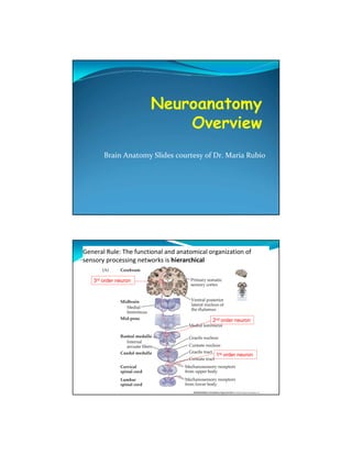
1. Brain Anatomy Slides courtesy of Dr. Maria Rubio
General Rule: The functional and anatomical organization of
sensory processing networks is hierarchical
3rd order neuron
2nd order neuron
1st order neuron
2. Gross Anatomy of the Spinal Cord
Spinal Nerves:
8 Cervical nerves
Neck, shoulder,
arms and hands
12 Thoracic nerves
Shoulders, chest,
and upper abdomen
5 lumbar nerves
Lower abdomen,
hips and legs
5 sacral nerves
Genitals and lower
digestive tract
Figure 16-1 M Figure 13-2
Gross Anatomy of the Spinal Cord
Cross Section:
Gray matter
Gray due to cell bodies
In CNS, gray matter also =
synapses
White Matter
White due to myelin; =
axons (both myelinated and
unmyelinated)
Dorsal Horn
Ventral Horn
Ventral Root
Dorsal Root
Cell bodies of unipolar
sensory neurons
No synapses
Dorsal Root Ganglion
3. Note functional
Sectional organization of gray matter
Anatomy of the
Spinal Cord
See 16.4M
Figure 6-41 V; 16.3M
Dorsal Columns Fiber Tracts:
Fasciculus cuneatus
Fasciculus gracilis
Ascending (Sensory)
Anterolateral columns Tracts
Note functional
E.g., spinothalamic tracts
organization of WHITE
Spinocerebellar tracts matter
4. Fiber Tracts: Descending (Motor) Tracts
Ventromedial System
Reticulospinal
Vestibulospinal
Tectospinal
Anterior Corticospinal
Dorsolateral System
Rubrospinal
Lateral Corticospinal
Pyramidal; extrapyramidal
terminology is outdated!
Spinal Nerves
Peripheral Nerve Structure
5. Dermatomes
Figure 16.6 M
Nerve Plexuses
A network of
interweaving anterior
rami of spinal nerves
Rami (pl.) ramus –
primary division of a
nerve or blood vessel
Cervical plexus
Brachial plexus
Lumbar plexus
Sacral plexus
6. Cervical Plexus
C1 – C4
Innervates neck
(sensory and
motor)
Phrenic nerve
(C3 - 4)
Innervates
diaphragm (so
you can
breathe!)
Figure 16.8 M
Brachial Plexus
C5 – T1
Sensory/motor
innervation of upper
extremity
More complex than
cervical plexuses
Anterior rami
Trunks
Divisions
Cords
Figure 16.9 M
7. Lumbar Plexus
L1 – L4
Supply lower limb of
each side
Less complex than
brachial plexus
Figure 16.10 M
Sacral Plexus
L4 – S4
Supply gluteal region,
plevis, perineum, and
lower limb of each
side
Together with
Lumbar plexus as
lumbosacral plexus
Figure 16.11 M
8. Components of Reflex Arc
See also Figure 16.12 M
Example of Deep Tendon Reflex
Patellar tendon (“knee jerk”) reflex
Clinical usefulness
9. Monosynaptic (Knee Jerk) and Disynaptic
(Flexor Withdrawal) Reflexes
Figure 16.13 M
Clinical usefulness of reflexes –
see table 16.6 M
Muscle tone
17. Cortex: Correlate Lobes with Function
ridges
Shallow
separation
Frontal
Motor, speech, personality,
emotion
Frontal lobotomies
Parietal
Somatosensory cortex, voluntary
movement
Occipital
Vision
Temporal
Hearing, balance, visual processing Figure 15-1 M
Lateral View
18. White Fiber Tracts
Projection fibers
Commisural fibers
Association fibers
19. Human Brain: coronal sections
Frontal
Caudal
Basal Ganglia
Nuclei important in motor
control
Caudate, putamen, globus
pallidus
Subthalamic nuclei,
substantia nigra
20. Internal Capsule
Between
putamen-globus
pallidus and
thalamus
Major
projection of
fibers to/from
cortex
Common CVA
(stroke) site
Rhinencephalon
Phylogenetically
ancient cortex
Olfactory bulb + tract
Fornix
Limbic system
Amygdala
Hippocampus
21. Limbic System
Emotional brain
Shape of a ring
(around
diencephalon)
Cingulate gyrus
Parahippocampal
gyrus
Hippocampus
Amygdala
Olfactory bulbs
Fornix
Diencephalon nuclei
Corpus Callosum: connects cerebral
hemispheres (with commisural fibers)
Figure 15-3 M
Cerebral Hemispheres: Frontal section at a:
Gray matter (cell bodies – in CNS, also SYNAPSES)
White matter (axons)
22. Figure 15-3 M
Cerebellum and Brainstem (section at b):
Gray matter (cell bodies + synapses)
White matter (axons)
Figure 15-3 M
Medulla (lower brainstem; section at c):
Gray matter (cell bodies + synapses)
White matter (axons)
23. Figure 15-3 M
Spinal Cord (section at d):
Gray matter (cell bodies + synapses)
White matter (axons)
Cranial Nerves
See lecture outline for
functions of each.
Ventral View
24. Ventricles (with CSF)
Research Martin Styner
The brain is a hollow organ
Brain Ventricles
Figure 15-6 M
25. Cranial Meninges
Figure 15-5 M
Cranial Meninges
Dura mater Arachnoid
Arachnoid
Pia mater
26. Spinal Meninges
Figure 16.2 M Figure 13–3
Cerebral Spinal Fluid (CSF)
Surrounds brain & spinal
cord; circulates through
ventricles
Cushions; protection
Formed in choroid plexus by
ependymal cells
Blood-brain barrier limits the
flow of solutes into CSF
Materials which easily pass
across the BBB:
Glucose, AAs, certain ions,
fatty acids, nicotine, CO,
CO2
BBB restricts these materials: Recall role of ASTROCYTES and
Blood, wastes (e.g. urea), TIGHT JUNCTIONS in forming BBB
proteins, K+ ions
28. The major arterial supply to the brain
Middle
Cerebral A.
Anterior
Cerebral A. Posterior
Cerebral A.
Bas
ilar
a
Internal Carotid A.
Vertebral A.
Circle of Willis
collateral circulation