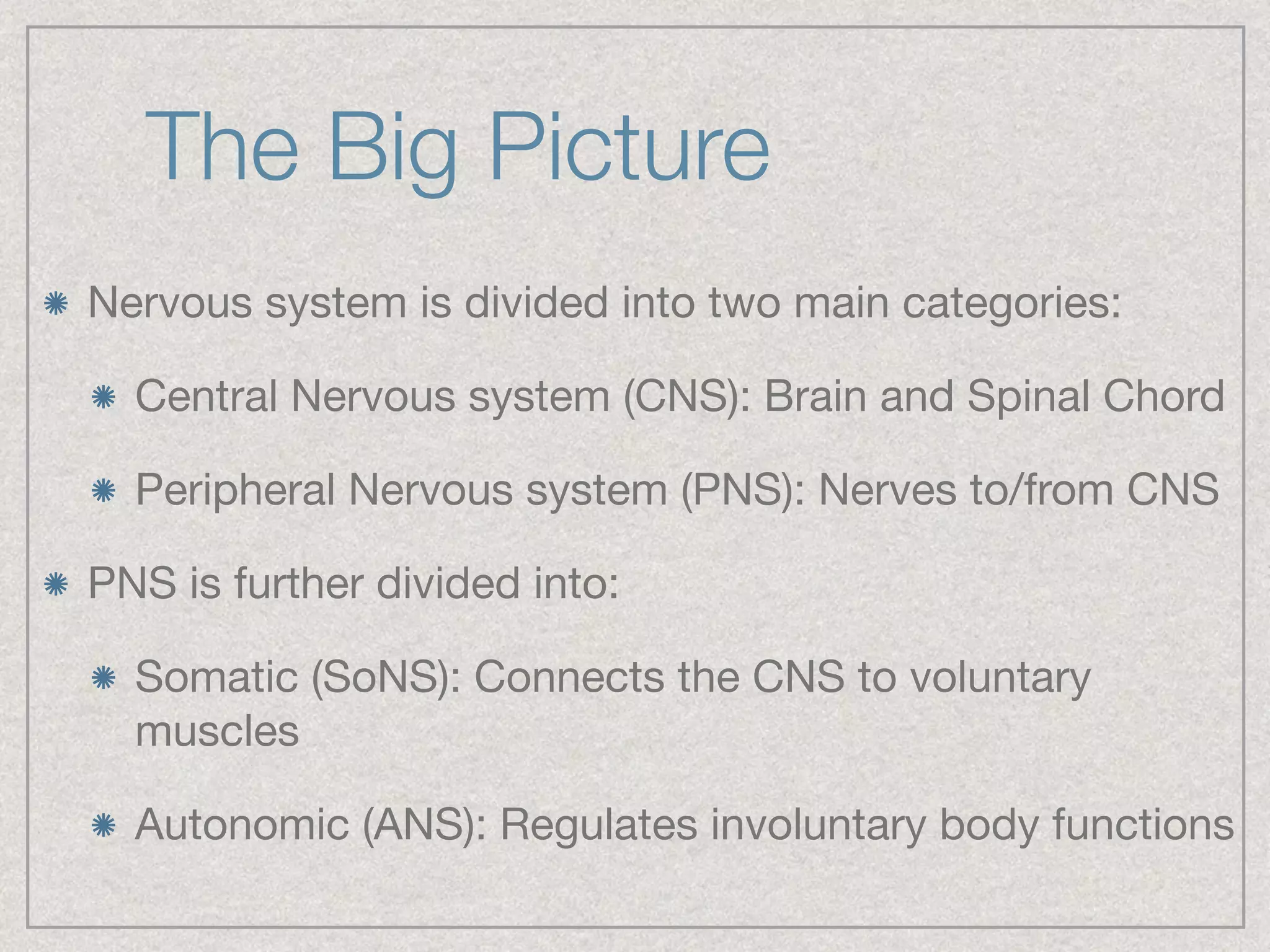
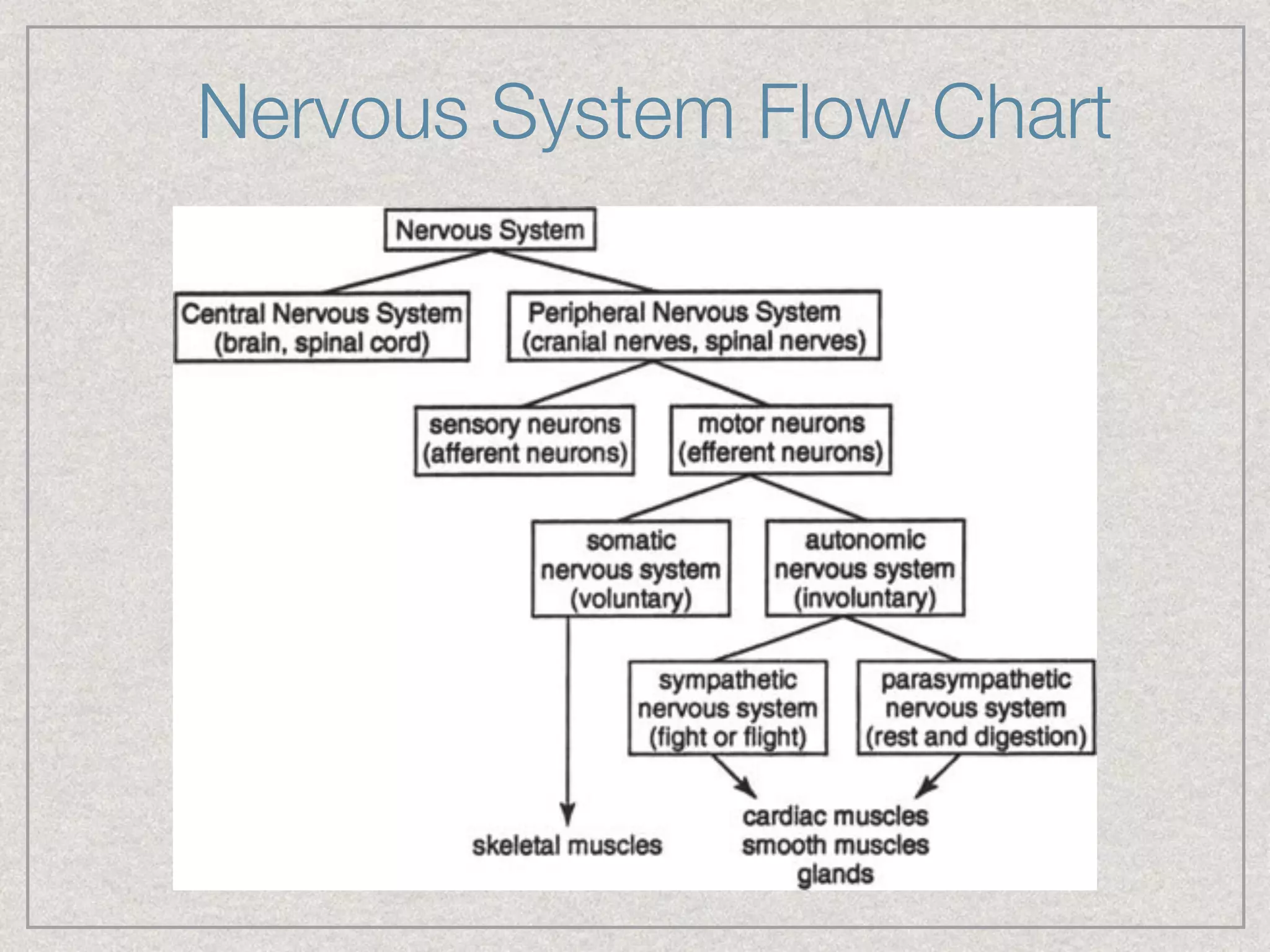
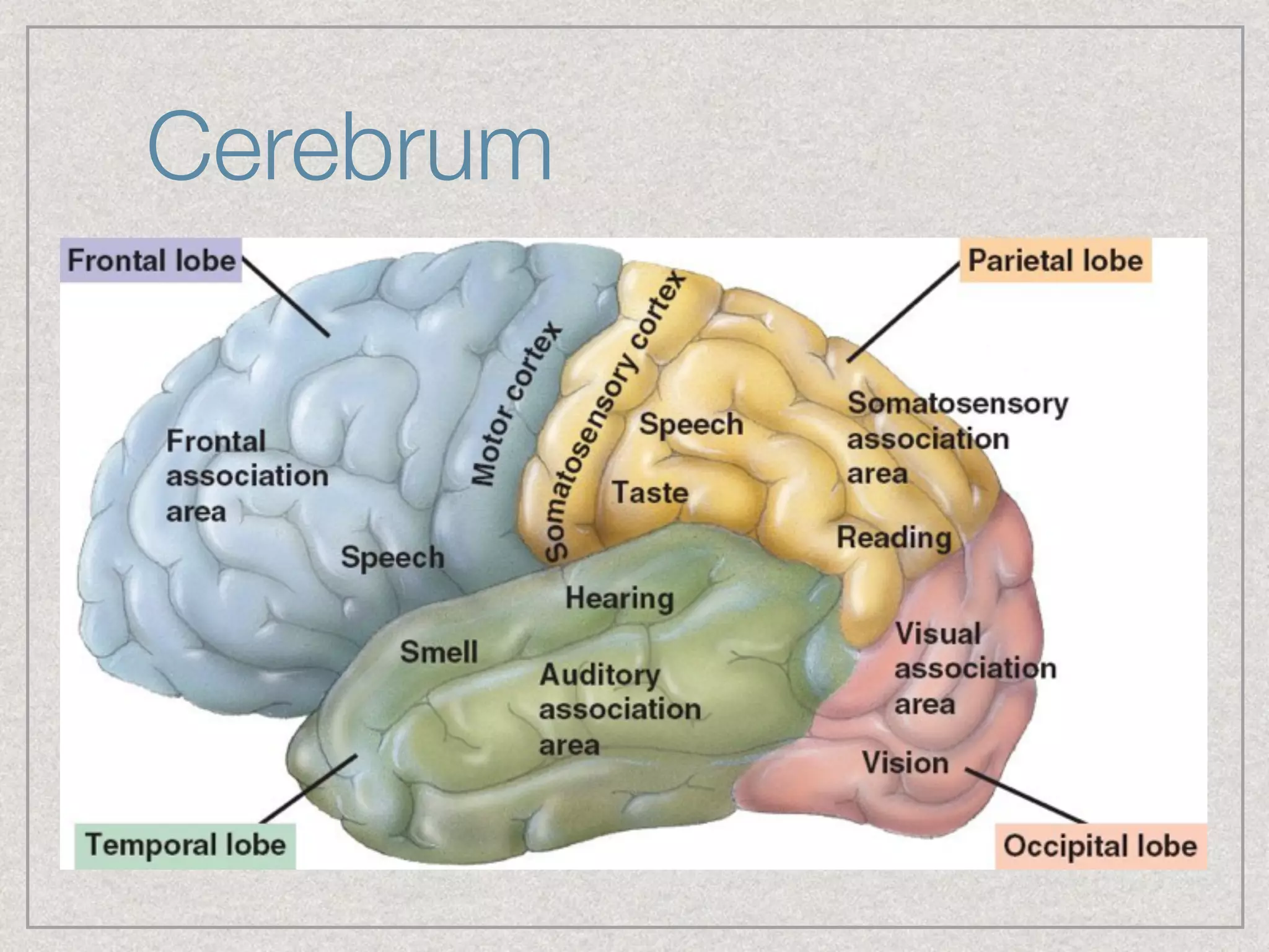
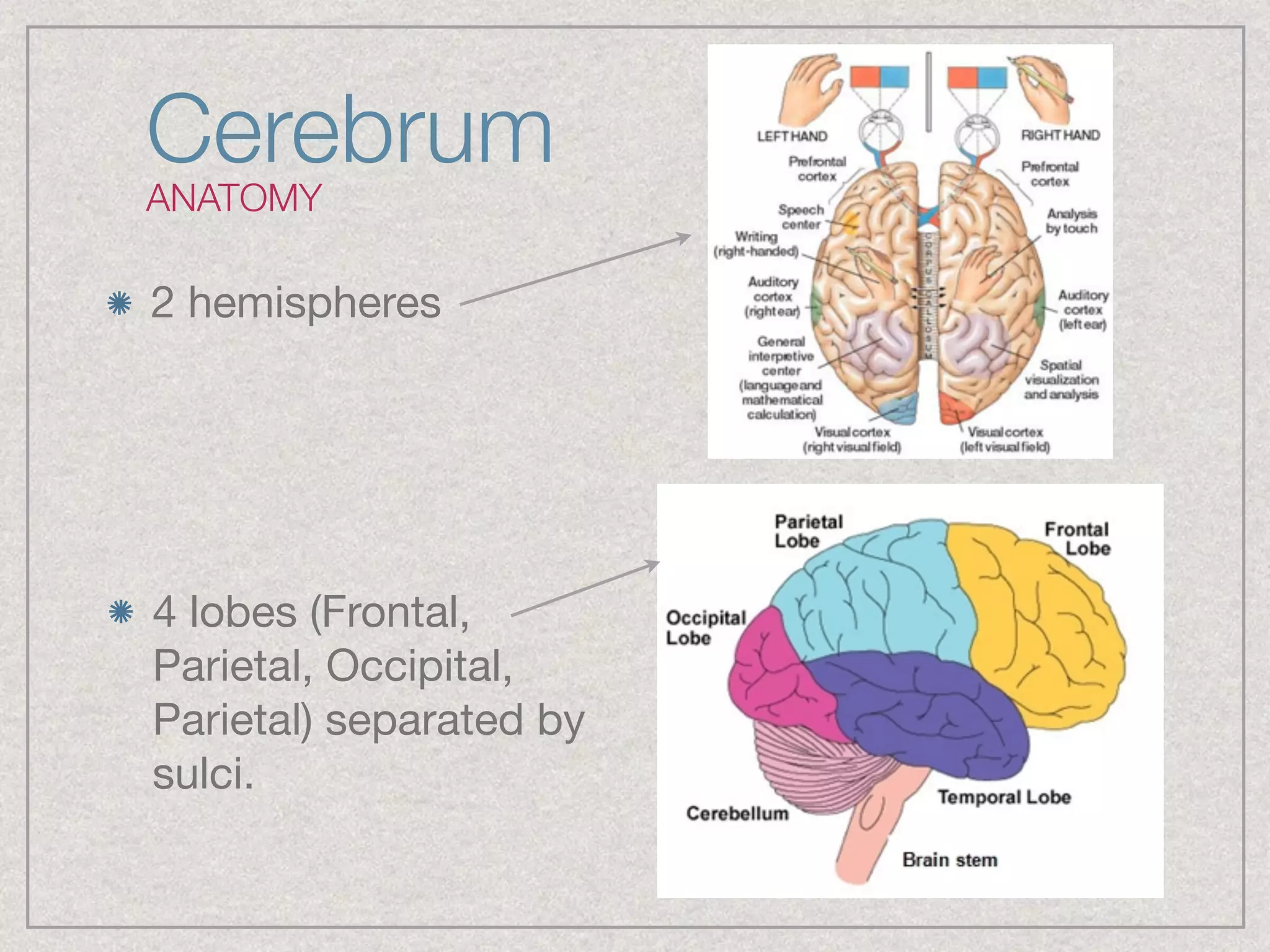
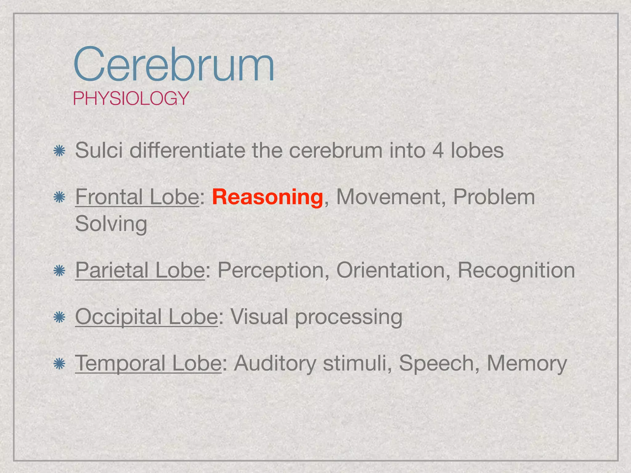
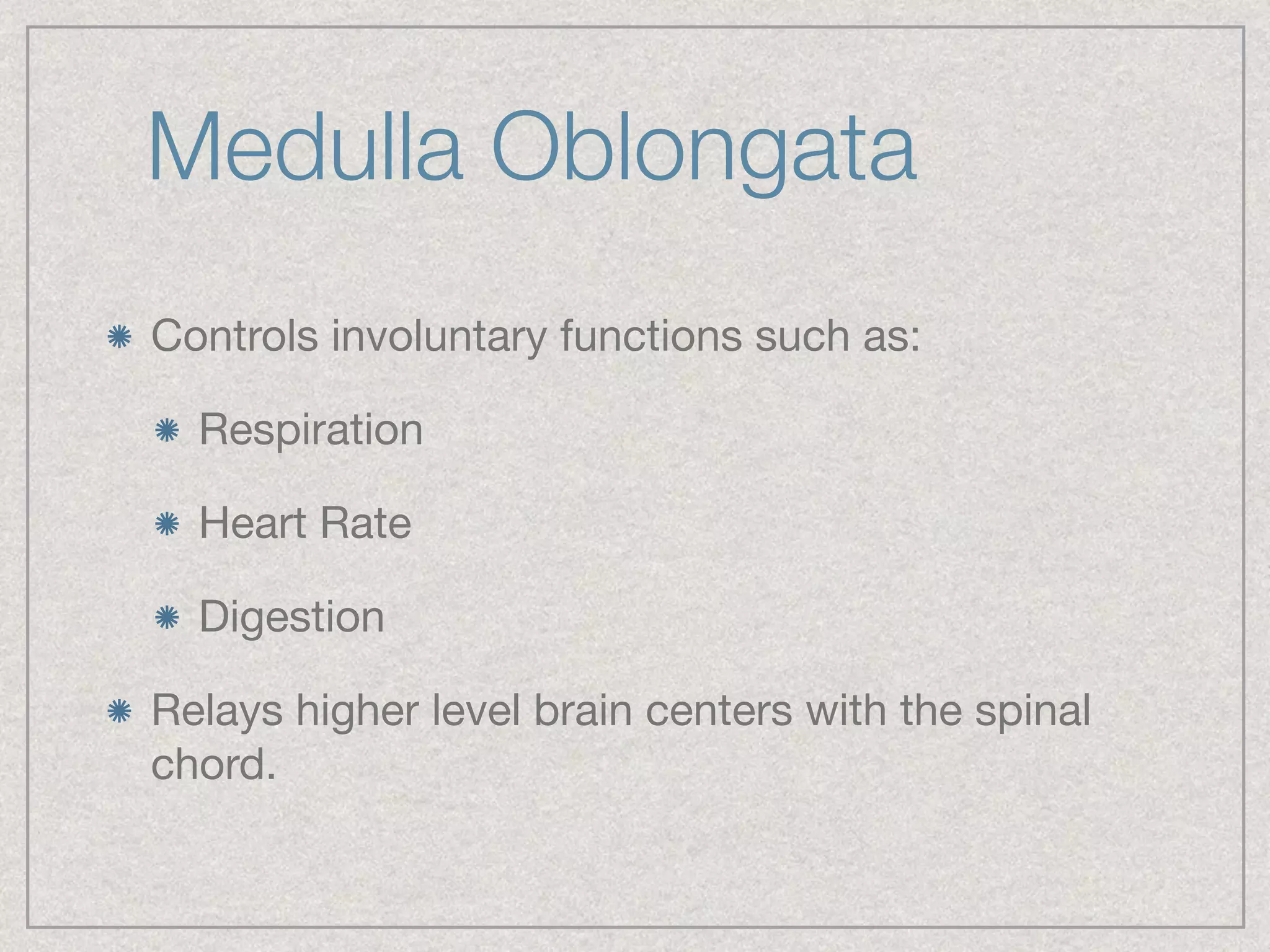
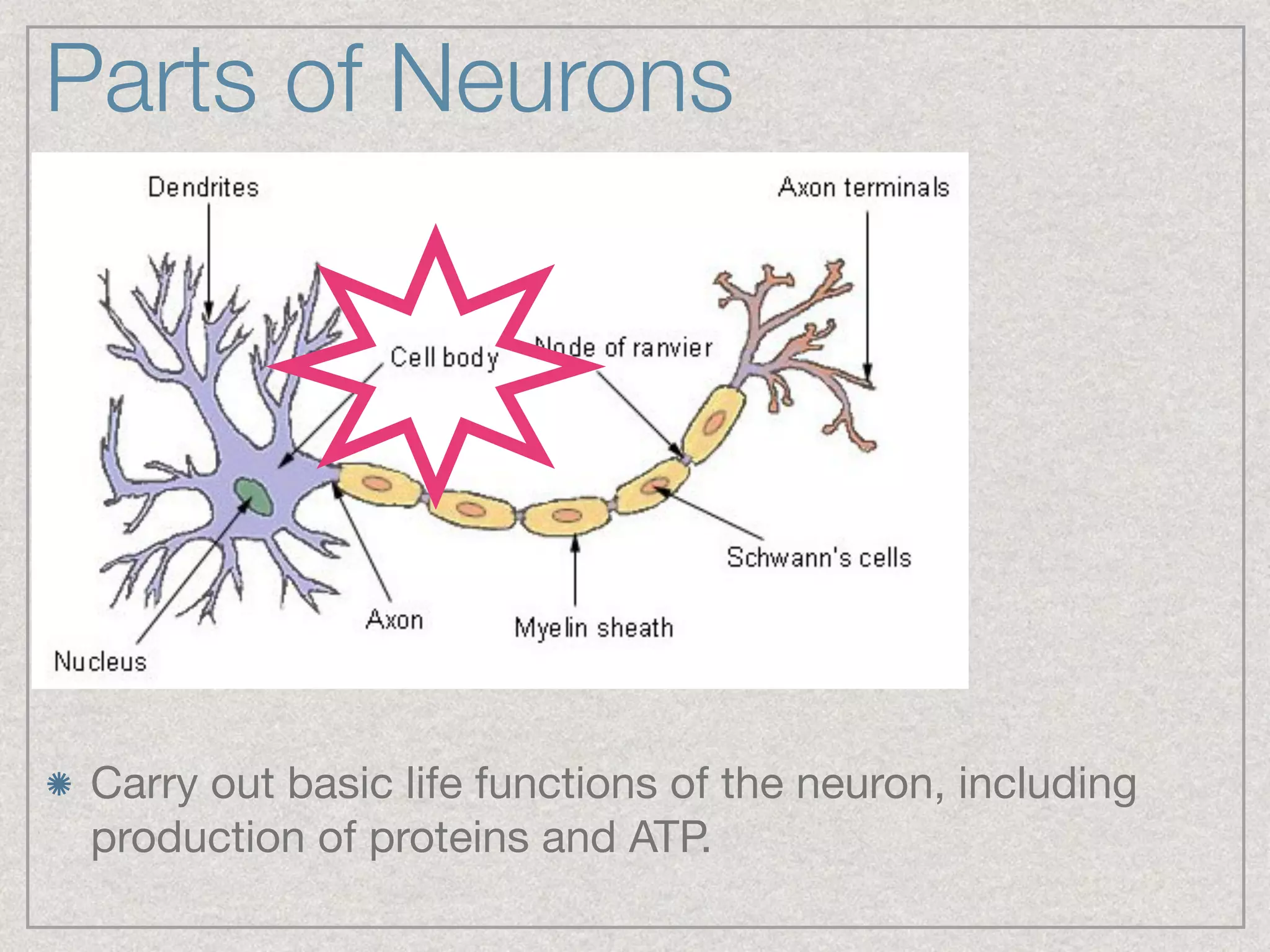
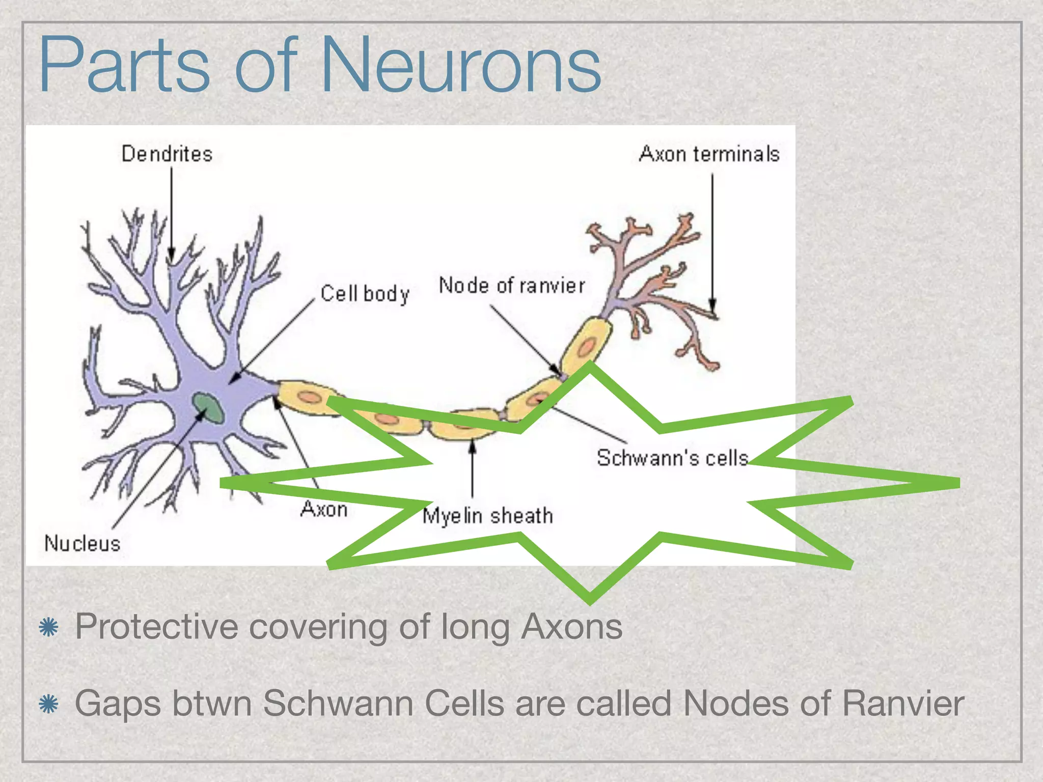
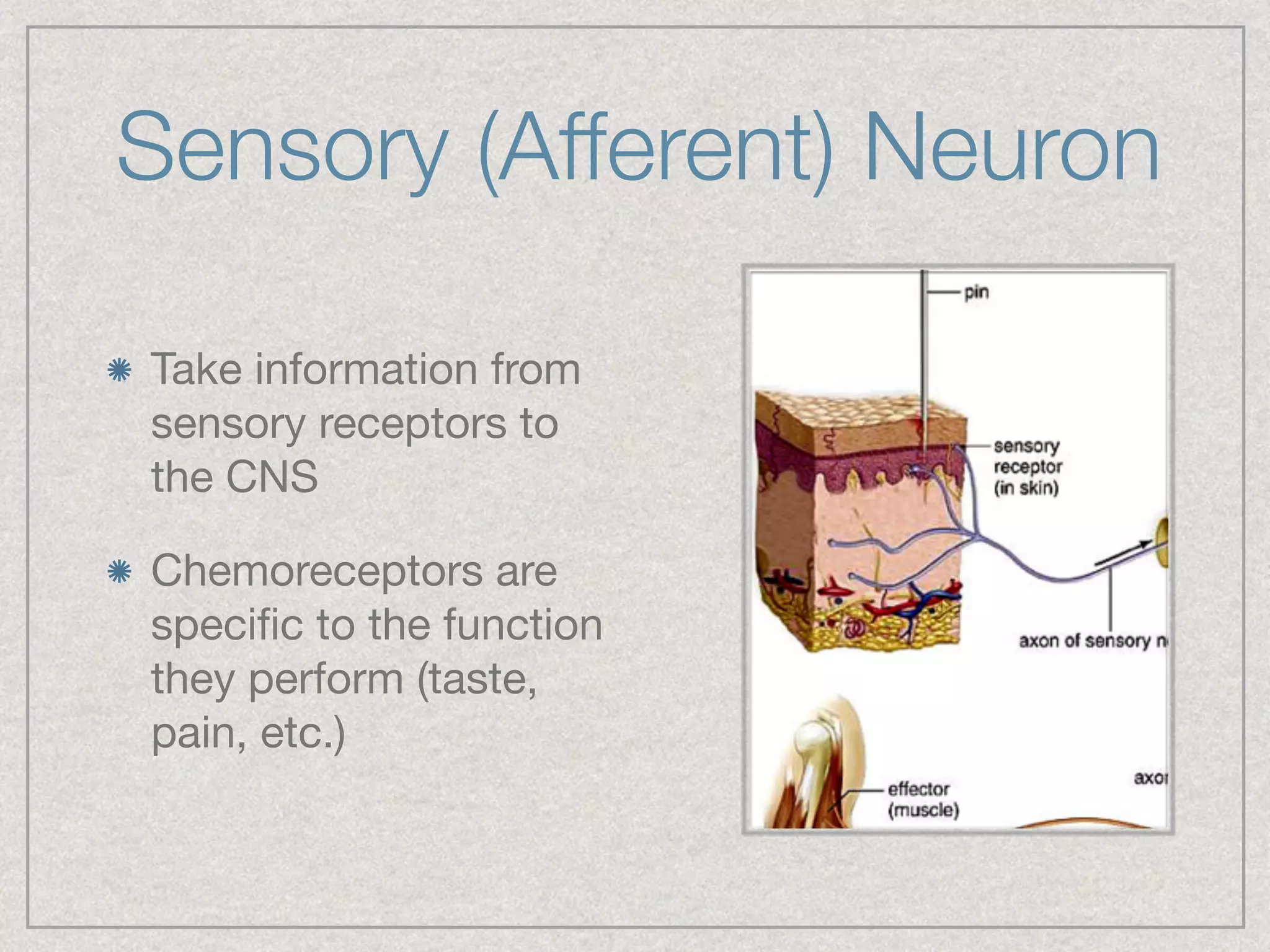
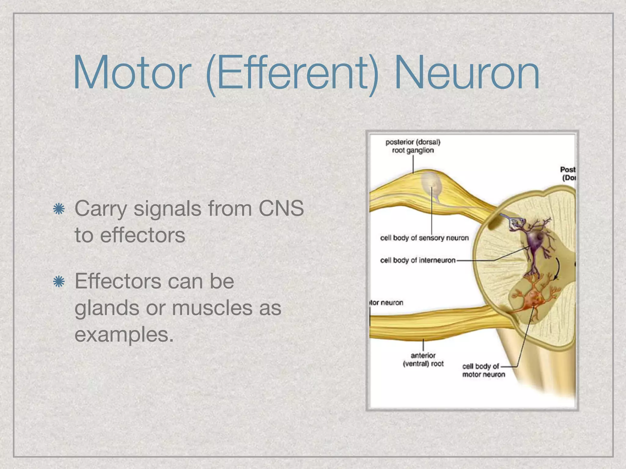
Unit 10 - The Nervous System
CNS, PNS, and associated components
The Big Picture
Nervous system is divided into two main categories:

Central Nervous system (CNS): Brain and Spinal Chord

Peripheral Nervous system (PNS): Nerves to/from CNS

PNS is further divided into:

Somatic (SoNS): Connects the CNS to voluntary
muscles

Autonomic (ANS): Regulates involuntary body functions
Big Picture Cont.
Autonomic is further divided into:

Sympathetic (SNS): “fight or flight”

Parasympathetic (PSNS): “rest and digest”
Nervous System Flow Chart
CNS
Parts and Functions
The Brain
The main components of the brain include:

cerebrum

corpus callosum

cerebellum

thalamus

hypothalamus

pituitary gland

medulla oblongata

meninges
Cerebrum
Cerebrum
ANATOMY
2 hemispheres
4 lobes (Frontal,
Parietal, Occipital,
Parietal) separated by
sulci.
Cerebrum
PHYSIOLOGY
Two hemispheres:

Right: creativity

Left: logic

Connected by the corpus callosum
CC is associated with integration of motor,
sensory, and cognitive functions between the
hemispheres.
Cerebrum
PHYSIOLOGY
Sulci differentiate the cerebrum into 4 lobes

Frontal Lobe: Reasoning, Movement, Problem
Solving

Parietal Lobe: Perception, Orientation, Recognition

Occipital Lobe: Visual processing

Temporal Lobe: Auditory stimuli, Speech, Memory
Cerebellum
ANATOMY
“Little Brain”

Two hemispheres with a highly folded surface.
Cerebellum
PHYSIOLOGY
Regulate eye movements

Coordinate limb movements

Maintain posture and balance

Motor decision making
Thalamus
Relays sensory
information from
the body to the
cerebral cortex.
Hypothalamus
Involved in: homeostasis, emotion, thirst, hunger,
circadian rhythms, and control of the autonomic nervous
system. 

Control of the pituitary gland
Pituitary Gland
“Master
Gland” of the
endocrine
system

Under control
of the
hypothalamus
Pituitary Gland
Medulla Oblongata
Medulla Oblongata
Controls involuntary functions such as:

Respiration

Heart Rate

Digestion

Relays higher level brain centers with the spinal
chord.
Meninges
3 Layers

Dura Mater: outer layer closest to skull

Arachnoid Mater: provides cushioning effect for CNS

Pia Mater: attached to brain, contains capillaries

Subdural haematoma (btwn dura and arachnoid)
resulting from traumatic event causes bleeding and
inter cranial pressure and brain trauma
Remember...
The Peripheral Nervous System is composed of:

Sensory Neurons (Afferent Neurons)

Motor Neurons (Efferent Neurons)

What are these made of and how do they transport
signals?
Parts of Neurons
Receive signals from other nerve cells

Covered in synapses
Parts of Neurons
Conveys electrical signals (range from 0.1mm to 2m)

Point of contact w/ cell body = Axon Hillock
Parts of Neurons
Carry out basic life functions of the neuron, including
production of proteins and ATP.
Parts of Neurons
Protective covering of long Axons

Gaps btwn Schwann Cells are called Nodes of Ranvier
Sensory (Afferent) Neuron
Take information from
sensory receptors to
the CNS

Chemoreceptors are
specific to the function
they perform (taste,
pain, etc.)
Interneuron
Act as middle men
between sensory and
motor neurons

Reside completely
within the CNS

Have many varieties
and are multipolar
(more than one
dendrite)
Motor (Efferent) Neuron
Carry signals from CNS
to effectors

Effectors can be
glands or muscles as
examples.
Signal Generation
Electrical activity is due to movement of ions past
the cell membrane (Na+ and K+)

Neurons have an Electrical Charge Different from
the Extracellular Fluid that surrounds them.  A
difference in electrical Charge between Two
Locations is called a POTENTIAL
Signal Generation
RESTING POTENTIAL
Positively charged Na+ pumped out of cell, K+ “leak” out
passively.

Causes a negative charge inside cell

Cell is now polarized and maintains this situation until
stimulated.
Signal Generation
THRESHOLD
Stimulation of the nerve is required to begin an
impulse down the nerve

One sufficient stimulation occurs we can say that a
THRESHOLD has been met

Once an impulse begins it can not be stopped,
hence an “all or none” system is employed by
neurons.
Signal Generation
ACTION POTENTIAL
Stimulation of the
nerve causes gates in
the neuron to open.

Gates allow positive
ions (Na+) to enter
neuron causing the
neuron to
DEPOLARIZE.
Signal Generation
ACTION POTENTIAL
After the impulse
passes K+ gates allow
K+ to leave cell

This causes
REPOLARIZATION of
the neuron.
Signal Generation
ACTION POTENTIAL
This depolarization
and repolarization is
what we call an action
potential or nerve
impulse.
Signal Generation
ACTION POTENTIAL
Na+/K+ pumps now
return the respective
ions to the proper side
of the cell membrane

This is called the
REFRACTORY period

No nerve signal can
propagate during this
period.
Signal Propagation
MYELINATION
Axons that are
myelinated propagate
signals faster

Signals jump between
the myelination
(nodes of ranvier)

Speeds of 200 m/s
can occur
The Synapse
Axons ends are slightly swollen and are called
AXON TERMINALS.

These terminals transmit signals with dendrites,
effectors (muscles) or receptors (sensors).

Small vesicles at the axon terminals contain
NEUROTRANSMITTERS.
The Synapse
NEUROTRANSMITTERS
Chemicals that are released into the synapse

Ex: Acetylcholine (ACh)

Join to receptors on neuron receiving the impulse

Cause membrane to become permeable to Na+ and a
depolarization of the neuron

Threshold is met, and a signal propagates along the
neuron.

Acetylocholinesterase rapidly breaks down ACh so that
the chemical signal stops
Reflex Arc
Reflex Arc
Sensory neurons are stimulated and send a signal
to the spinal chord

Interneurons relay the stimuli to motor neurons

Motor neurons send the signal to the effector
muscles

The Brain does not receive a signal before the
movement occurs and pain is not sensed until
after the effector has been stimulated
SYMPATHETIC
Fight or Flight

Increases HR

Inhibits digestion

Dilates pupils

Vasoconstriction

Increases respiration
PARASYMPATHETIC
Rest and Digest

Pupil constriction

Promotes digestion

Controls resting HR

Relaxes muscles
ANS BRANCHES

Nervous System Presentation