Downloaded 244 times

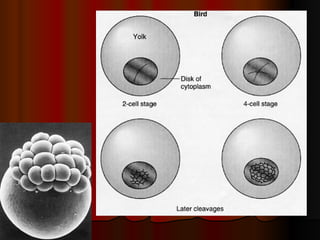
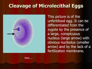
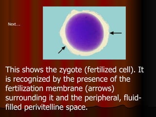
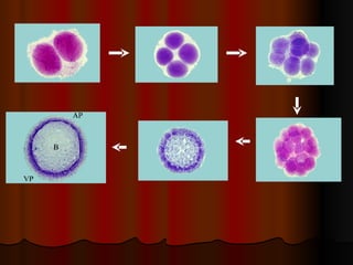
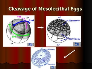
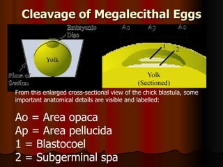
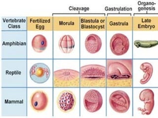
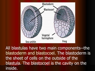


The document summarizes key stages in animal embryogenesis including fertilization, cleavage, blastulation, gastrulation, and neurulation. During fertilization, a sperm fuses with an egg to form a zygote. Cleavage involves cell divisions that form a ball of cells or blastula. Gastrulation establishes the three germ layers through cell movements. It occurs differently depending on egg characteristics like yolk content. Neurulation transforms the gastrula into a neurula by forming the neural tube from ectoderm.


