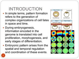
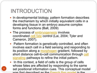
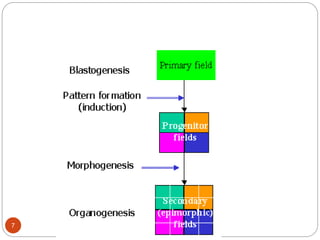
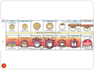
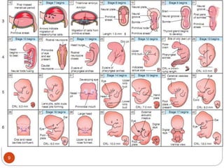
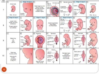
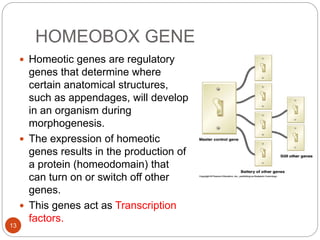
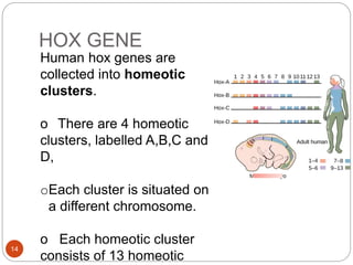
This document outlines a course on developmental biology and teratology. It discusses how pattern formation during embryogenesis is genetically controlled and involves cells responding to morphogen gradients and cell signaling pathways to develop spatial patterns. Key genes involved in pattern formation are homeobox genes, which help specify where anatomical structures will develop. In particular, Hox genes are organized in clusters and control patterning along the anteroposterior body axis. Mutations in genes of pattern formation can lead to various clinical congenital malformations and anomalies.
How 3 germ layers are formed in Chick that are endoderm, mesoderm and ectoderm.As Chick are polylecithal so cell movements are somewhat restricted and gastrulation is modified as compared to frog.
cell commitment and differentiation, stem cell,types of differentiationshallu kotwal
The commitment of cells to specific cell fates and their capacity to differentiate into particular kinds of cells.
Cellular differentiation is the process in which a cell changes from one cell type to another. Usually, the cell changes to a more specialized type. Differentiation occurs numerous times during the development of a multicellular organism as it changes from a simple zygote to a complex system of tissues and cell types. Differentiation continues in adulthood as adult stem cells divide and create fully differentiated daughter cells during tissue repair and during normal cell turnover.
Introduction
About Drosophila
Genome of Drosophila
Life cycle
Differentiation
Development of Drosophila
* Embryonic development
* Dorsal -ventral and
* Anterior posterior development
* Body segmentation
* Homeotic gene
Conclusion
Reference
INTRODUCTION
DEFINATION
GAMETES
STRUCTURE OF GAMETES
SPERM
OVUM
RECOGNITION OF EGG AND SPERM
CAPACITATION
ACROSOME REACTION
SPECIES-SPECIFIC RECOGNITION
GAMETE BINDING AND RECOGNITION
GAMETE FUSION
PREVENTION OF POLYSPERMY
ACTIVATION OF GAMETE METABOLISM
FUSION OF THE GENETIC MATERIAL
SIGNIFICANCE OF FERTILIZATION
CONCLUSIONS
REFERENCES
Welcome to the world of Homeotic genes. In this presentation I talk about the interesting history behind homeotic genes as to how it was discovered. Also, the various deformities in Drosophila related to mutations in homeotic genes and the characteristics of homeotic genes. I also talk about hox genes in humans and their function.
cell lineage , cell fate - diverse class of cell fate, cell fate in plant meristem, mammalian development cell fate, nutritional effects on epigenetics, epigenetics of plants,
control of cell fate.
Here is a presentation of development biology. For further assistance contact at my email. It's a pleasure to me.if my work is helpful for anyone of you. Need your appreciation and criticism to improve my work. Thanks.
Anomalies of the first and second branchial archesDr Medical
https://userupload.net/8n9v7tg9jkl1
Anomalies of the branchial arches are the second most common congenital lesions of the head and neck in children [1]. They may present as cysts, sinus tracts, fistulae or cartilaginous remnants and present with typical clinical and radiological patterns dependent on which arch is involved. The course of a particular branchial anomaly is caudal to the structures derived from the corresponding arch and dorsal to the structures that develop from the following arch. Branchial anomalies are further typed into cysts, sinuses, and fistulas.
3. INTRODUCTION
3
In simple terms, pattern formation
refers to the generation of
complex organizations of cell fates
in space and time.
During embryogenesis,
information encoded in the
genome is translated into cell
proliferation, morphogenesis, and
early stages of differentiation.
Embryonic pattern arises from the
spatial and temporal regulation
and coordination of these events.
4. INTRODUCTION
4
In developmental biology, pattern formation describes
the mechanism by which initially equivalent cells in a
developing tissue in an embryo assume complex
forms and functions (Ball, 2009)
The process of embryogenesis involves
coordinated cell fate control (Lai, 2004; Tyler and
Cameron, 2007).
Pattern formation is genetically controlled, and often
involves each cell in a field sensing and responding to
its position along a morphogen gradient, followed by
short distance cell-to-cell communication through cell
signaling pathways to refine the initial pattern.
In this context, a field of cells is the group of cells
whose fates are affected by responding to the same
set positional information cues. This conceptual model
5. Why Pattern Formation?
5
The reliable development of highly complex
organisms is an intriguing and fascinating
problem. The genetic material is, as a rule, the
same in each cell of an organism. How do then
cells, under the influence of their common genes,
produce spatial patterns ?
Development of an organism is, of course, under
genetic control but the genetic information is
usually the same in all cells.
A crucial problem is therefore the generation of
spatial patterns that allow a different fate of some
cells in relation to others
(Koch and Meinhardt, 1994).
6. DEFINITION OF TERMS
6
Induction is the stimulation of a cell to differentiate in
response to a stimulus produced by another cell. It is
mediated by inducer substances that diffuse from
one cell to another. It results in cell determination.
Determination is the commitment of a cell to undergo
differentiation. It is an irreversible process but is not
accompanied by morphological changes.
Determinants are the cytoplasmic effector molecules
that mediate determination.
Differentiation is the variation in the pattern of
expression of a common set of genes to form cells of
diverse morphology and function.
11. Time-table of landmarks in early
human development
11
Day 1 - cleavage
Days 2-4 - morula; free-
floating conceptus in uterine
tube
Days 5-6 - formation of
the blastocyst and
embryoblast;
- implantation
Week 2 (days 7-14)
- formation of the
bilaminar embryo 0.1 mm
Week 3 (days 15-20) -
formation of the trilaminar
embryo 1.0 mm
Week 4 (days 21-28)
Day 21 - formation of neural
tube 2.0 mm
Day 22 - formation of the heart
Day 23 - formation of eye and
ear rudiments
Day 25 - formation of branchial
arches
Day 26 - formation of upper
limb bud
Day 28 - formation of the lower
limb bud 5.0 mm
Weeks 5 to 9 (2nd month) - Period of
organogenesis
Week
6 1.0
cm
Week 9
4.0 cm
12. GENES OF PATTERN
FORMATION
12
Every organism has a unique body pattern.
This patterning is controlled and influenced by the
HOMEOBOX genes.
These specify how different areas of the body
develop their individual structures, e.g. Arms, legs
etc.
13. HOMEOBOX GENE
13
Homeotic genes are regulatory
genes that determine where
certain anatomical structures,
such as appendages, will develop
in an organism during
morphogenesis.
The expression of homeotic
genes results in the production of
a protein (homeodomain) that
can turn on or switch off other
genes.
This genes act as Transcription
factors.
14. HOX GENE
14
Human hox genes are
collected into homeotic
clusters.
o There are 4 homeotic
clusters, labelled A,B,C and
D,
oEach cluster is situated on
a different chromosome.
o Each homeotic cluster
consists of 13 homeotic
15. The RNA expression pattern of three mouse Hox genes in the
vertebral column of a sectioned 12.5-day-old mouse embryo:
the anterior limit of each of the expression pattern is different
Each Hox gene is expressed in a continuous block beginning at a
Specific anterior limit and running posteriorly to the end of the
developing vertebral column
16. HOX GENE
16
The four numerically corresponding genes for the four
different clusters form a paralogous group.
o The hox genes are responsible for patterning along
the antero-posterior axis.
o The genes are expressed sequentially beginning with
the paralogous group 1, which is expressed first
o The sequential genes specify different segments in
cranio-caudal sequence extending from paralogous
group 1, which specifies the most cranial structures, to
paralogous group 13, which specifies the most caudal
structures.
o Thus the first genes to be expressed specify the most
cranial structures while the last to be expressed specify
the most caudal structures. This is responsible for the
cranio-caudal sequence of development, where the
more cranial segments develop slightly before the
more caudal structures. Consequently the upper limb
develops ahead of the lower limb.
17. Clinical Correlates
17
Mutations in genes of pattern formation leads to a
lot of clinical important congenital malformations
and anomalies
Aniridia
Synpolydactyly
Axenfeld-Rieger syndrome
Branchiootorenal syndrome
Coloboma
Langer mesomelic dysplasia
Léri-Weill dyschondrosteosis
Microphthalmia
Mowat-Wilson syndrome
Amelia
Limb deformities
21. REFERENCES
21
• A. J. Koch and H. Meinhardt (1994). Biological
Pattern Formation : from Basic Mechanisms to
Complex Structures. Rev. Modern Physics 66,
1481-1507
• Ball, (2009). Shapes, pp. 261–290.
• Eric C. Lai (2004). "Notch signaling: control of
cell communication and cell fate" 131 (5). pp.
965–73. doi:10.1242/dev.01074
• Melinda J. Tyler, David A. Cameron (2007).
"Cellular pattern formation during retinal
regeneration: A role for homotypic control of cell
fate acquisition". Vision Research 47 (4): 501–
time-table of landmarks in early human development
Day 1 - cleavage
Days 2-4 - morula; free-floating conceptus in uterine tube
Days 5-6 - formation of the blastocyst and embryoblast;
- implantation
Week 2 (days 7-14)
- formation of the bilaminar embryo 0.1 mm
Week 3 (days 15-20)
-formation of the trilaminar embryo 1.0 mm
Week 4 (days 21-28)
Day 21 - formation of neural tube 2.0 mm
Day 22 - formation of the heart
Day 23 - formation of eye and ear rudiments
Day 25 - formation of branchial arches
Day 26 - formation of upper limb bud
Day 28 - formation of the lower limb bud 5.0 mm
Weeks 5 to 9 (2nd month)
- Period of organogenesis
Week 6 1.0 cm
Week 9 4.0 cm
End of embryonic period