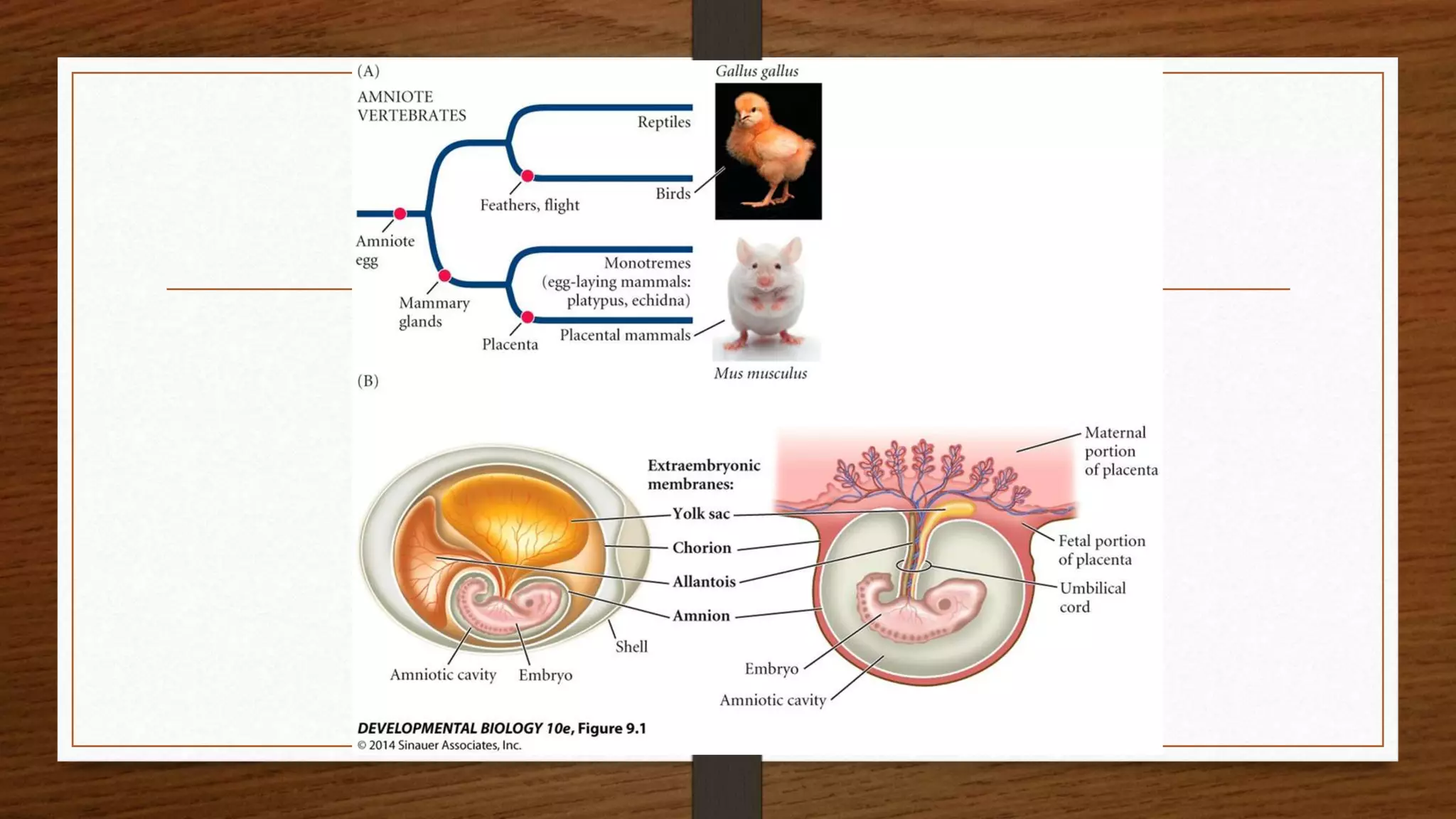
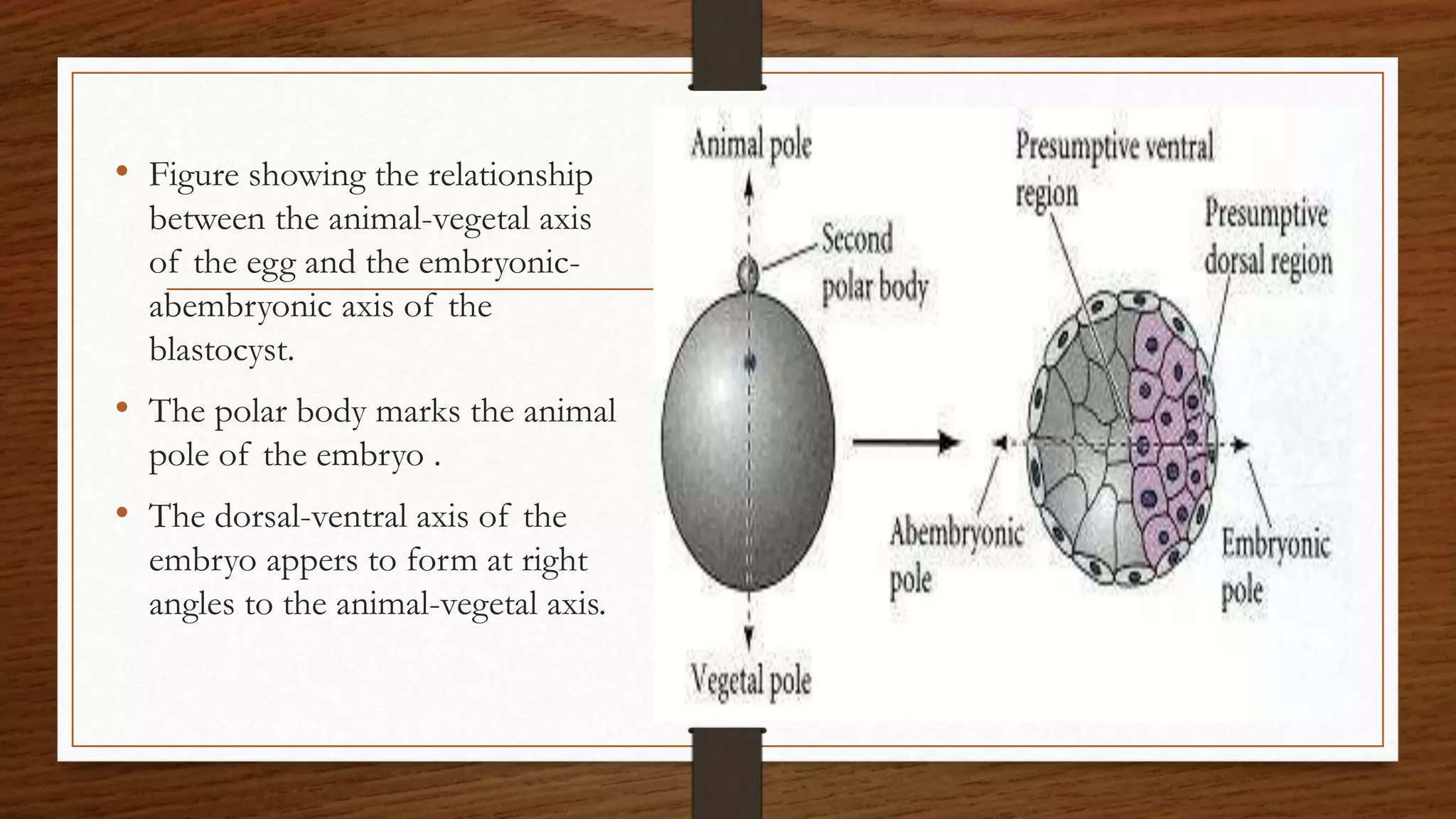
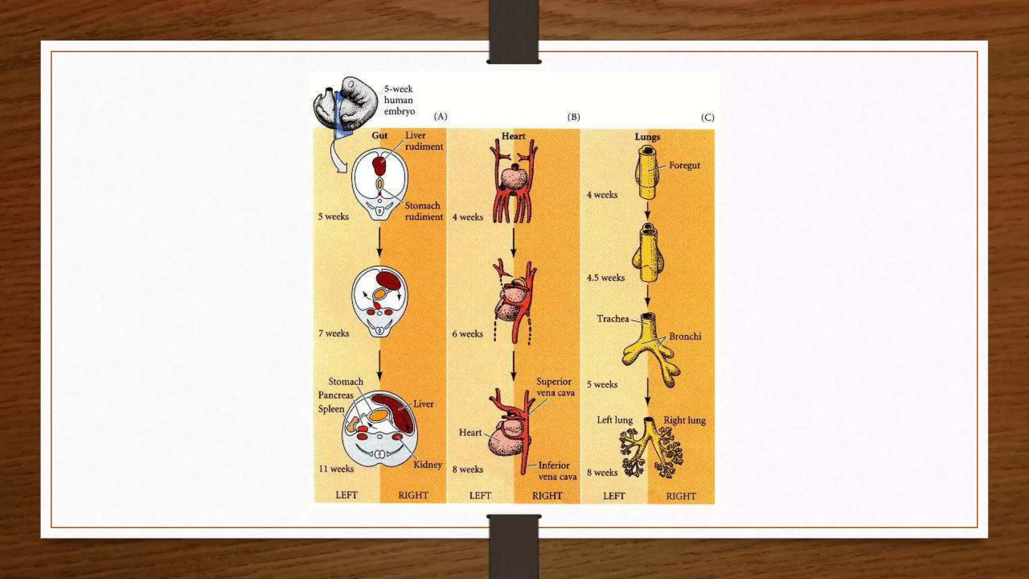
Polyspermy describes an egg that has been fertilized by more than one sperm. Diploid organisms normally contain two copies of each chromosome, one from each parent. The cell resulting from polyspermy
The first issue that an egg and a sperm of any organism type face in successfully producing an embryo is the possibility of polyspermy. Polyspermy is the fertilization of an egg by multiple sperm, and the results of such unions are lethal.
If multiple sperm fertilize an egg, the embryo inherits multiple paternal centrioles. This causes competition for extra chromosomes and results in the disruption of the creation of the cleavage furrow, thus causing the zygote to die. As an important model organism in the study of fertilization and embryonic development, polyspermy in sea urchins has been studied in detail. The sea urchin’s methods of polyspermy prevention have been broken down into two main pathways. These two primary pathways are known as the fast block and the slow block to polyspermy
After the sperm’s receptors come into contact with the egg’s jelly layer and the acrosomal enzymes are released and break down the jelly layer, the sperm head comes into contact with the vitelline and plasma membranes of the egg. When the two plasma membranes contact one another, signals in the egg are initiated.
First, Na+ channels in the egg open, allowing Na+ to flood into the egg. This causes a depolarization of the egg from it’s normal resting potential of -70 mV.
While depolarization is occurring, the remainder of the jelly layer is dissolving. With the dissolution of the jelly layer and the depolarization of the plasma membrane, the first block to preventing fertilization by multiple sperm is put into place.
These two simple changes are part of the first block to polyspermy, known as the fast block. Within 1/10th of a second of contact, the fast block t
cell commitment and differentiation, stem cell,types of differentiationshallu kotwal
The commitment of cells to specific cell fates and their capacity to differentiate into particular kinds of cells.
Cellular differentiation is the process in which a cell changes from one cell type to another. Usually, the cell changes to a more specialized type. Differentiation occurs numerous times during the development of a multicellular organism as it changes from a simple zygote to a complex system of tissues and cell types. Differentiation continues in adulthood as adult stem cells divide and create fully differentiated daughter cells during tissue repair and during normal cell turnover.
Introduction
About Drosophila
Genome of Drosophila
Life cycle
Differentiation
Development of Drosophila
* Embryonic development
* Dorsal -ventral and
* Anterior posterior development
* Body segmentation
* Homeotic gene
Conclusion
Reference
How 3 germ layers are formed in Chick that are endoderm, mesoderm and ectoderm.As Chick are polylecithal so cell movements are somewhat restricted and gastrulation is modified as compared to frog.
It describes the gamete fusion and early development in mammals.
Compaction,cavitation,Blastocyst, gastrula formation, Extra embryonic membranes development in mammals. Formation of twins, difference between monozygotic and dizygotic twins.
This is a slide for complete development in chick ,as chick is a vertebrate so with the help of the development in a chick we can we can understand development in vertebrates .
This topic explains the whole process of growth and development in animal the processes include
Fertilization and incubation
Cleavage
Morula
Blastula
Gastrulation
Notochord And Mesoderm Formation
Neurulation
Professional air quality monitoring systems provide immediate, on-site data for analysis, compliance, and decision-making.
Monitor common gases, weather parameters, particulates.
Slide 1: Title Slide
Extrachromosomal Inheritance
Slide 2: Introduction to Extrachromosomal Inheritance
Definition: Extrachromosomal inheritance refers to the transmission of genetic material that is not found within the nucleus.
Key Components: Involves genes located in mitochondria, chloroplasts, and plasmids.
Slide 3: Mitochondrial Inheritance
Mitochondria: Organelles responsible for energy production.
Mitochondrial DNA (mtDNA): Circular DNA molecule found in mitochondria.
Inheritance Pattern: Maternally inherited, meaning it is passed from mothers to all their offspring.
Diseases: Examples include Leber’s hereditary optic neuropathy (LHON) and mitochondrial myopathy.
Slide 4: Chloroplast Inheritance
Chloroplasts: Organelles responsible for photosynthesis in plants.
Chloroplast DNA (cpDNA): Circular DNA molecule found in chloroplasts.
Inheritance Pattern: Often maternally inherited in most plants, but can vary in some species.
Examples: Variegation in plants, where leaf color patterns are determined by chloroplast DNA.
Slide 5: Plasmid Inheritance
Plasmids: Small, circular DNA molecules found in bacteria and some eukaryotes.
Features: Can carry antibiotic resistance genes and can be transferred between cells through processes like conjugation.
Significance: Important in biotechnology for gene cloning and genetic engineering.
Slide 6: Mechanisms of Extrachromosomal Inheritance
Non-Mendelian Patterns: Do not follow Mendel’s laws of inheritance.
Cytoplasmic Segregation: During cell division, organelles like mitochondria and chloroplasts are randomly distributed to daughter cells.
Heteroplasmy: Presence of more than one type of organellar genome within a cell, leading to variation in expression.
Slide 7: Examples of Extrachromosomal Inheritance
Four O’clock Plant (Mirabilis jalapa): Shows variegated leaves due to different cpDNA in leaf cells.
Petite Mutants in Yeast: Result from mutations in mitochondrial DNA affecting respiration.
Slide 8: Importance of Extrachromosomal Inheritance
Evolution: Provides insight into the evolution of eukaryotic cells.
Medicine: Understanding mitochondrial inheritance helps in diagnosing and treating mitochondrial diseases.
Agriculture: Chloroplast inheritance can be used in plant breeding and genetic modification.
Slide 9: Recent Research and Advances
Gene Editing: Techniques like CRISPR-Cas9 are being used to edit mitochondrial and chloroplast DNA.
Therapies: Development of mitochondrial replacement therapy (MRT) for preventing mitochondrial diseases.
Slide 10: Conclusion
Summary: Extrachromosomal inheritance involves the transmission of genetic material outside the nucleus and plays a crucial role in genetics, medicine, and biotechnology.
Future Directions: Continued research and technological advancements hold promise for new treatments and applications.
Slide 11: Questions and Discussion
Invite Audience: Open the floor for any questions or further discussion on the topic.
Observation of Io’s Resurfacing via Plume Deposition Using Ground-based Adapt...Sérgio Sacani
Since volcanic activity was first discovered on Io from Voyager images in 1979, changes
on Io’s surface have been monitored from both spacecraft and ground-based telescopes.
Here, we present the highest spatial resolution images of Io ever obtained from a groundbased telescope. These images, acquired by the SHARK-VIS instrument on the Large
Binocular Telescope, show evidence of a major resurfacing event on Io’s trailing hemisphere. When compared to the most recent spacecraft images, the SHARK-VIS images
show that a plume deposit from a powerful eruption at Pillan Patera has covered part
of the long-lived Pele plume deposit. Although this type of resurfacing event may be common on Io, few have been detected due to the rarity of spacecraft visits and the previously low spatial resolution available from Earth-based telescopes. The SHARK-VIS instrument ushers in a new era of high resolution imaging of Io’s surface using adaptive
optics at visible wavelengths.
Seminar of U.V. Spectroscopy by SAMIR PANDASAMIR PANDA
Spectroscopy is a branch of science dealing the study of interaction of electromagnetic radiation with matter.
Ultraviolet-visible spectroscopy refers to absorption spectroscopy or reflect spectroscopy in the UV-VIS spectral region.
Ultraviolet-visible spectroscopy is an analytical method that can measure the amount of light received by the analyte.
This pdf is about the Schizophrenia.
For more details visit on YouTube; @SELF-EXPLANATORY;
https://www.youtube.com/channel/UCAiarMZDNhe1A3Rnpr_WkzA/videos
Thanks...!
Deep Behavioral Phenotyping in Systems Neuroscience for Functional Atlasing a...Ana Luísa Pinho
Functional Magnetic Resonance Imaging (fMRI) provides means to characterize brain activations in response to behavior. However, cognitive neuroscience has been limited to group-level effects referring to the performance of specific tasks. To obtain the functional profile of elementary cognitive mechanisms, the combination of brain responses to many tasks is required. Yet, to date, both structural atlases and parcellation-based activations do not fully account for cognitive function and still present several limitations. Further, they do not adapt overall to individual characteristics. In this talk, I will give an account of deep-behavioral phenotyping strategies, namely data-driven methods in large task-fMRI datasets, to optimize functional brain-data collection and improve inference of effects-of-interest related to mental processes. Key to this approach is the employment of fast multi-functional paradigms rich on features that can be well parametrized and, consequently, facilitate the creation of psycho-physiological constructs to be modelled with imaging data. Particular emphasis will be given to music stimuli when studying high-order cognitive mechanisms, due to their ecological nature and quality to enable complex behavior compounded by discrete entities. I will also discuss how deep-behavioral phenotyping and individualized models applied to neuroimaging data can better account for the subject-specific organization of domain-general cognitive systems in the human brain. Finally, the accumulation of functional brain signatures brings the possibility to clarify relationships among tasks and create a univocal link between brain systems and mental functions through: (1) the development of ontologies proposing an organization of cognitive processes; and (2) brain-network taxonomies describing functional specialization. To this end, tools to improve commensurability in cognitive science are necessary, such as public repositories, ontology-based platforms and automated meta-analysis tools. I will thus discuss some brain-atlasing resources currently under development, and their applicability in cognitive as well as clinical neuroscience.
Richard's entangled aventures in wonderlandRichard Gill
Since the loophole-free Bell experiments of 2020 and the Nobel prizes in physics of 2022, critics of Bell's work have retreated to the fortress of super-determinism. Now, super-determinism is a derogatory word - it just means "determinism". Palmer, Hance and Hossenfelder argue that quantum mechanics and determinism are not incompatible, using a sophisticated mathematical construction based on a subtle thinning of allowed states and measurements in quantum mechanics, such that what is left appears to make Bell's argument fail, without altering the empirical predictions of quantum mechanics. I think however that it is a smoke screen, and the slogan "lost in math" comes to my mind. I will discuss some other recent disproofs of Bell's theorem using the language of causality based on causal graphs. Causal thinking is also central to law and justice. I will mention surprising connections to my work on serial killer nurse cases, in particular the Dutch case of Lucia de Berk and the current UK case of Lucy Letby.
1. Axis formation in Birds and
Mammals
PG Biotechnology
Department-Plant Biology and Biotechnology
Cell and developmental biology
2. Introduction
• A multicellular organism develops from a single cell (the zygote) into a
collection of many different cell types, organized into tissues and organs.
• Development involves cell division, body axis formation, tissue and organ
development, and cell differentiation (gaining a final cell type identity).
3. Amniotic Eggs-Found in birds , reptiles
and mammals
Amniotic eggs contain membranes that are designed to allow survival on land:
amnion – the membrane that allows the embryo to float in a fluid environment to
avoid desiccation
yolk sac – enables nutrient uptake and development of the circulatory system
chorion – contains blood vessels that exchange gasses with the external environment.
4.
5. Types of Cleavage
• Meroblastic Cleavage: Eggs having high yolk content (telolecithal eggs) the
cytoplasm and nucleus are restricted to animal pole region and the cleavage
plane continuously divides this region . Yolk region does not get divided.
When viewed from the pole a disc is appeared (as the mass of cells float
around the yolk)this disc is known as the blastodisc and the cleavage is
known as meroblastic cleavage. Example : Found in eggs of birds
• Holoblastic Cleavage: The cleavage planes divides the zygote completely
then it is known as holoblastic cleavage. Example : found in eggs of
mammals.
6. Early development in Birds (Chick)
• Domestic chicken is used in embryological studies
• Easily accessible and easily raised
• Meroblastic cleavage- Blastodisc seen
• First cleavage furrow appears in the blastodisc
• Other cleavage form the single layered blastoderm
• Equatorial and vertical cleavages:- five- six layered blastoderm
7. Dicoidal Meroblastic
Cleavage in birds
Area Pellucida - Deep cells present
in the centre of the blastoderm that
are shed and disc leaving behind is
area pellucida .
Area Opaca –the peripheral ring of
blastodermal cells that has not shed
their deep cells constitute the area
opaca.
Marginal Zone or Marginal Belt:
a thin layer between area pellucida
and area opaca is the marginal zone.
They become important in
determining the cell fate of early
chick development.
8. Gastrulation in the Avian (chick) Embryo
• The embryo of the chick blastula is two layered
1. Upper layer- epiblast 2. Lower layer – primary hypoblast
• Formation of the Secondary Hypoblast
Koller’s Sickle : In avian gastrulation, Koller's sickle is a local thickening of cells that
acts as a margin separating sheets of cells.
Primitive streak :After 3-4 hrs of incubation the process of condensation and
convergence of cells at the posterior end of the epiblast gastrulation and germ layer
formation begins. After 12 hrs of incubation a streak of cell layer is seen which is
known as the primitive streak.
9. • After 16 hrs of incubation the streak acquires a definite shape and this stage is
known as primitive streak stage .
• A groove appears within the streak cephalocaudally known as the primitive
groove .
• At the anterior end the streak cells condense and the region becomes very thick
and this region is known as the Hensen’s node or primitive knot. It acts a
organizer for gastrulation and starts regressing when formation og head starts.
• Migration of cells from primitive streak: after the formation of primitive
streak the epiblast cells migrate through it into the blastocoel. In this way the
continuous flow of migratory cells through the node down into the blastocoel
and migrate anteriorly forming the endoderm, the notochord and cephalic
mesoderm.
• After cell migration the primitive streak is almost disappeared and a portion of it
is seen in tail bud and partly with the cloaca.
10.
11. Axis formation in the Avian (chick) embryo
• The role of pH in forming the dorsal-ventral axis: Dorsal Ventral axis is critical to
the formation of the hypoblast and to the further development. This axis is established
when the cleaving cells of the blastoderm establish a barrier between the basic(pH-
9.5) albumin above the blastodosc and acidic(ph-6.5) subgerminal space below it. A
potential difference of 25mV across the epiblast cell layer due to the transportation of
water and sodium ions from the albumin to the subgerminal cavity.
• This distinguishes the two sides of epiblast
1. The dorsal side(side facing the negative and basic albumin)
2. The ventral side (side facing the positive and the acidic subgerminal space fluid)
12. • The role of gravity in forming the anterior posterior axis: The
conversion of the radially symmetrical blastoderm into a bilaterally
symmetrical structure is determination by gravity.
13. Left-right axis formation
• The distinction between left and right sides is regulated by two proteins :the
paracrine factor Nodal and the transcription factor Pitx2.
• Right side: the transcription of sonic hedgehog gene ceases due to the
expression activin on the right side of the embryo. This in turn activates the
expression of fgf8 which in turn prevents the transcription of the caronte
gene. In the absence of caronte, bone morphogenetic proteins (BMP’s)
which block the expression of the nodal and lefty-2. This activates the snail
gene (cSNR) that is characteristics of the right side of the avain embryonic
organs.
14. Left side: the lefty-I protein blocks the
expression of fgf8 while sonic
hedgehog activates caronte. Caronte is
a paracrine factor that prevents BMPs
from expressing the nodal and lefty-2
genes , and also inhibits BMPs from
blocking the expressin of lefty-1 on the
ventral midline structures. Nodal and
Lefty-2 activate Pitx2 and repress snail
(cSNR) Pitx2 is crucial in directing the
asymmetry of embryonic structures.
The left side structures starts forming.
15. Early development in Mammals
• Mammals undergo holoblastic cleavage which is rotational and the slowest
amongst the other members of the animal kingdom.
• The first cleavage is a normal meridional division; however, in the second
cleavage, one of the two blastomeres divides meridionally and the other
divides equatorially . This type of cleavage is known as rotational cleavage.
16. Compaction: 8cell stage -
tight junctions between outside cells seal
off inside of sphere.
Morula – 16cell stage small group of
internal cells; inner cell mass (ICM) . ICM
will form the embryo proper -
larger group of external cells;
trophoblast (trophectoderm) trophoblast
will form extraembryonic structures -
secretes hormones causing uterus to retain
foetus.
Cavitation – trophoblast secretes fluid into
morula (via Na + pumps) creates blastocoel
hydrostatic pressure pushes ICM to one end
17. Blastocyst Hatching & Implantation
• Zona pellucida prevents adhesion to uterine wall (premature adhesion =
ectopic pregnancy)
• Trophoblast attaches to uterine wall forms the chorion –
embryonic portion of the placenta
• Trophoblast secretes proteases digests uterine ECM - blastocyst implants
• ICM – forms the embryo proper also, the yolk sac, allantois, and amnion
18. Mammalian anterior-posterior axis formation
• Two Signalling Centres: one in the node “the organizer” and the other in the
anterior visceral endoderm.
• The node is responsible for the formation of all the body parts.
• The two signalling centres are said to be responsible for the formation of the brain.
• The node produces the chordin and noggin ,while the anterior visceral endoderm
expresses several genes that are necessary for head formation. These include the
genes for transcription factors Hesx-1,Lim-1, and Otx-2 as well as the gene for the
paracrine factor Cerberus. The anterior visceral endoderm is established before the
node, and primitive streak always forms on the side of the epiblast opposite this
anterior site.
19. Patterning the Anterior-Posterior Axis: The
Hox Code Hypothesis
• It is specified by the expression of hox genes once gastrulation begins. These genes are homologous
to the homeotic gene complex (Hom-C) of the fruit fly .
• The Hom-C are arranged in the same order (as in Drosophila) as their expression pattern along the
anterior-posterior , the most 3´ gene (labial) being required for producing the most anterior structures,
and the most 5´ gene (AbdB) specifying the development of the posterior abdomen.
• Mouse and human genomes contain four copies of Hox complex per haploid set ,located o four
different chromosomes (Hoxa through Hoxd in the mouse , HOXA through HOXD in humans).
• The mammalian Hox/HOX genes are numbered from 1 to 13 , starting from that end of each
complex that is expressed most anteriorly.
• The equivalent genes in each mouse complex such as Hoxa-1, Hoxb-1 and Hoxd-1 are called
paralogous chromosomes.
20. Expression of Hox gene along the dorsal axis
• Hox gene expression can be seen along the
dorsal axis ( in the neural tube, neural crest,
paraxial mesoderm, and surface
ectoderm)from the anterior boundary of
the hindbrain through tail.
• The hox gene expression is said to be the
reason for specifying the different regions.
• In general, the genes of paralogous group 1
are expressed from the tip of that tail to the
most anterior border of the hindbrain.
• 2 genes are expressed throughout the
spinal cord, but the anterior limit of
expression stops two segments more
caudally than that of paralogue 1 genes.
• The higher numbered Hox paralogues
are expressed solely in the posterior
regions of the neural tube, where they
also form a “nested” set.
21. The Dorsal-Ventral Axis
• Very little is known about the mechanisms.
• In mice and human, the hypoblast forms on the side of the ICM that in
contact with the trophoblast.
• Dorsal – ventral axis is defined by the embryonic-Abembryonic axis of the
blastocyst.
22. • Figure showing the relationship
between the animal-vegetal axis
of the egg and the embryonic-
abembryonic axis of the
blastocyst.
• The polar body marks the animal
pole of the embryo .
• The dorsal-ventral axis of the
embryo appers to form at right
angles to the animal-vegetal axis.
23. The Left-Right Axis
• Mammalian body is not similar
• Heart formation begins in the
midline left side of chest and loops
in the right.
• The spleen in the left side of
abdomen.
• Major lobe of the liver is seen in
the right side of abdomen
• Large intestinal loops right to left as it
transverses the abdominal cavity.
• Right lung has one more lobe than the
left lung
• The distinction between left and right
sides of the axis formation begins in
the ciliary cells of the node.