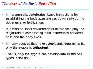
1. The document describes key stages in animal development from fertilization through organogenesis.
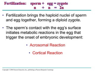
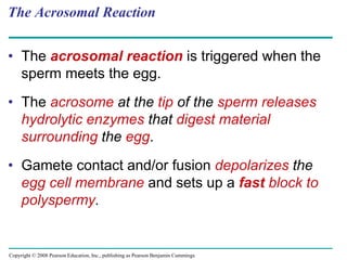
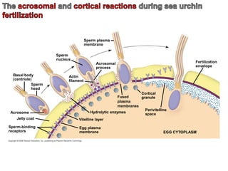
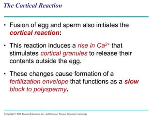
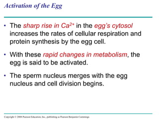
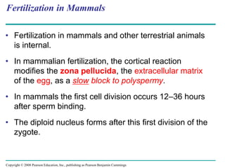
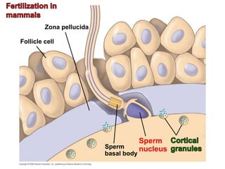
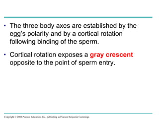
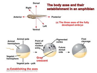
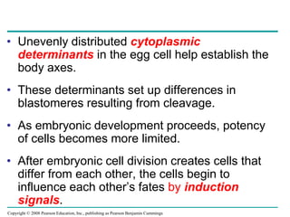
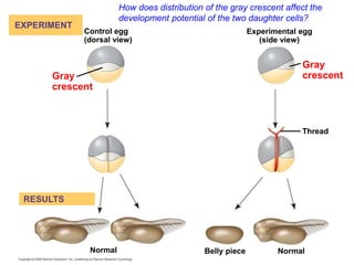
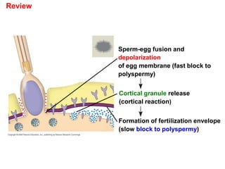
2. It explains that fertilization involves the acrosomal reaction and cortical reaction fusing the sperm and egg nuclei, activating embryonic development.
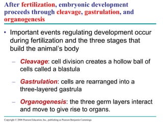
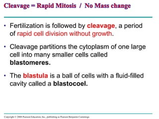
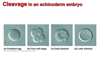
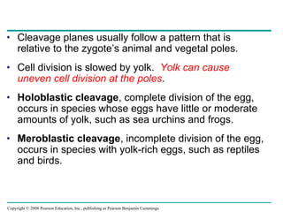
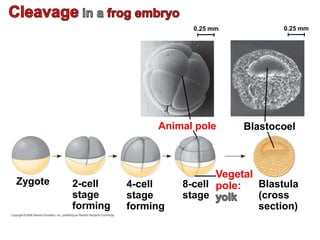
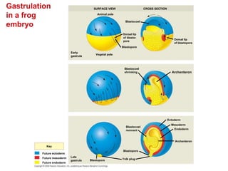
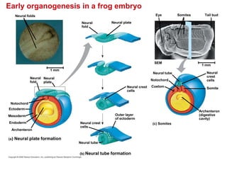
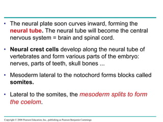
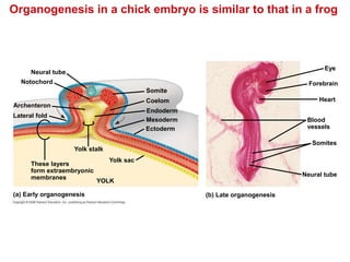
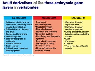
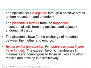
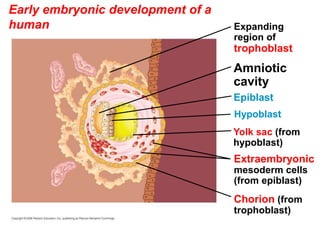
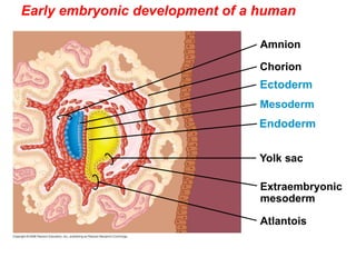
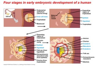
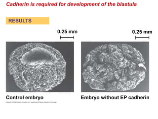
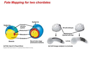
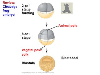
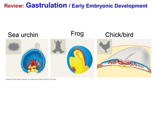
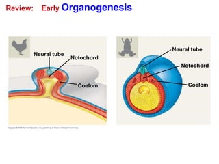
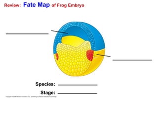
3. Gastrulation then rearranges the blastula cells into three germ layers - ectoderm, endoderm and mesoderm - forming the gastrula stage. Organogenesis follows as the germ layers develop into rudimentary organs.