The document summarizes key stages in early chick development:
1) Fertilization occurs internally in the oviduct, followed by cleavage and the beginning of gastrulation even before egg laying.
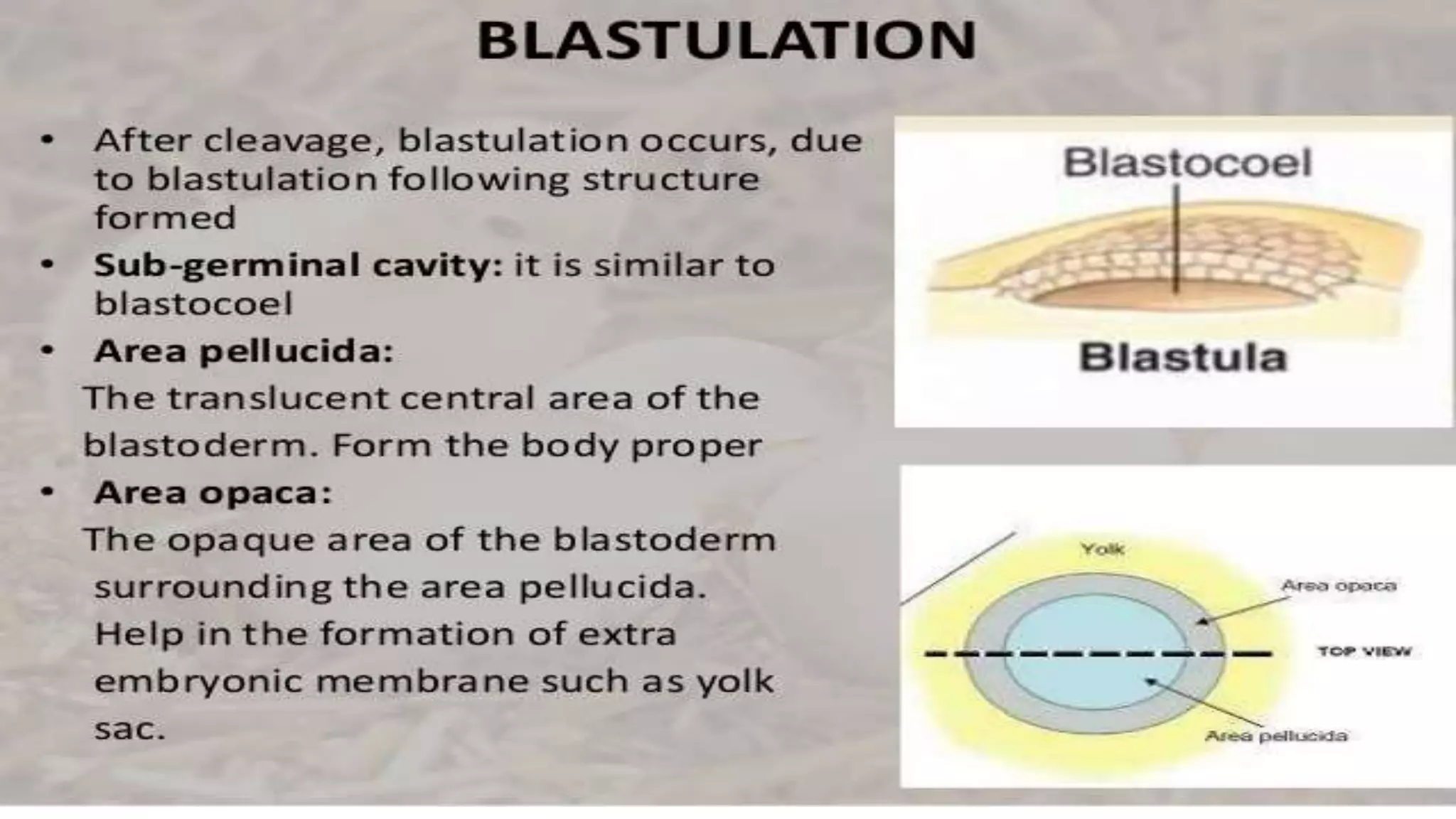
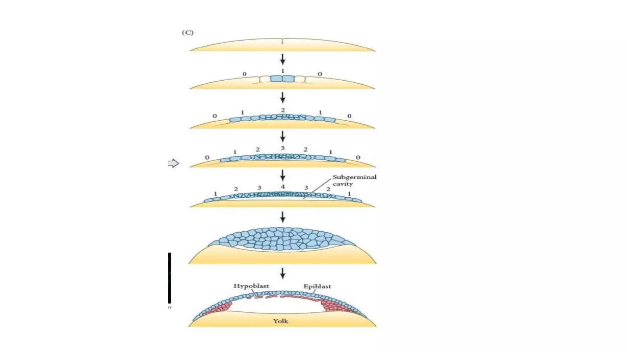
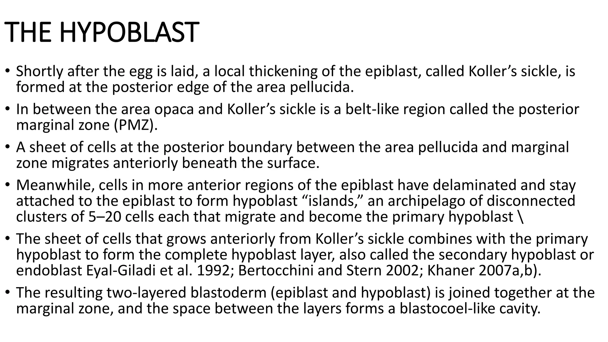
2) Blastulation involves the formation of the blastoderm layers and subgerminal cavity.
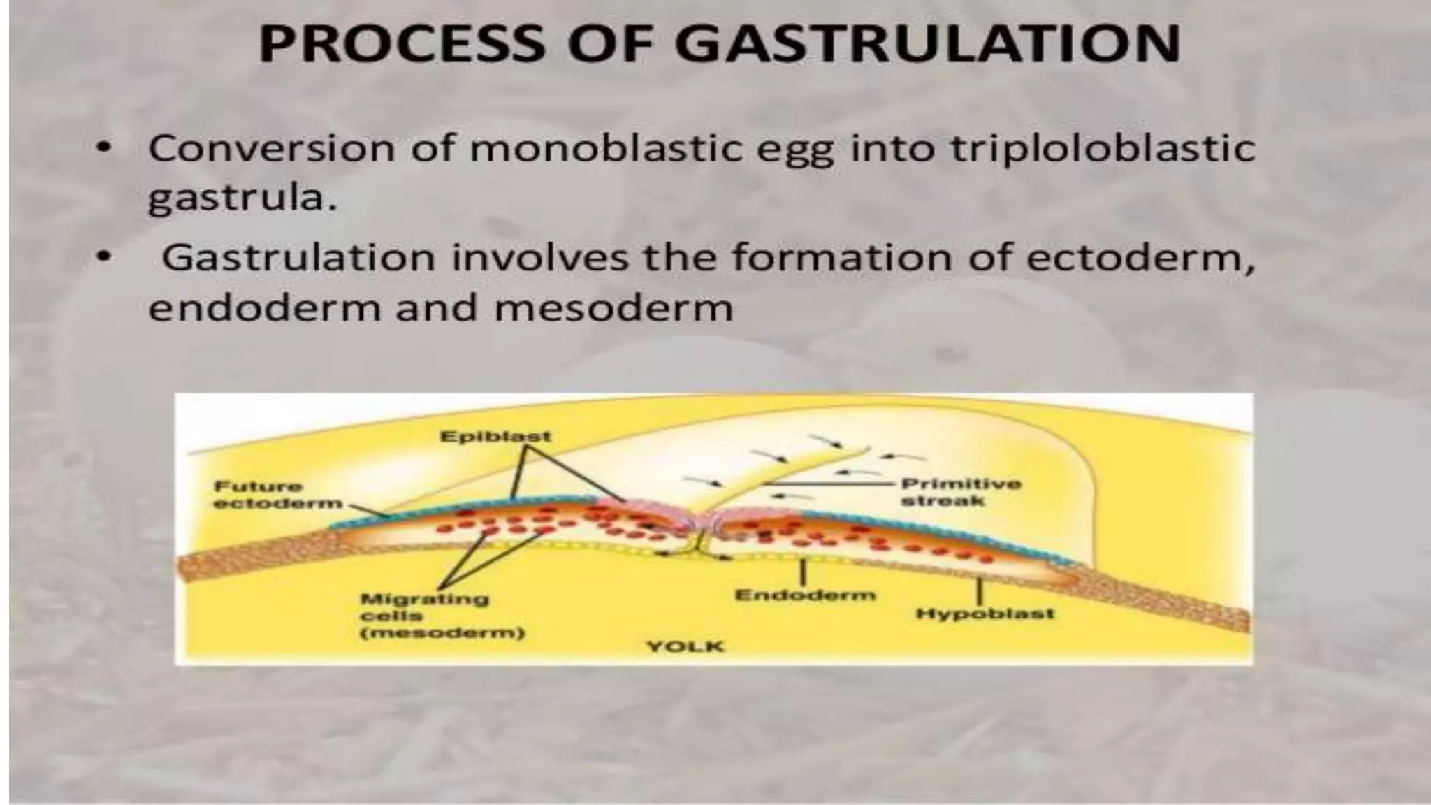
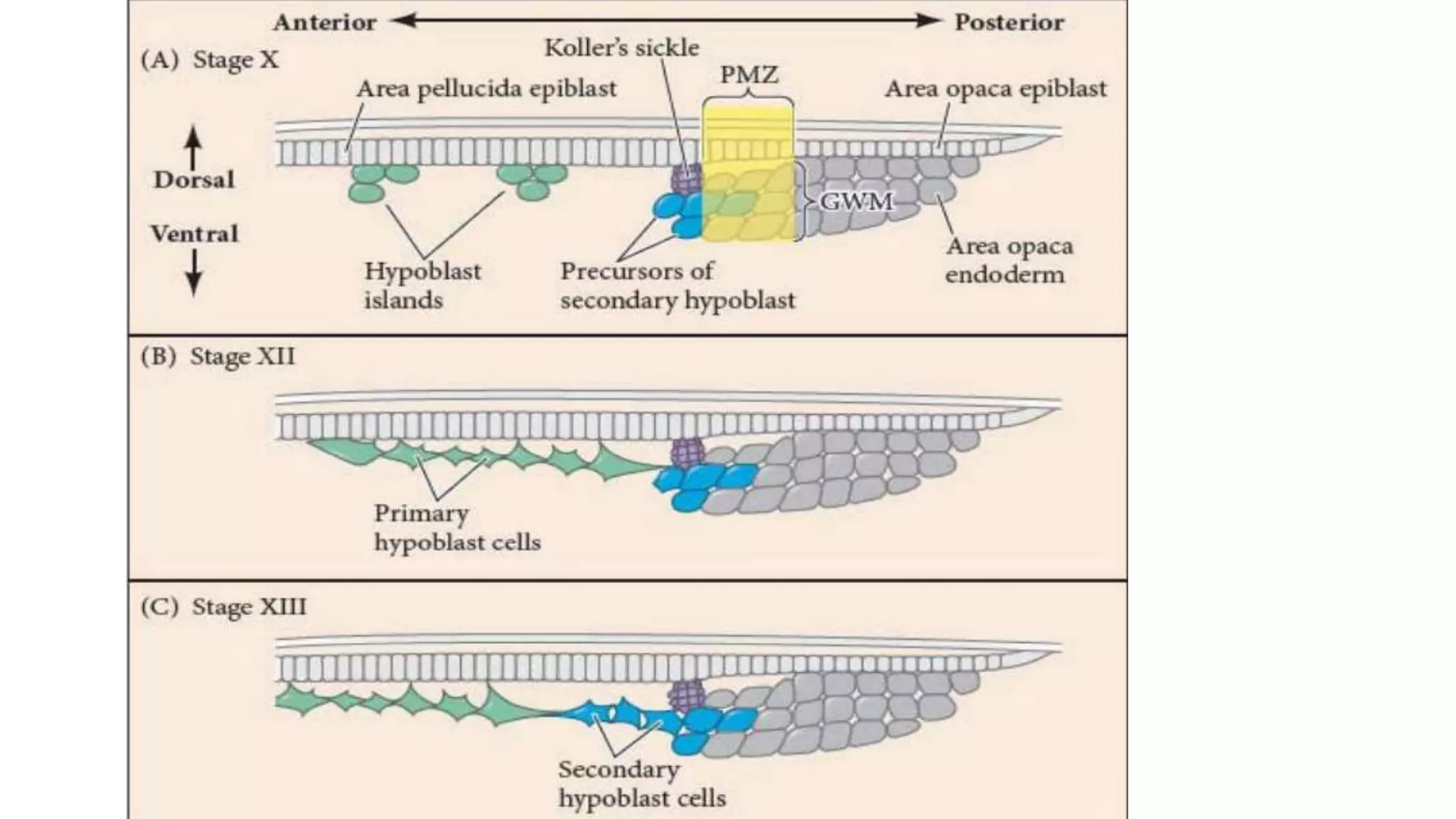
3) Gastrulation continues after laying, forming the primitive streak which elongates toward the head region through cell migration.
4) Cells migrate through the streak to form the endoderm and mesoderm, and the ectoderm covers the embryo through epiboly. Axis specification establishes bilateral symmetry.
1. DEFINITION
These are the membranes which do not form any part of
the embryo proper but performs various functions which
assist in the development of the embryo . These are
discarded at the time of hatching. These membranes
formed outside the embryo.
2. Types of Extra Embryonic Membranes
Yolk Sac
Amnion
Chorion
Allantois
3.Discussed Their
At Time of ORIGIN
It's FUNCTION
After HATCHING
4. AMNIOTIC CAVITY
............................END......................................................
Polyspermy describes an egg that has been fertilized by more than one sperm. Diploid organisms normally contain two copies of each chromosome, one from each parent. The cell resulting from polyspermy
The first issue that an egg and a sperm of any organism type face in successfully producing an embryo is the possibility of polyspermy. Polyspermy is the fertilization of an egg by multiple sperm, and the results of such unions are lethal.
If multiple sperm fertilize an egg, the embryo inherits multiple paternal centrioles. This causes competition for extra chromosomes and results in the disruption of the creation of the cleavage furrow, thus causing the zygote to die. As an important model organism in the study of fertilization and embryonic development, polyspermy in sea urchins has been studied in detail. The sea urchin’s methods of polyspermy prevention have been broken down into two main pathways. These two primary pathways are known as the fast block and the slow block to polyspermy
After the sperm’s receptors come into contact with the egg’s jelly layer and the acrosomal enzymes are released and break down the jelly layer, the sperm head comes into contact with the vitelline and plasma membranes of the egg. When the two plasma membranes contact one another, signals in the egg are initiated.
First, Na+ channels in the egg open, allowing Na+ to flood into the egg. This causes a depolarization of the egg from it’s normal resting potential of -70 mV.
While depolarization is occurring, the remainder of the jelly layer is dissolving. With the dissolution of the jelly layer and the depolarization of the plasma membrane, the first block to preventing fertilization by multiple sperm is put into place.
These two simple changes are part of the first block to polyspermy, known as the fast block. Within 1/10th of a second of contact, the fast block t
A chart showing the fate of each part of an early embryo, in a particular blastula stage is called fate maps. It is done because the correct interpretation of gastrulation is impossible without the knowledge of the position which are the presumptive germinal layers (Ectoderm, Mesoderm and Endoderm) occupy in blastula.
Fate mapping is a method used in developmental biology to study the embryonic origin of various adult tissues and structures. The "fate" of each cell or group of cells is mapped onto the embryo, showing which parts of the embryo will develop into which tissue. When carried out at single-cell resolution, this process is called cell lineage tracing. It is also used to trace the development of tumors.
This is a slide for complete development in chick ,as chick is a vertebrate so with the help of the development in a chick we can we can understand development in vertebrates .
This topic explains the whole process of growth and development in animal the processes include
Fertilization and incubation
Cleavage
Morula
Blastula
Gastrulation
Notochord And Mesoderm Formation
Neurulation
It describes the gamete fusion and early development in mammals.
Compaction,cavitation,Blastocyst, gastrula formation, Extra embryonic membranes development in mammals. Formation of twins, difference between monozygotic and dizygotic twins.
This presentation is about gastrulation, formation of primitive streak and neurulation (i.e. formation of brain and spinal cord).
Hope you like it.
Thank You
Synthetic Fiber Construction in lab .pptxPavel ( NSTU)
Synthetic fiber production is a fascinating and complex field that blends chemistry, engineering, and environmental science. By understanding these aspects, students can gain a comprehensive view of synthetic fiber production, its impact on society and the environment, and the potential for future innovations. Synthetic fibers play a crucial role in modern society, impacting various aspects of daily life, industry, and the environment. ynthetic fibers are integral to modern life, offering a range of benefits from cost-effectiveness and versatility to innovative applications and performance characteristics. While they pose environmental challenges, ongoing research and development aim to create more sustainable and eco-friendly alternatives. Understanding the importance of synthetic fibers helps in appreciating their role in the economy, industry, and daily life, while also emphasizing the need for sustainable practices and innovation.
Palestine last event orientationfvgnh .pptxRaedMohamed3
An EFL lesson about the current events in Palestine. It is intended to be for intermediate students who wish to increase their listening skills through a short lesson in power point.
Unit 8 - Information and Communication Technology (Paper I).pdfThiyagu K
This slides describes the basic concepts of ICT, basics of Email, Emerging Technology and Digital Initiatives in Education. This presentations aligns with the UGC Paper I syllabus.
Instructions for Submissions thorugh G- Classroom.pptxJheel Barad
This presentation provides a briefing on how to upload submissions and documents in Google Classroom. It was prepared as part of an orientation for new Sainik School in-service teacher trainees. As a training officer, my goal is to ensure that you are comfortable and proficient with this essential tool for managing assignments and fostering student engagement.
Acetabularia Information For Class 9 .docxvaibhavrinwa19
Acetabularia acetabulum is a single-celled green alga that in its vegetative state is morphologically differentiated into a basal rhizoid and an axially elongated stalk, which bears whorls of branching hairs. The single diploid nucleus resides in the rhizoid.
Francesca Gottschalk - How can education support child empowerment.pptxEduSkills OECD
Francesca Gottschalk from the OECD’s Centre for Educational Research and Innovation presents at the Ask an Expert Webinar: How can education support child empowerment?
Embracing GenAI - A Strategic ImperativePeter Windle
Artificial Intelligence (AI) technologies such as Generative AI, Image Generators and Large Language Models have had a dramatic impact on teaching, learning and assessment over the past 18 months. The most immediate threat AI posed was to Academic Integrity with Higher Education Institutes (HEIs) focusing their efforts on combating the use of GenAI in assessment. Guidelines were developed for staff and students, policies put in place too. Innovative educators have forged paths in the use of Generative AI for teaching, learning and assessments leading to pockets of transformation springing up across HEIs, often with little or no top-down guidance, support or direction.
This Gasta posits a strategic approach to integrating AI into HEIs to prepare staff, students and the curriculum for an evolving world and workplace. We will highlight the advantages of working with these technologies beyond the realm of teaching, learning and assessment by considering prompt engineering skills, industry impact, curriculum changes, and the need for staff upskilling. In contrast, not engaging strategically with Generative AI poses risks, including falling behind peers, missed opportunities and failing to ensure our graduates remain employable. The rapid evolution of AI technologies necessitates a proactive and strategic approach if we are to remain relevant.
Operation “Blue Star” is the only event in the history of Independent India where the state went into war with its own people. Even after about 40 years it is not clear if it was culmination of states anger over people of the region, a political game of power or start of dictatorial chapter in the democratic setup.
The people of Punjab felt alienated from main stream due to denial of their just demands during a long democratic struggle since independence. As it happen all over the word, it led to militant struggle with great loss of lives of military, police and civilian personnel. Killing of Indira Gandhi and massacre of innocent Sikhs in Delhi and other India cities was also associated with this movement.
How to Make a Field invisible in Odoo 17Celine George
It is possible to hide or invisible some fields in odoo. Commonly using “invisible” attribute in the field definition to invisible the fields. This slide will show how to make a field invisible in odoo 17.
1. P G DEPARTMENT OF ZOOLOGY
MINOR 2
SEM4
TOPIC: EARLY DEVELPOMENT IN CHICK
SUBMITTED TO:R K PANDITA
SUBMITTED BY: VAISHALI SHARMA
R.NO:01
2. Steps Involved in the Development of Chick
I. Fertilization:
Ova leave the ovary (ovulation) as primary oocytes.
They are released in coelom and caught by the expanded funnel-like opening of
oviduct.
They are fertilized in the upper part of oviduct which also receives sperms from the
male bird during copulation.
Fertilization is thus internal in birds
II. Cleavage:
Cleavage begins about 3 hours after fertilization.
Cleavage and early gastrulation is completed by the time the egg is laid.
3. • III. Blastulation:
• The free margin of blastoderm grows rapidly over the surface of yolk. A
small fluid-filled space appears just beneath the central mass of cells, this
is the sub-germinal cavity, often called blastocoel, though not real.
• IV. Gastrulation:
• Gastrulation begins even before laying of eggs. It involves the formation of
endoderm so that the monoblastic embryo or blastula is converted into
diploblastic or two-layered gastrula. There is no invagination of
prospective endoderm through a blastopore as found in frog.
4. • V. Incubation:
• Egg is warmed during incubation by the bird’s breast.
• In artificial incubation, the required temperature (37°C to 40°C) is
maintained in an incubator.
• With the resumption of development under heat of incubation, there
occurs mesoderm formation, notogenesis (formation of notochord),
neurogenesis (formation of neural tube), formation of mesoblastic somites
and organogeny.
5.
6.
7.
8. BLASTULATION:
• Between the blastoderm and the yolk of avian eggs is a space called the
subgerminal cavity, which is created when the blastoderm cells absorb water
from the albumin and secrete the fluid between themselves and the yolk (New
1956).
• At this stage, the deep cells in the center of the blastoderm appear to be shed
and die, leaving behind a 1-cell-thick area pellucida; this part of the blastoderm
forms most of the actual embryo.
• The peripheral ring of blastoderm cells that have not shed their deep cells
constitutes the area opaca.
• Between the area pellucida and the area opaca is a thin layer of cells called the
marginal zone (Eyal-Giladi 1997; Arendt and Nübler-Jung 1999).
• Some marginal zone cells become very important in determining cell fate during
early chick development
9.
10.
11. • The blastoderm will form an upper layer called the epiblast and a lower layer
called the hypoblast
• The avian embryo comes entirely from the epiblast; the hypoblast does not
contribute any cells to the developing embryo (Rosenquist 1966, 1972).
• Rather, the hypoblast cells form portions of the extraembryonic membranes
especially the yolk sac and the stalk linking the yolk mass to the endodermal
digestive tube.
• Hypoblast cells also provide chemical signals that specify the migration of
epiblast cells.
• However, the three germ layers of the embryo proper (plus the amnion, chorion,
and allantois extraembryonic membranes) are formed solely from the epiblast
(Schoenwolf 1991)
12. THE HYPOBLAST
• Shortly after the egg is laid, a local thickening of the epiblast, called Koller’s sickle, is
formed at the posterior edge of the area pellucida.
• In between the area opaca and Koller’s sickle is a belt-like region called the posterior
marginal zone (PMZ).
• A sheet of cells at the posterior boundary between the area pellucida and marginal
zone migrates anteriorly beneath the surface.
• Meanwhile, cells in more anterior regions of the epiblast have delaminated and stay
attached to the epiblast to form hypoblast “islands,” an archipelago of disconnected
clusters of 5–20 cells each that migrate and become the primary hypoblast
• The sheet of cells that grows anteriorly from Koller’s sickle combines with the primary
hypoblast to form the complete hypoblast layer, also called the secondary hypoblast or
endoblast Eyal-Giladi et al. 1992; Bertocchini and Stern 2002; Khaner 2007a,b).
• The resulting two-layered blastoderm (epiblast and hypoblast) is joined together at the
marginal zone, and the space between the layers forms a blastocoel-like cavity.
13.
14.
15. FORMATION OF PRIMITIVE STREAK:
• Avian and mammalian gastrulation takes place through the primitive streak.
• This can be considered the equivalent of an elongated blastopore lip of
amphibian embryos (Alev et al. 2013; Bertocchini et al. 2013; Stower et al. 2015).
• the primitive streak first arises from Koller’s sickle and the epiblast above it
(Bachvarova et al. 1998; Lawson and Schoenwolf 2001a,b; Voiculescu et al. 2007).
• As cells converge to form the primitive streak, a depression called the primitive
groove forms within the streak.
• Most migrating cells pass through the primitive groove, which serves as a
gateway into the deep layers of the embryo (FIGURE 12.4; Voiculescu et al. 2014).
• Thus, the primitive groove is homologous to the amphibian blastopore, and the
primitive streak is homologous to the blastopore lip
16.
17.
18.
19.
20. ELONGATION OF THE PRIMITIVE STREAK
• The streak elongates toward the future head region as more anterior cells migrate
toward the center of the embryo.
• Convergent extension is responsible for the progression of the streak Cell division adds
to the length produced by convergent extension, and some of the cells from the
anterior portion of the epiblast contribute to the formation of Hensen’s node (Streit et
al. 2000; Lawson and Schoenwolf 2001b).
• At the same time, the secondary hypoblast (endoblast) cells continue to migrate
anteriorly from the posterior marginal zone of the blastoderm
• The elongation of the primitive streak appears to be coextensive with the anterior
migration of these secondary hypoblast cells, and the hypoblast directs the movement
of the primitive streak (Waddington 1933; Foley et al. 2000; Voiculescu et al. 2007,
2014).
• The streak eventually extends to 60%–75% of the length of the area pellucid
21.
22. FORMATION OF ENDODERM AND MESODERM
• As soon as the primitive streak has formed, epiblast cells begin to migrate through it
and into the space between epiblast and hypoblast (reminiscent of the amphibian
blastocoel).
• Cells migrating through the anterior end pass down into the embryonic space and
migrate anteriorly, forming the endoderm, head mesoderm, and notochord;
• Cells passing through the more posterior portions of the primitive streak give rise to
the majority of mesodermal tissues ; Rosenquist 1966; Schoenwolf et al. 1992).
• The first cells to migrate through Hensen’s node are those destined to become the
pharyngeal endoderm of the foregut.
• Once deep within the embryo, these endodermal cells migrate anteriorly and
eventually displace the hypoblast cells, causing the hypoblast cells to be confined to a
region in the anterior portion of the area pellucida.
• This anterior region, the germinal crescent, does not form any embryonic structures,
but it does contain the precursors of the germ cells, which later migrate through the
blood vessels to the gonads
23.
24. •REGRESSION OF THE PRIMITIVE STREAK AND EPIBOLY OF
THE ECTODERM
• As mesodermal ingression continues, the primitive streak starts to regress,
moving Hensen’s node from near the center of the area pellucida to a more
posterior position
• The regressing streak leaves in its wake the dorsal axis of the embryo, including
the notochord.
• The notochord is laid down in a head-to-tail direction, starting at the level where
the ears and hindbrain form and extending caudally to the tailbud.
• the pharyngeal endoderm and head mesendoderm will largely induce the
anterior parts of the brain, while the notochord will induce the hindbrain and
spinal cord.
• By this time, all the presumptive endodermal and mesodermal cells have entered
the embryo and the epiblast is composed entirely of presumptive ectodermal
cells.
25.
26. Axis specification and the avian “organizer”
• Gastrulating avian (and mammalian) embryos exhibit a distinct anterior-to-
posterior gradient.
• While cells of the posterior portions of the embryo are still part of a
primitive streak and entering the inside of the embryo, cells at the anterior
end are already starting to form organs (see Darnell et al. 1999).
• For the next several days, the anterior end of the embryo is more advanced
in its development (having had a “head start,” if you will) than the posterior
end.
27. • The role of gravity and the PMZ The conversion of the radially symmetrical
blastoderm into a bilaterally symmetrical structure appears to be
determined by gravity.
• As the ovum passes through the hen’s reproductive tract, it is rotated for
about 20 hours in the shell gland.
• This spinning, at a rate of 15 revolutions per hour, shifts the yolk such that
its lighter components (probably containing stored maternal determinants
for development) lie beneath one side of the blastoderm.
• This imbalance tips up one end of the blastoderm, and that end becomes
the posterior marginal zone, where primitive streak formation begins
(Figure 12.9; Kochav and Eyal-Giladi 1971; Callebaut et al. 2004).
28. Left-right axis formation
• The vertebrate body has distinct right and left sides.
• The heart and spleen, for instance, are generally on
the left side of the body, whereas the liver is usually
on the right.
• The distinction between the sides is primarily
regulated by the left-sided expression of two proteins:
the paracrine factor Nodal and the transcription
factor Pitx2.