Downloaded 821 times


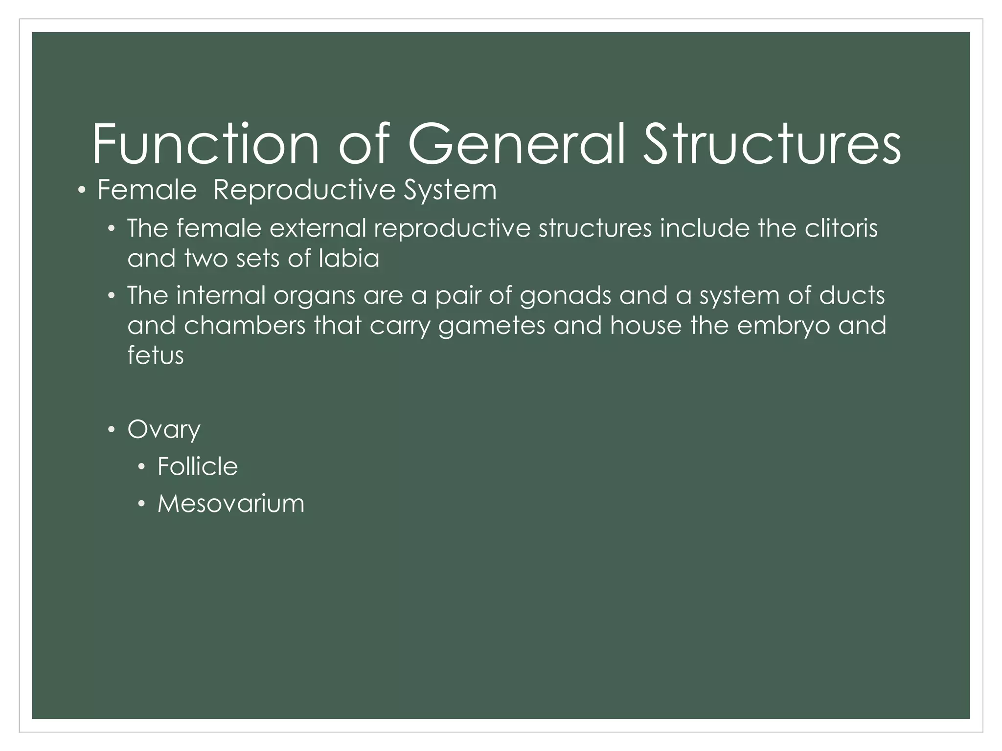
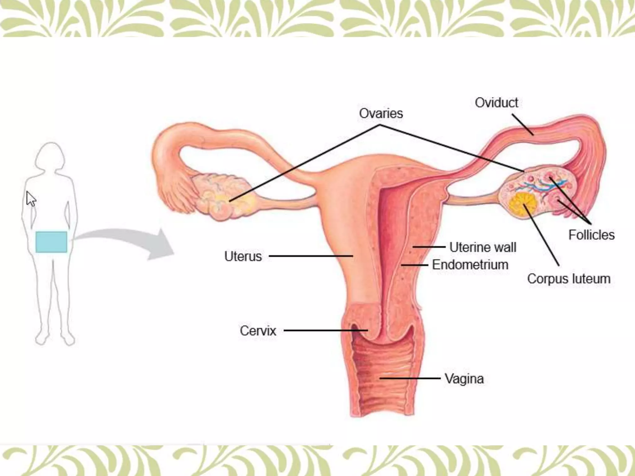
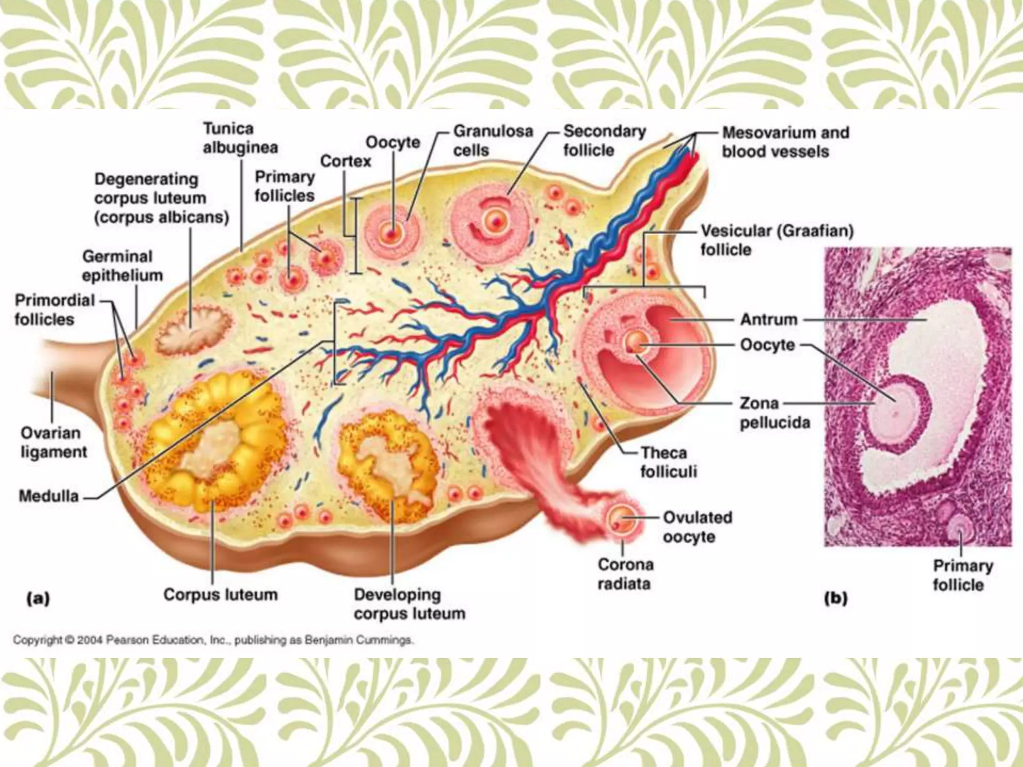
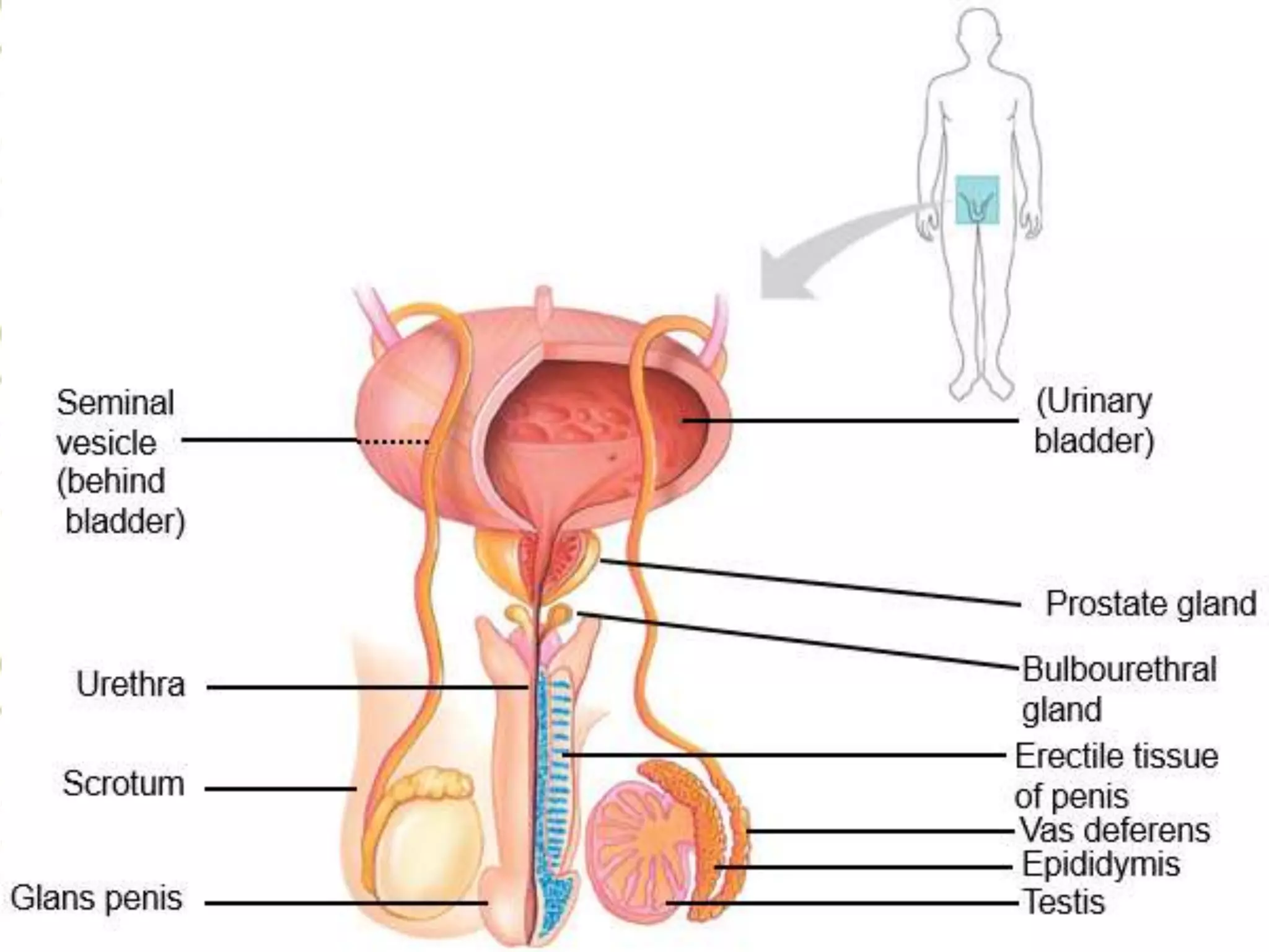
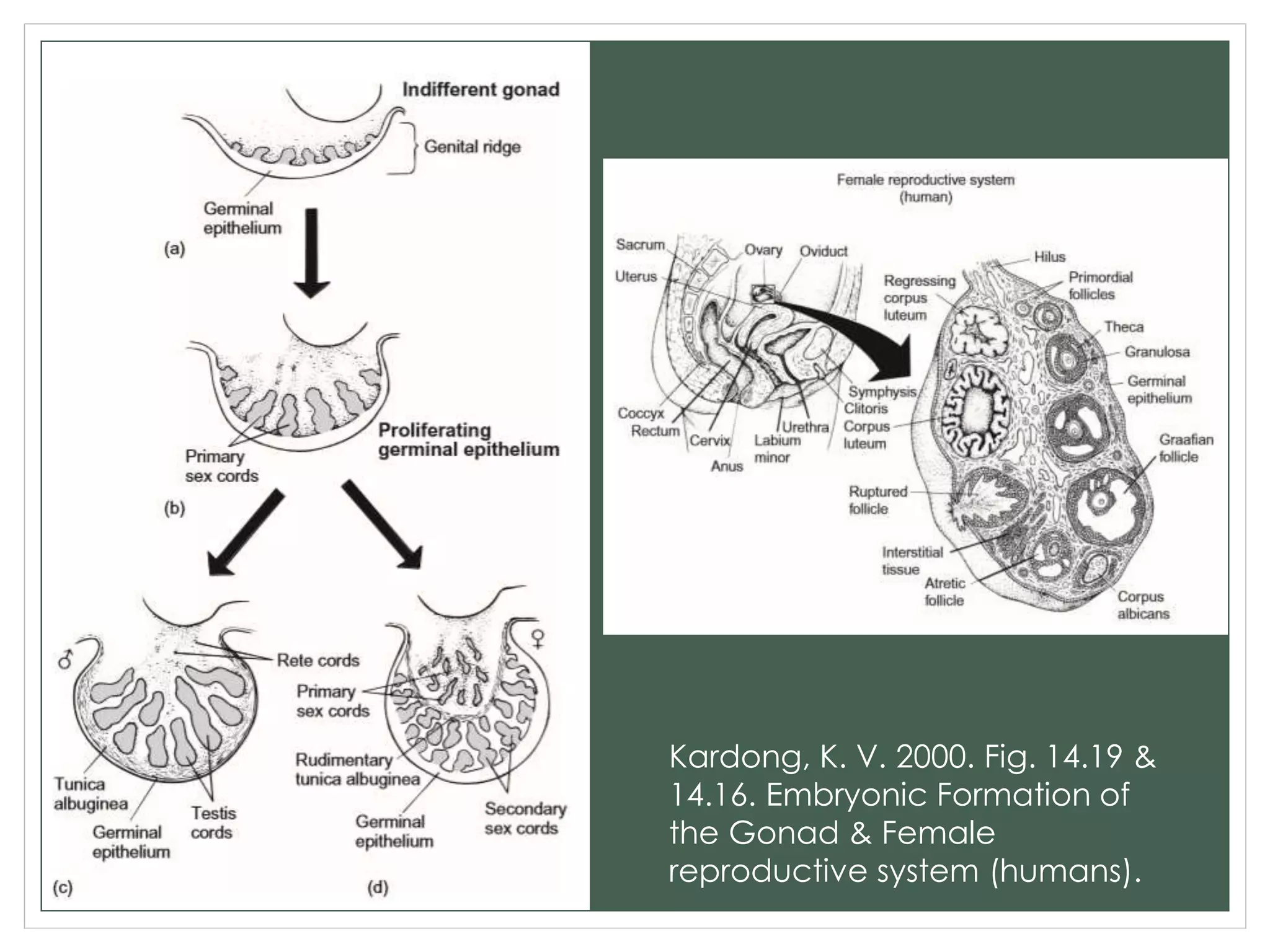
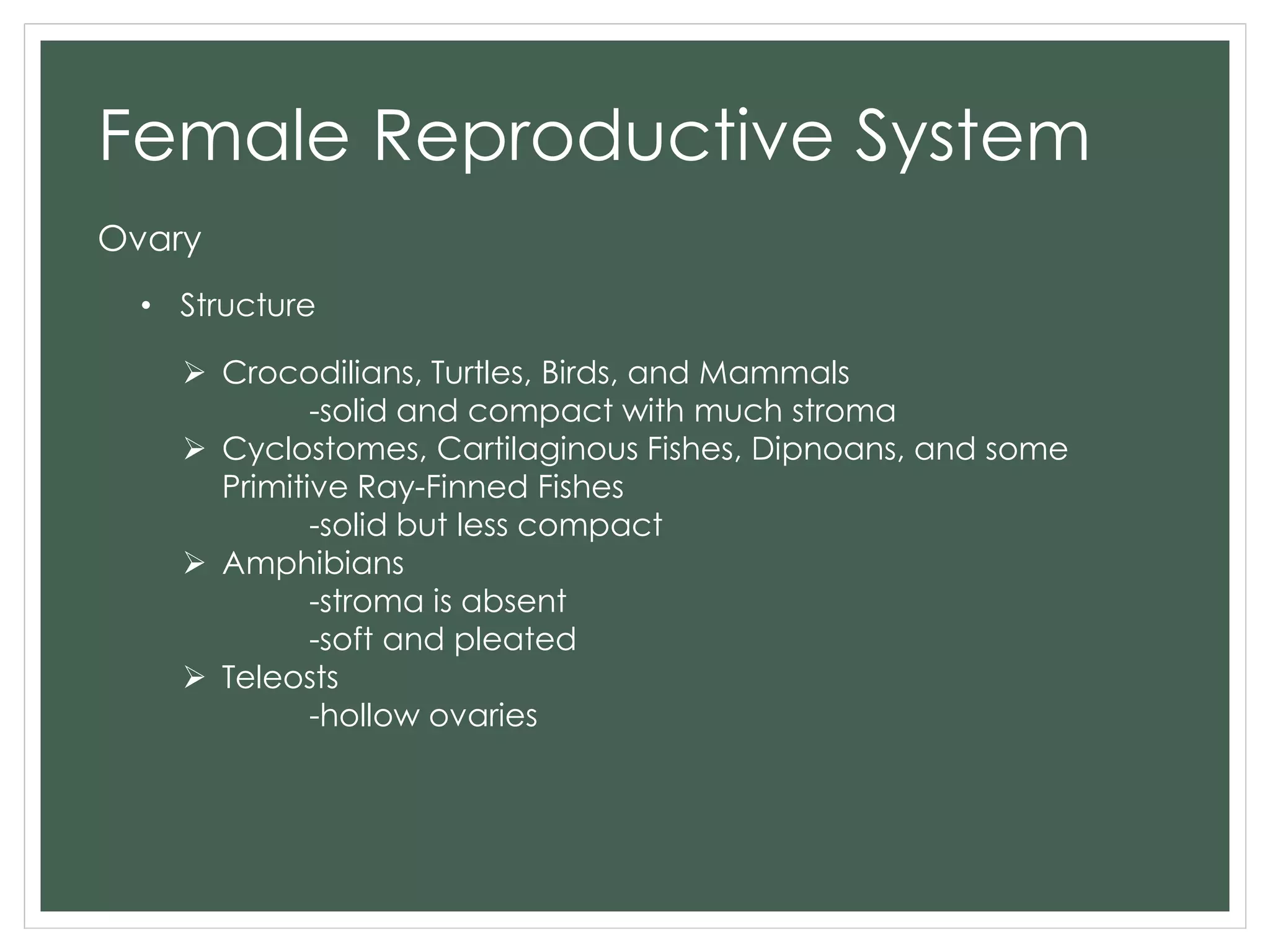
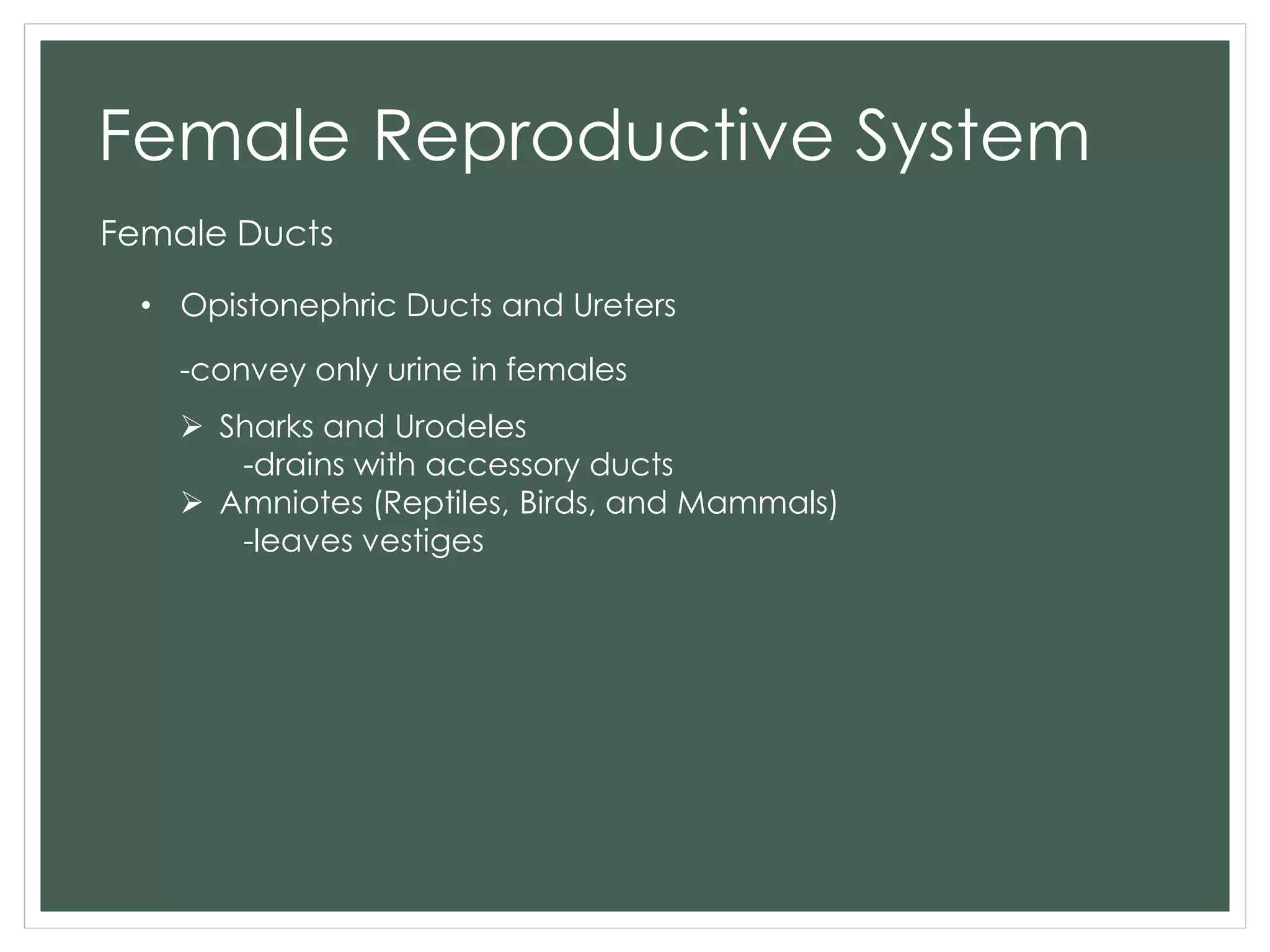
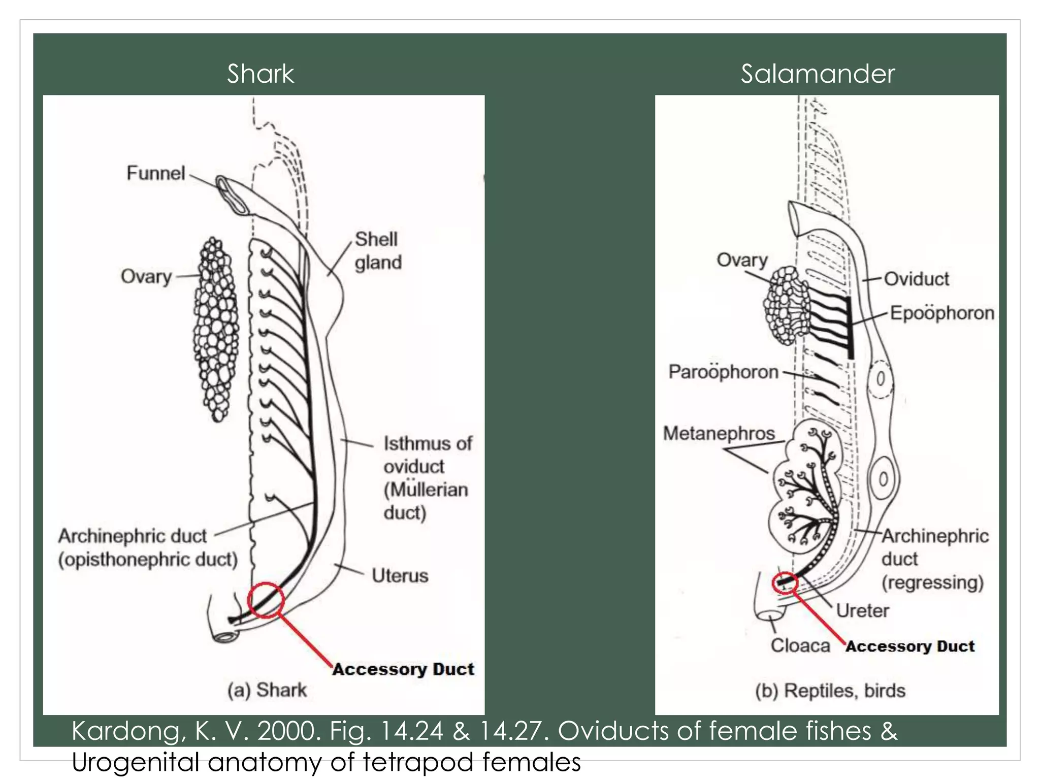
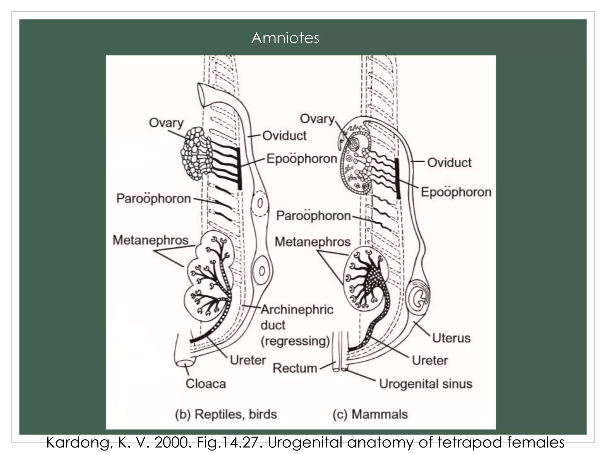
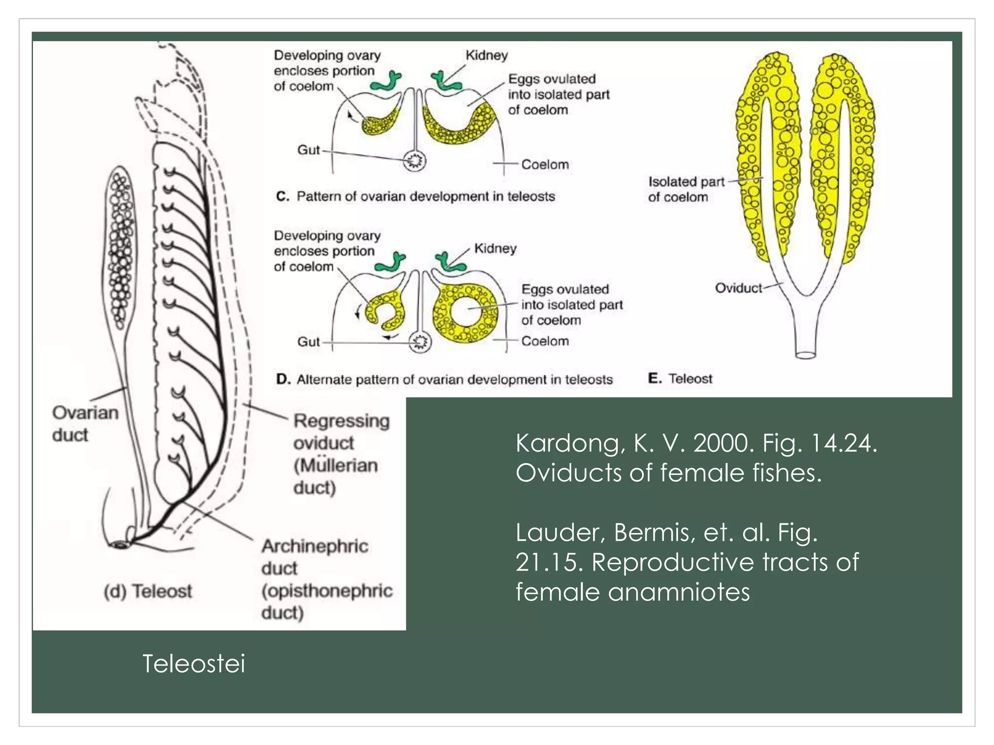

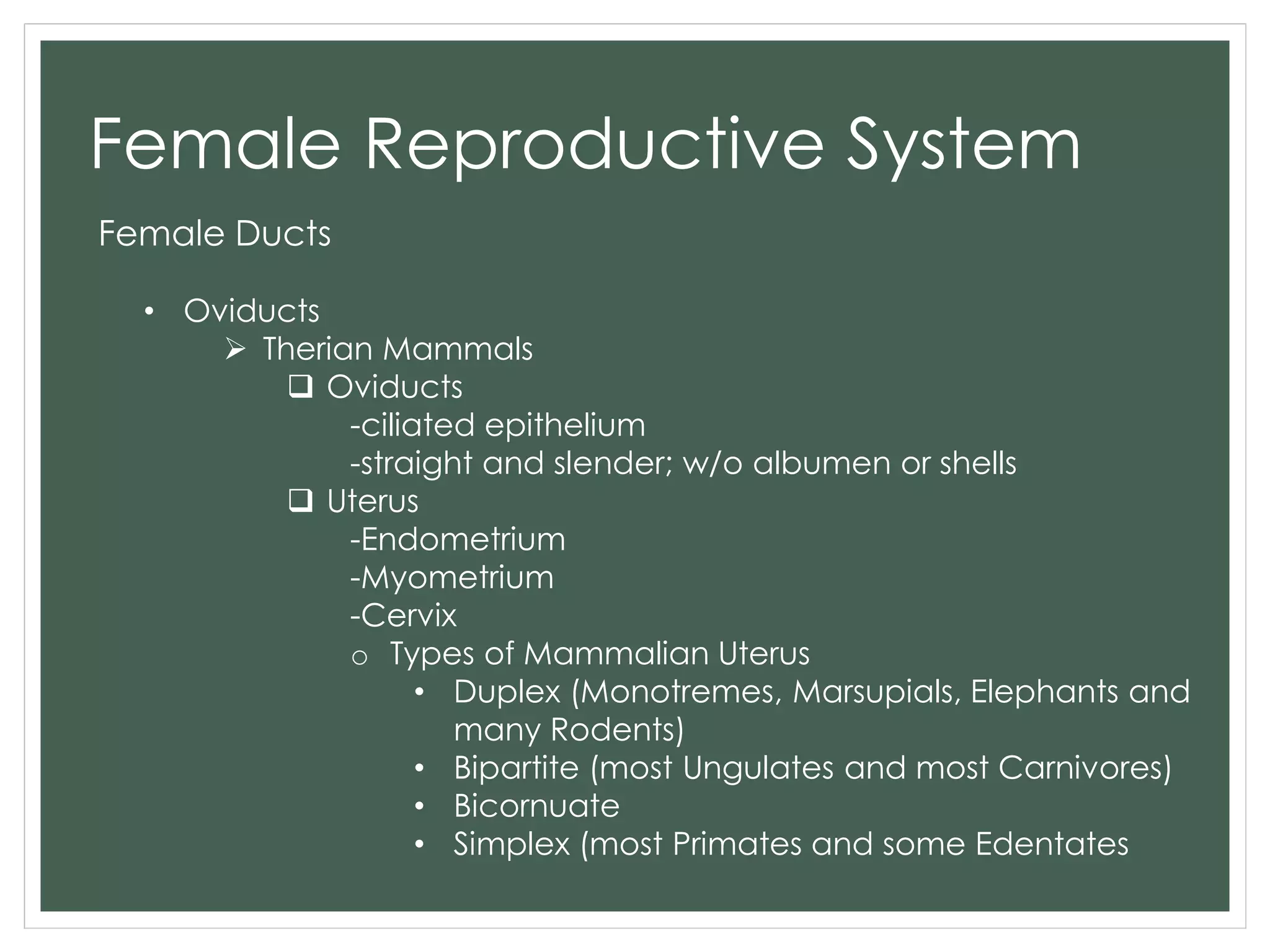
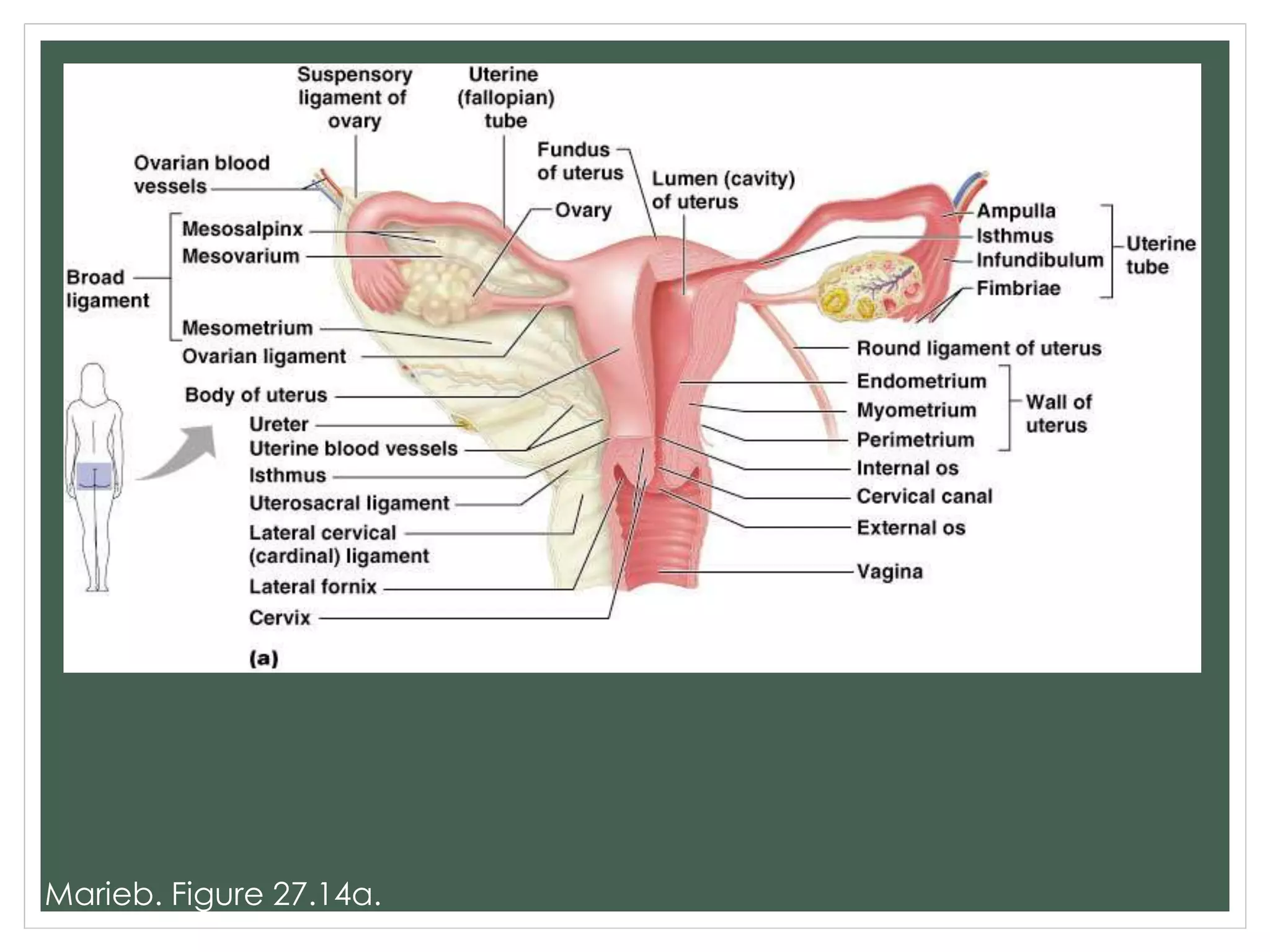
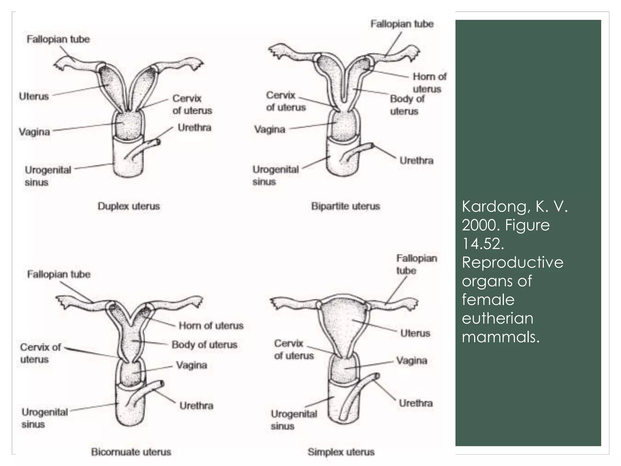
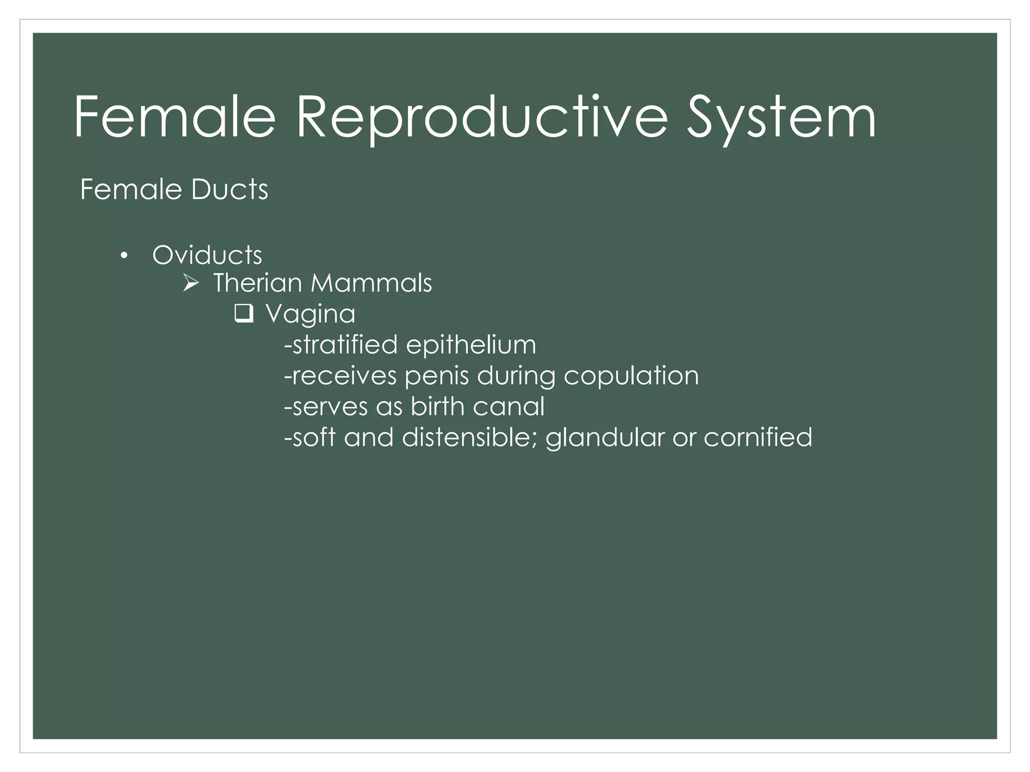
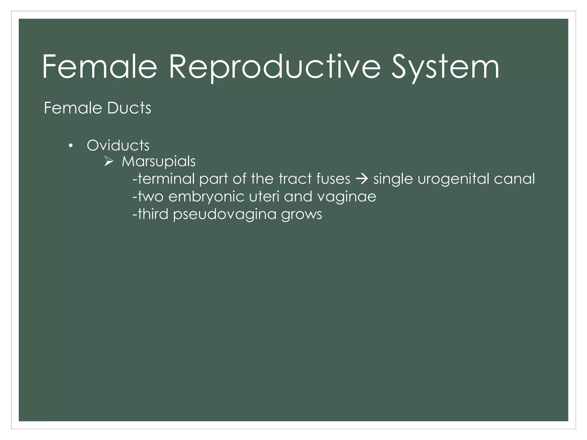
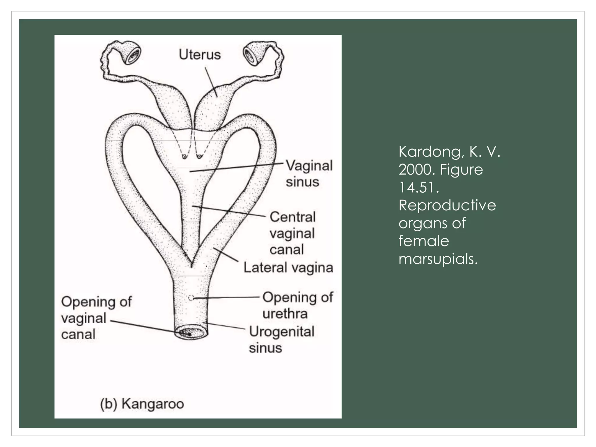
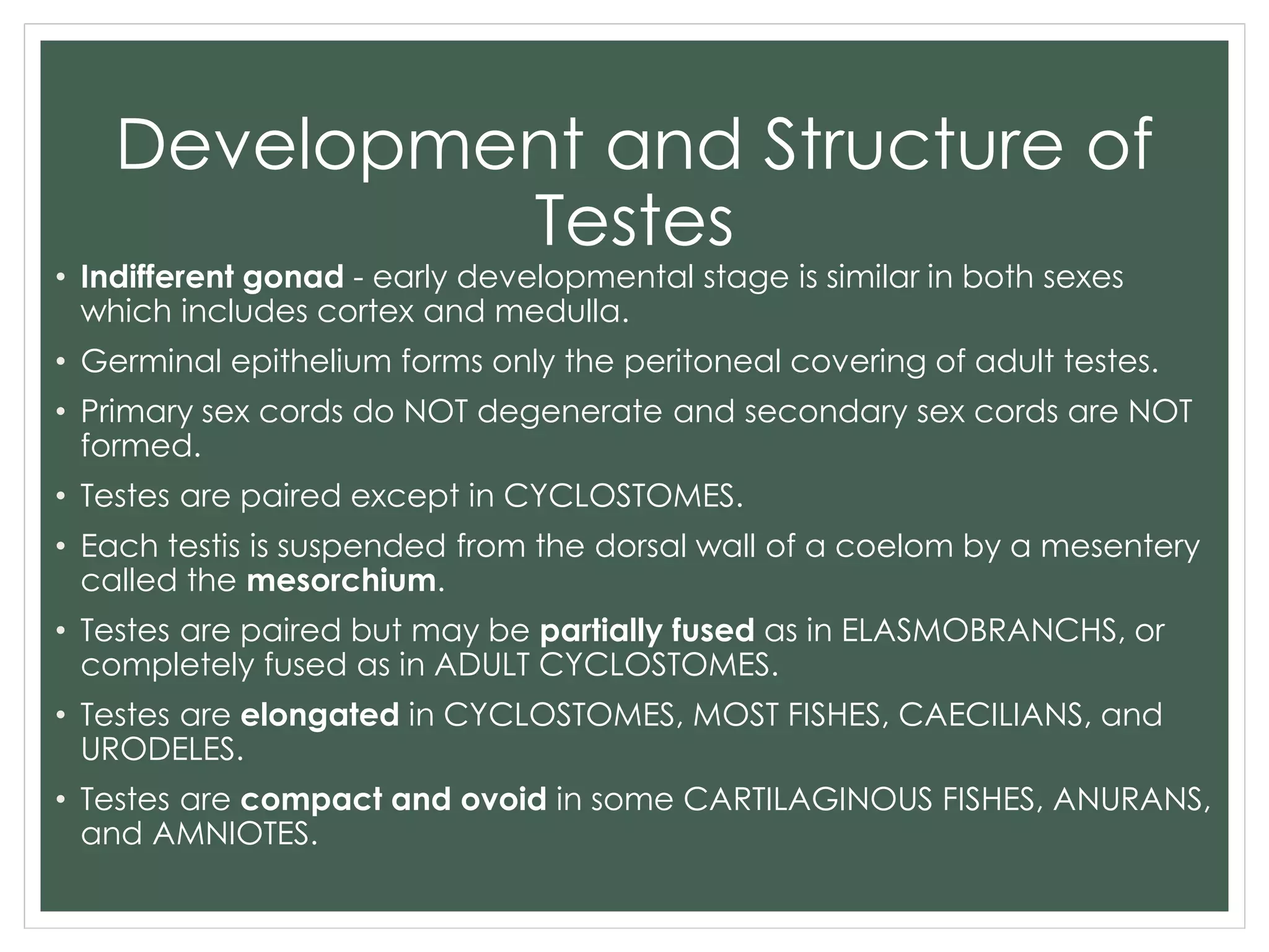
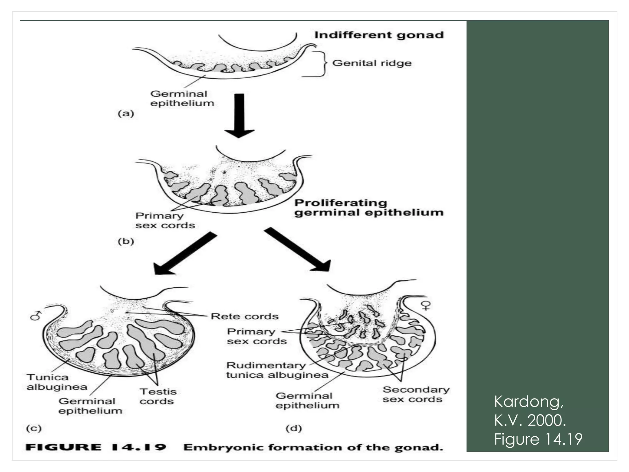
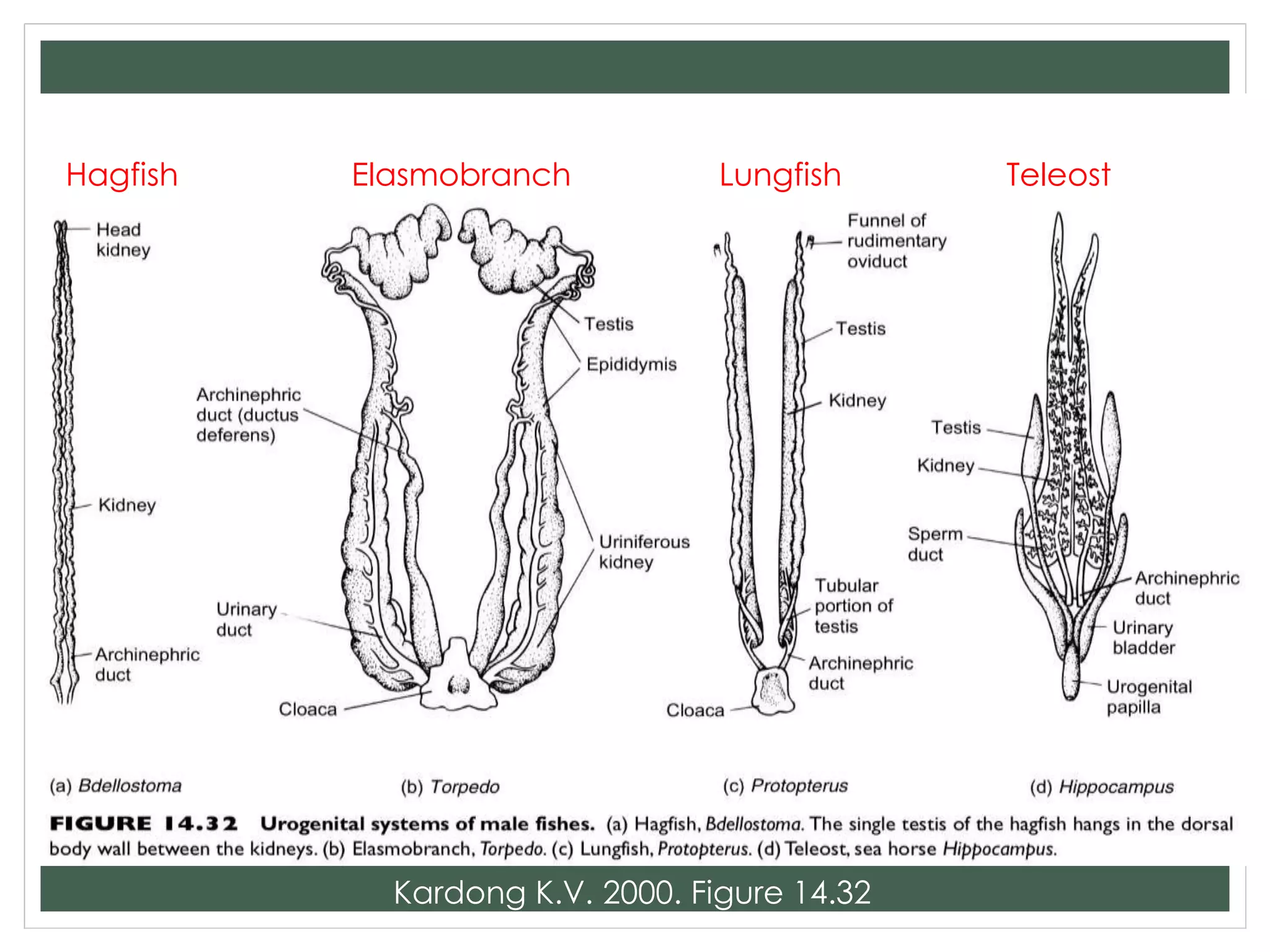
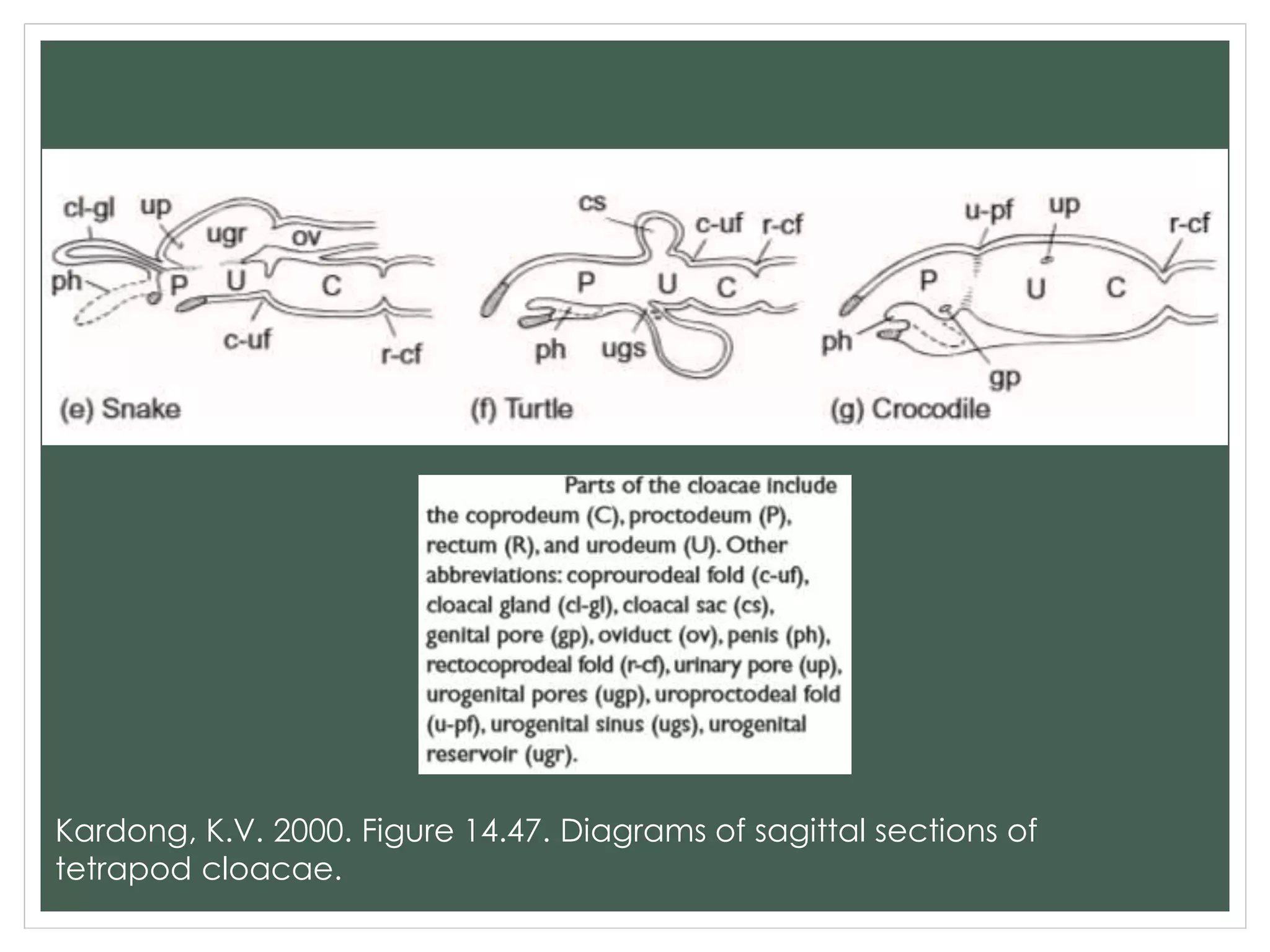
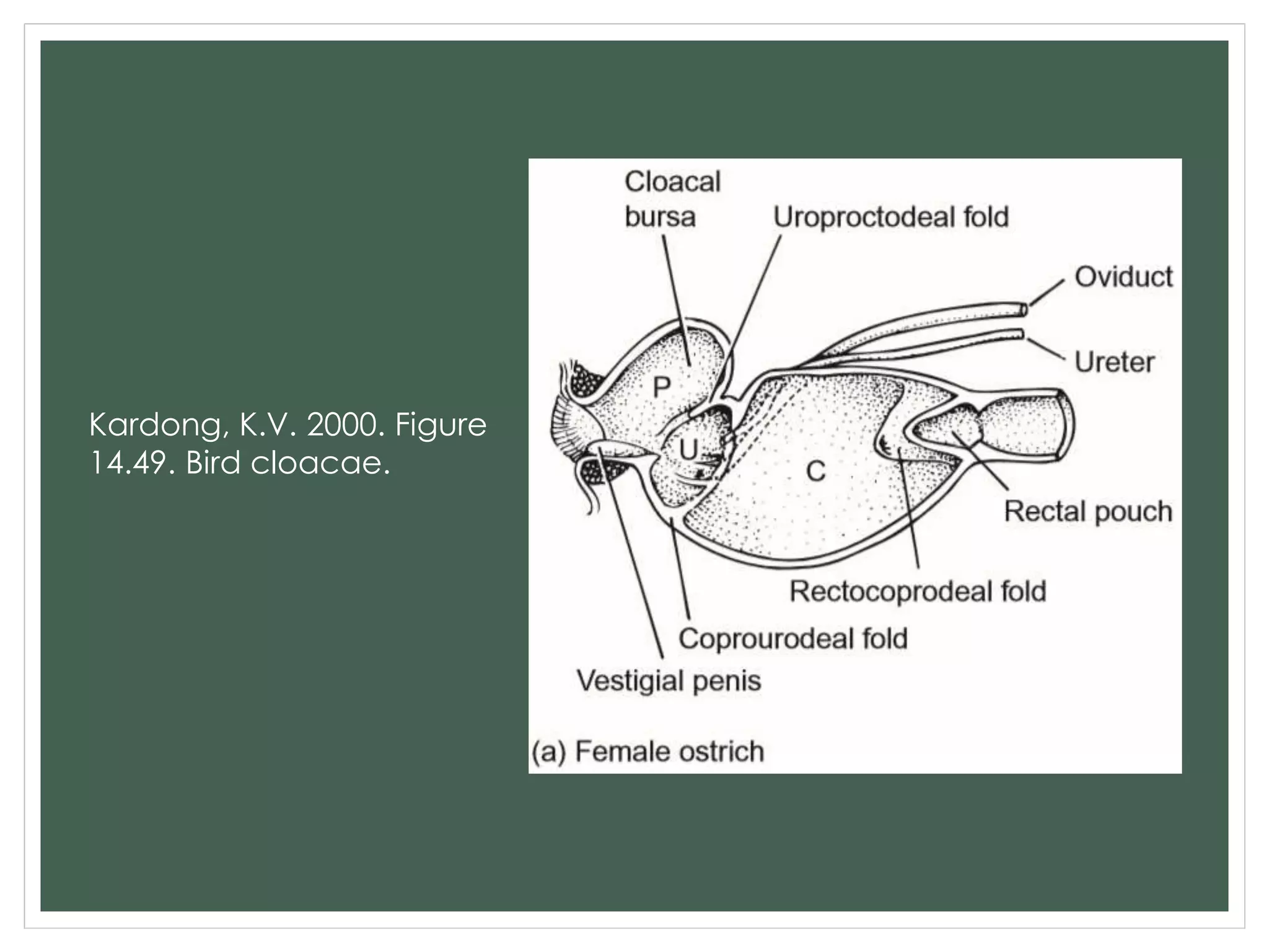
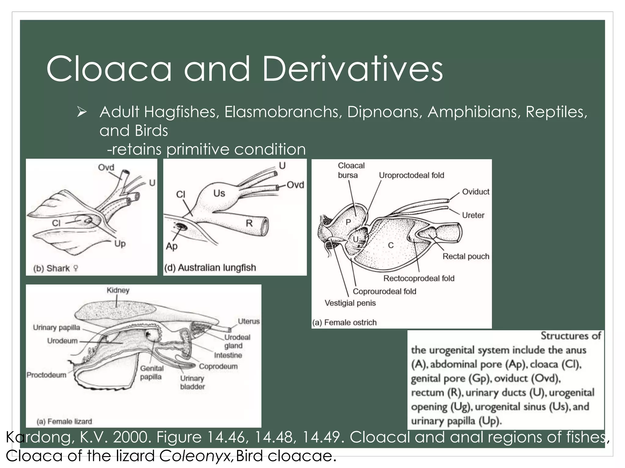
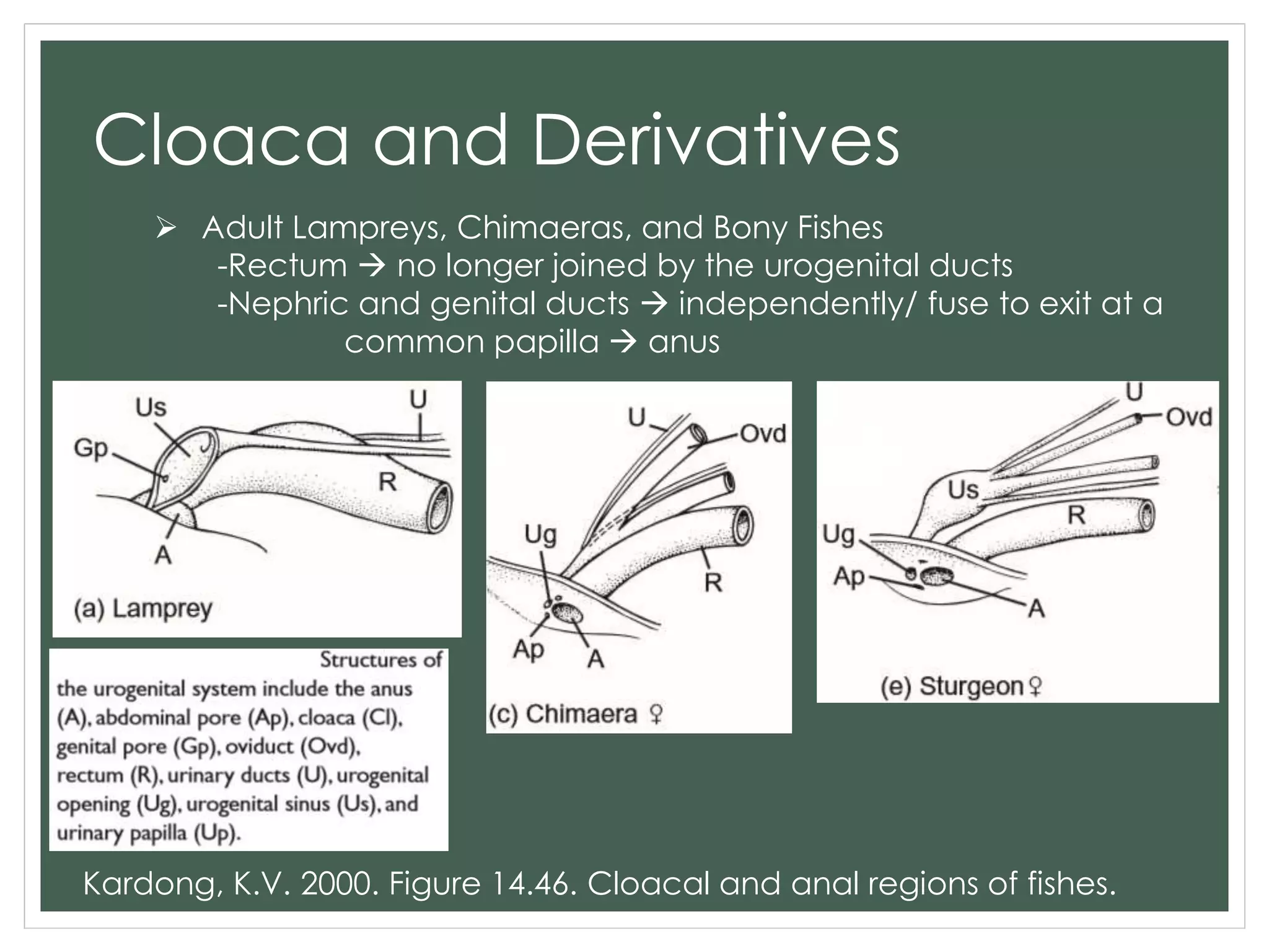
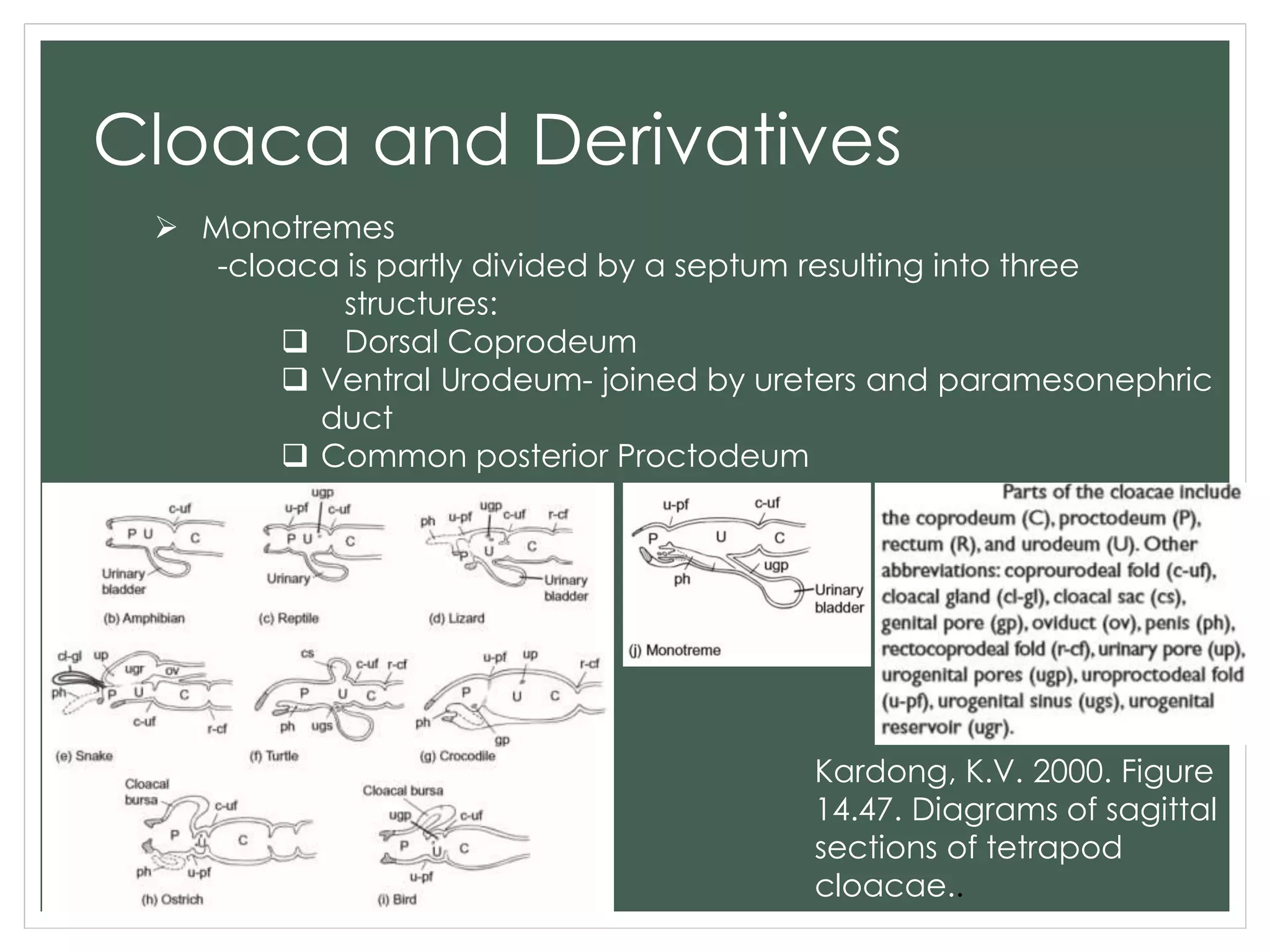
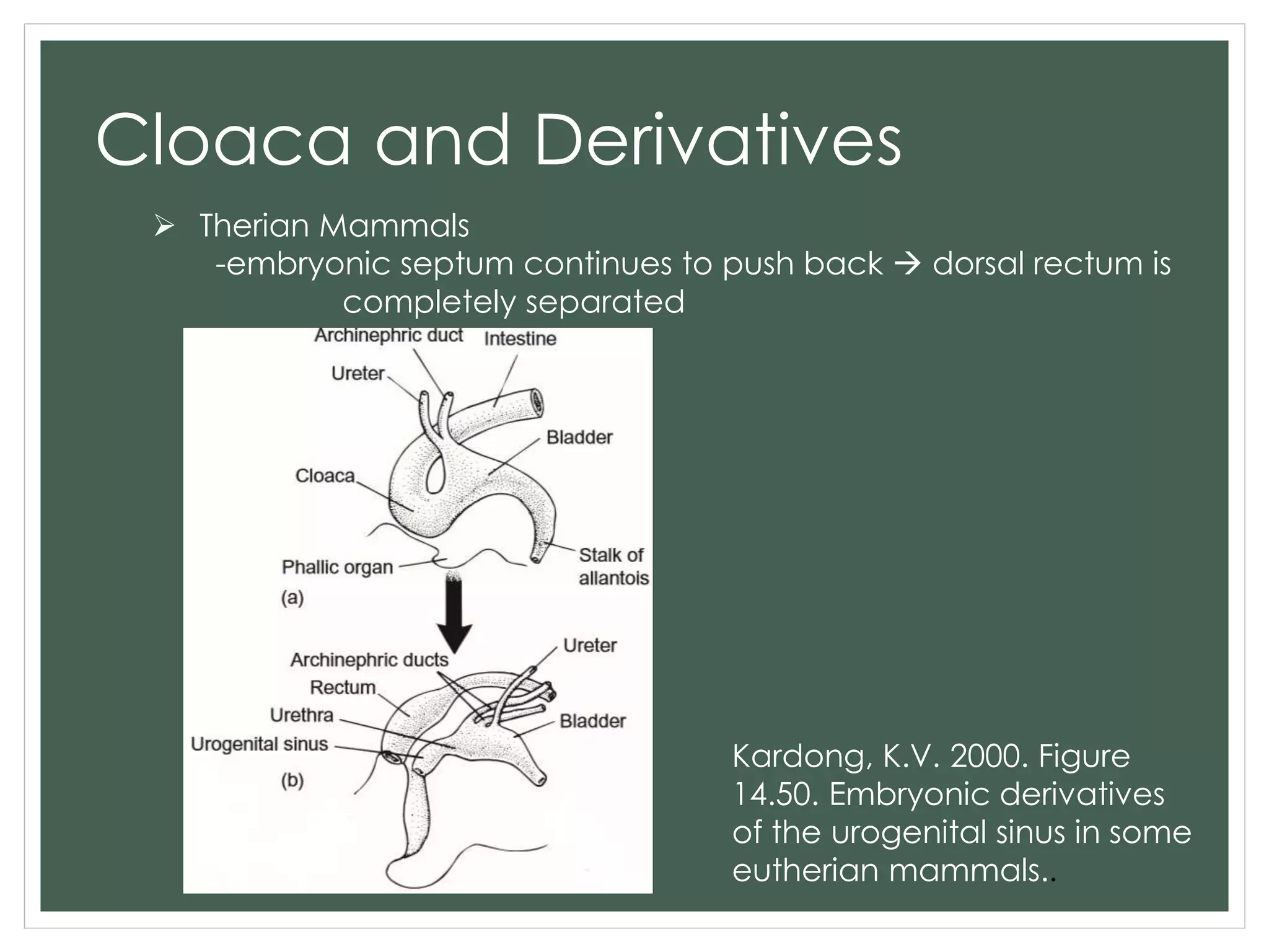
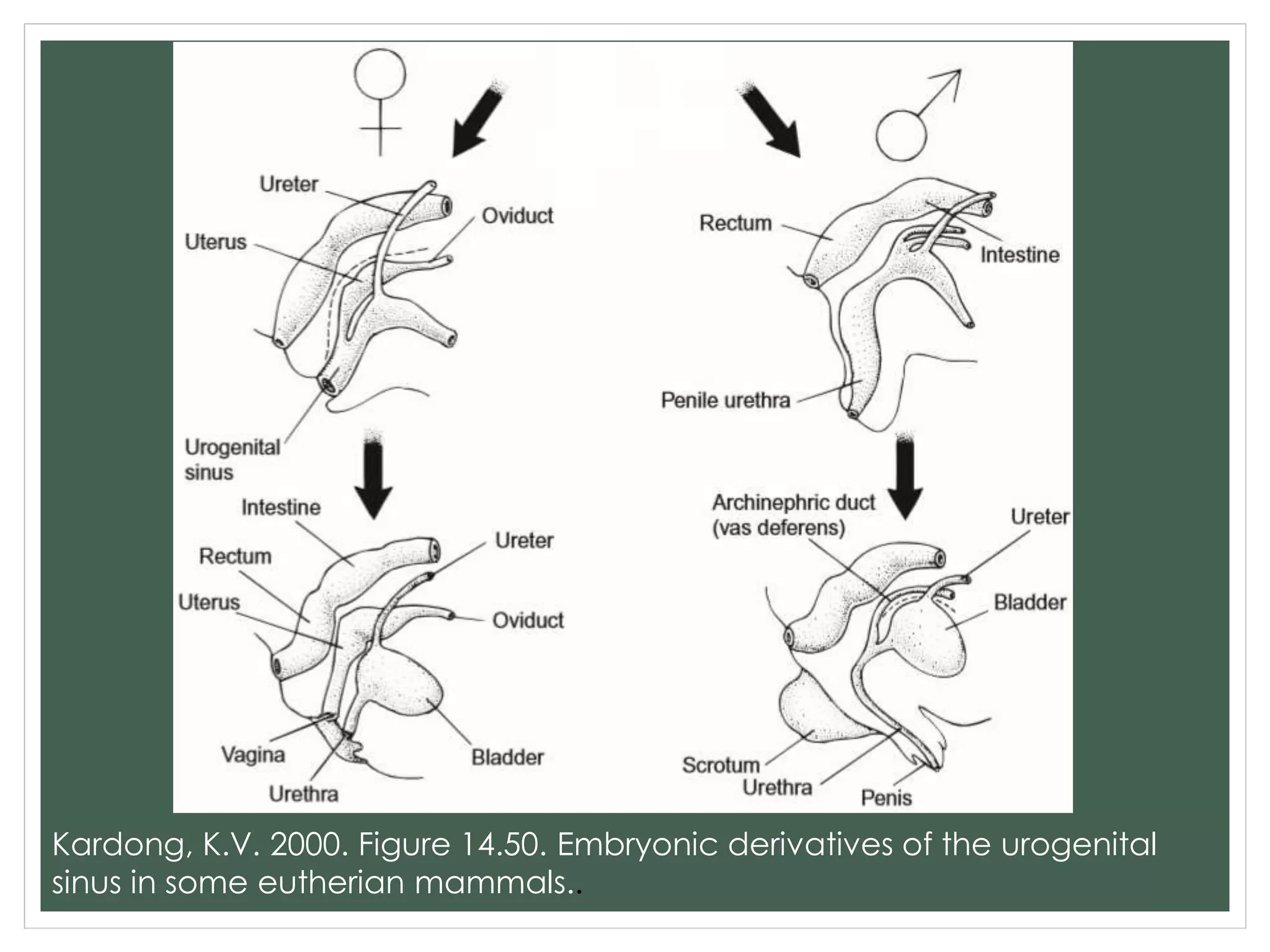

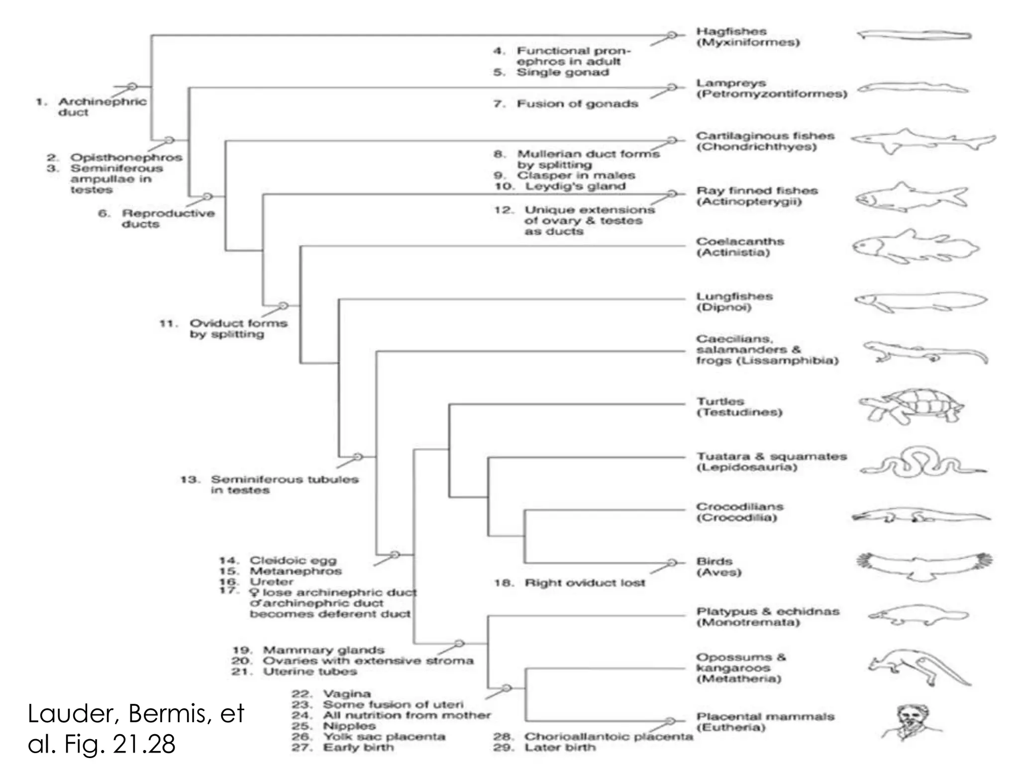


The document provides an overview of the reproductive systems of vertebrates. It describes the general structures and functions of the male and female reproductive systems. Key points include: 1) The female system produces eggs and houses the embryo, while the male system produces sperm. 2) Structures such as ovaries, oviducts, uterus, and vagina are present across vertebrate classes with some variations. 3) Testes, ducts and accessory glands are the main male structures. 4) Fertilization and copulation methods vary between external in frogs to internal in mammals. 5) The cloaca is modified in different groups, remaining primitive in some and separating functions in others like mammals.


