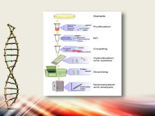
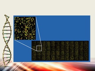
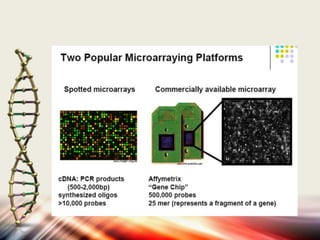
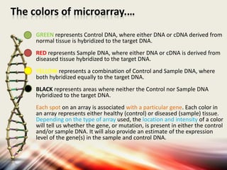
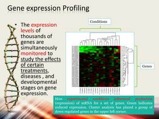
Microarrays allow researchers to analyze gene expression levels across thousands of genes simultaneously. DNA microarrays work by hybridizing fluorescently-labeled cDNA or cRNA to complementary DNA probes attached to a solid surface. This technology has applications in gene expression profiling, disease diagnosis, drug discovery, and toxicology research. While microarrays provide high-throughput analysis, their limitations include not reflecting true protein levels, complex data analysis, expense, and short shelf life of DNA chips.
4. history
Microarray technology evolved from Southern
blotting.
The concept of microarrays was first proposed in the
late 1980s by Augenlicht and his colleagues.
They spotted 4000 cDNA sequences on nitrocellulose
membrane and used radioactive labeling to analyze
differences in gene expression patterns among different
types of colon tumors in various stages of malignancy.
5. PRINCIPLE
• The principle of DNA microarrays lies on the
hybridization between the nucleotide. Using this
technology the presence of one genomic or cDNA
sequence in 1,00,000 or more sequences can be
screened in a single hybridization.
Fluorescent labeled target sequences that bind to a
probe sequence generate a signal that depends on
the strength of the hybridization determined by the
number of paired bases.
6. why use this technic?
• By using an array containing many DNA
samples, scientists can determine, in a
single experiment, the expression levels
of hundreds or thousands of genes within
a cell by measuring the amount of mRNA
bound to each site on the array.
7. DNA MICROARRAY TECHNOLOGY
DNA microarray technology defined as a high-throughput
and versatile technology used for parallel gene expression
analysis for thousands of genes of known and unknown
functions.
A DNA microarray is a collection of microscopic DNA
spots on solid surface. Each spot contains picomoles of a
specific DNA sequence, known as probes or reporters.
A microarray is a pattern of ssDNA probes which are
immobilized on a surface called a chip or a slide.
Microarrays use hybridization to detect a specific DNA or
RNA in a sample.
16. The colors of microarray.…
GREEN represents Control DNA, where either DNA or cDNA derived from
normal tissue is hybridized to the target DNA.
RED represents Sample DNA, where either DNA or cDNA is derived from
diseased tissue hybridized to the target DNA.
YELLOW represents a combination of Control and Sample DNA, where
both hybridized equally to the target DNA.
BLACK represents areas where neither the Control nor Sample DNA
hybridized to the target DNA.
Each spot on an array is associated with a particular gene. Each color in
an array represents either healthy (control) or diseased (sample) tissue.
Depending on the type of array used, the location and intensity of a color
will tell us whether the gene, or mutation, is present in either the control
and/or sample DNA. It will also provide an estimate of the expression
level of the gene(s) in the sample and control DNA.
17. - Clustering
Idea: Groups of genes that share similar function have similar expression
patterns
Hierarchical clustering
k-means
Bayesian approaches
Projection techniques
Principal Component Analysis
Independent Component Analysis
- Classification
Idea: A cell can be in one of several states
(Diseased vs. Healthy, Cancer X vs. Cancer Y vs. Normal)
Can we train an algorithm to use the gene expression patterns to
determine which state a cell is in?
Support Vector Machines
Decision Trees
Neural Networks
K-Nearest Neighbors
19. MICROARRAY AS A GENE EXPRESSION
PROFILING TOOL
The principle aim of using microarray technology as a
gene expression profiling tool is to answer some of the
fundamental questions in biology such as "when,
where, and to what magnitude genes of interest are
expressed.
Microarray analysis measure changes in the multigene
patterns of expression to better understand about
regulatory mechanisms and broader bioactivity
functions of genes.
20. Gene expression Profiling
• The expression
levels of
thousands of
genes are
simultaneously
monitored to
study the effects
of certain
treatments,
diseases , and
developmental
stages on gene
expression.
Conditions
Genes
How experimental conditions influenced production
(expression) of mRNA for a set of genes. Green indicates
reduced expression. Cluster analysis has placed a group of
down regulated genes in the upper left corner.
21. MICROARRAY AS A COMPARATIVE
GENOMICS TOOL
Microarray technology have widespread use in
comparative gene mutation analysis to analyse
genomic alterations such as sequence and single
nucleotide polymorphisms.
In microbiology microarray gene mutation
analysis is directed to characterisation of genetic
differences among microbial isolates,
particularly closely related species.
22. DISEASE DIAGNOSIS
Different types of cancer have been classified on the
basis of the organs in which the tumors develop.
Now, with the evolution of microarray technology, it
will be possible for the researchers to further classify
the types of cancer on the basis of the patterns of gene
activity in the tumor cells.
• Use in diagnosis (presence)
• Example: characterizing acute lymphoblastic leukemia. Also
breast cancer.
• Use in prognosis
• Example: assessing the likelihood of metastasis in
medulloblastoma (brain tumor in children)
23. TOXICOLOGICAL RESEARCH
Microarray technology provides a robust platform for
the research of the impact of toxins on the cells and
their passing on to the progeny.
Toxicogenomics establishes correlation between
responses to toxicants and the changes in the genetic
profiles of the cells exposed to such toxicants.
The microarray permits researchers to examine
thousands of different genes in the same experiment
and thus to obtain a good understanding of the
relative levels of expression between different genes in
an organism.
24. Advantages of DNA Microarrays.…
DNA microarrays are better than other profiling
methods ,it has features such as :
Easier to use
Are high-throughput (can analyze thousands of genes
or markers at a time)
Generate large amounts of data in little time
Do not require large-scale sequencing
- Allow the quantitation of thousands of genes from
many samples
25. Limitations of array.…
• Do not necessarily reflect true levels of
proteins - protein levels are regulated by
translation initiation & degradation as well
• The production of too many results at a
time requires long time for analysis, which
is quite complex in nature.
• Expensive! $20-$100K to make your own /
buy enough to get publishable data
• The DNA chips do not have very long shelf
life, which proves to be another major
disadvantage of the technology.
Why ?
To analyze the expression of thousands of genes in single reaction, very quickly and in an efficient manner.
To understand the genetic causes for the abnormal functioning of the human body.
To understand which genes are active and which genes are inactive in different cell types.
ﺗﻌﺮﻳﻒ رﻳﺰ اراﻳﻪ
Microarray
ﺑﻄﻮر ﺳﺎده رﻳﺰ اراﻳﻪ ﻋﺒﺎرت اﺳﺖ از ﻓﻨﺎوري ﺑﺮرﺳﻲ ﻓﻌﺎﻟﻴﺖ ده ﻫﺎ ، ﺻﺪﻫﺎ و ﻫﺰاران ژن و ﻳﺎ ﭘـﺮوﺗﺌﻴﻦ در ﻳﻚ ﺳﻄﺢ ﻛﻮﭼﻚ ) ﻳﻚ ﻻم ﻣﻴﻜﺮوﺳﻜﻮﭘﻲ و ﻳﺎ ﻳﻚ رﻳﺰ اراﻳﻪ در ﺣـﺪ ﭼﻨـﺪ ﺳـﺎﻧﺘﻲ ﻣﺘـﺮ ﻣﺮﺑـﻊ ( ﺟﻬـﺖ ﻣﻘﺎﻳﺴﻪ، ﻣﺸﺎﺑﻬﺖ و ﻳﺎ ﺑﺮرﺳﻲ ﺗﻐﻴﻴﺮات ﺷﺎﻣﻞ، ﺗﻐﻴﻴﺮ، ﻛﺎﻫﺶ ، اﻓـﺰاﻳﺶ و ﻳـﺎ ﻋـﺪم ﺗﻐﻴﻴـﺮ در ﺳـﺎﺧﺘﺎر و ﻓﻌﺎﻟﻴﺖ ژن ﻫﺎ و ﻳﺎ ﭘﺮوﺗﺌﻴﻦ ﻫﺎي ﻧﻤﻮﻧﻪ ﺑﺎ ﻧﻤﻮﻧﻪ ﻫﺎي ﺷﺎﻫﺪ ﺣﺎﺻﻞ اﻳﻦ ﺑﺮرﺳﻲ ارزش ﺑﺴﻴﺎري در ﺗﺸﺨﻴﺺ، ﺗﻔﻜﻴﻚ و ژﻧﻮﺗﺎﻳﭗ ﻋﻮاﻣﻞ ﻋﻔﻮﻧﻲ؛ ﺗﻐﻴﻴﺮات ﻣﻮﻟﻜﻮﻟﻲ ﺳﻠﻮلﻫﺎ و ﺑﺎﻓﺖ ﻫﺎ و ﺷﻨﺎﺳﺎﻳﻲ ﻋﻮاﻣﻞ ﻣﻮﻟﻜﻮﻟﻲ ﺗﻐﻴﻴﺮ ﻳﺎﻓﺘﻪ در ﻳﻚ ﺳﻠﻮل، ﺑﺎﻓﺖ و ارﮔﺎن اﺳﻴﺐ دﻳـﺪه ﺑـﺎ ﻧﻤﻮﻧـه ﺳﺎﻟﻢ و در ﻧﻬﺎﻳﺖ اﺳﺘﻔﺎده ﮔﺴﺘﺮده از اﻳﻦ ﻓﻨﺎوري در ﺗﺤﻘﻴﻖ ، ﺗﻮﺳﻌﻪ، ﭘﻴﺸﮕﻴﺮي ، ﺗـﺸﺨﻴﺺ و درﻣـﺎن را ﻋﻤﻠﻲ ﻣﻲ ﺳﺎزد
واﺿﺢ اﺳﺖ ﮐﻪ ھﺰاران ژن و ﻣﺤﺼﻮﻻت آﻧﮫﺎ(ﻣﺎﻧﻨﺪ RNA و ﭘﺮوﺗﺌﯿﻦ) ھﻤﮕﯽ در ﯾﮏ ﻣﺴﯿﺮ ﭘﯿﭽﯿﺪه و ﻣﻨﻈﻢ در ﺑﺪن ﯾﮏ ﻣﻮﺟﻮد زﻧﺪه ﺑﻪ ﻓﻌﺎﻟﯿﺖ ﻣﺸﻐﻮﻟﻨﺪ. ﺑﺎ اﯾﻦ ﺣﺎل روش ھﺎی ﻗﺪﯾﻤﯽ در زﯾﺴﺖ ﺷﻨﺎﺳﯽ ﻣﻮﻟﮑﻮﻟﯽ ﺑﺮ اﺳﺎس "ﯾﮏ ژن در ﯾﮏ آزﻣﺎﯾﺶ" ﮐﺎر ﻣﯽ ﮐﻨﻨﺪ ﮐﻪ در اﯾﻦ ﺻﻮرت ﺑﺪﺳﺖ آوردن ﺗﺼﻮﯾﺮ ﺟﺎﻣﻊ از ﻋﻤﻠﮑﺮد ھﻤﻪ ژن ھﺎ ﺳﺨﺖ و ﻏﯿﺮ ﻣﻤﮑﻦ ﺑﻨﻈﺮ ﻣﯽ رﺳﺪ.
در ﭼﻨﺪ ﺳﺎل اﺧﯿﺮ ﻓﻦ آوری ﺟﺪﯾﺪی، ﺑﻪ ﻧﺎم microarray ﺗﻮﺟﻪ داﻧﺸﻤﻨﺪان ﺑﯿﻮﻟﻮژی ﻣﻮﻟﮑﻮﻟﯽ و دﯾﮕﺮ رﺷﺘﻪ ھﺎی ﻋﻠﻤﯽ را ﺑﻪ ﺧﻮد ﺟﻠﺐ ﮐﺮده اﺳﺖ. اﯾﻦ ﻓﻦ آوری اﻣﮑﺎن ﻧﺸﺎن دادن ھﻤﻪ ی ژﻧﻮم ﯾﮏ ارﮔﺎﻧﯿﺴﻢ را ﺑﺮ روی ﯾﮏ chip ﻓﺮاھﻢ آورده اﺳﺖ و ﻟﺬا ﺑﻪ ﻣﺤﻘﻘﯿﻦ ﮐﻤﮏ ﻣﯽ ﮐﻨﺪ ﺗﺎ ﺑﺘﻮاﻧﻨﺪ دﯾﺪ ﺑﮫﺘﺮ و ﮔﺴﺘﺮده ﺗﺮی از ﺗﻌﺎﻣﻞ ﻣﯿﺎن ھﺰاران ژن ﺑﻪ طﻮر ھﻤﺰﻣﺎن داﺷﺘﻪ ﺑﺎﺷﻨﺪ..
آراﯾﻪ ﯾﮏ آراﯾﺶ ﻣﻨﻈﻢ از وﯾﮋﮔﯽ ھﺎ ﻣﯽ ﺑﺎﺷﺪ.
ﺑﻪ طﻮر ﮐﻠﯽ، آراﯾﻪ ھﺎ ﺑﻪ ﻋﻨﻮان macroarrays ﯾﺎ Microarrays ھﺴﺘﻨﺪ و ﺗﻔﺎوت در اﻧﺪازه spotھﺎی ﻧﻤﻮﻧﻪ اﺳﺖ.
Macroarrays ﺷﺎﻣﻞ ﻧﻤﻮﻧﻪ ھﺎﯾﯽ ﺑﺎ اﻧﺪازه spot در ﺣﺪود ٣٠٠ ﻣﯿﮑﺮون ﻗﻄﺮ ﯾﺎ ﺑﺰرﮔﺘﺮ اﺳﺖ و ﺑﻪ راﺣﺘﯽ ﻣﯽ ﺗﻮان ﺑﺎ ژل ھﺎی ﻣﻮﺟﻮد و اﺳﮑﻨﺮ ﺑﻼت ﺗﺼﻮﯾﺮﺑﺮداری اﻧﺠﺎم ﻧﻤﻮد.
اﻧﺪازهspot ﻧﻤﻮﻧﻪ در Microarray ﺑﻪ طﻮر ﻣﻌﻤﻮل ﮐﻤﺘﺮ از ٢٠٠ ﻣﯿﮑﺮون ﻗﻄﺮ دارد و در اﯾﻦ آراﯾﻪ ھﺎ ﻣﻌﻤﻮﻻ ﺣﺎوی ھﺰاران spot اﺳﺖ.
به دست آوردن تصوير كلي از عملكرد هاي ژن كار آساني نيست ازاشكال مختلف ميكروآري به موارد زير مي توان اشاره كرد:
DNA ميكروآري،
پروتئين ميكروآري،
ميكروآري سلول وبافتي به تنهايي يا به صورت تلفيقي با ديگر روش ها در تحقيقات زيستي، تشخيص پزشكي، تحويل دارو ) و توكسيكوژنوميكس استفاده شده است.
انواع ميكرواري را مي توان بر اساس نوع پروب هاي تثبيت شده به دو نوع كلي تقسيم بندي كرد:
DNA ميكرواري كه عمدتا با DNA و به كارگيري روش PCR توليد مي شود.
( اوليگو نوكلئوتيد ميكرواري كه با توالي هايي اوليگو نوكلئوتيدي كوتاهتر ) 11 تا 01 واحد( يا بلندتر )بيش از 121 واحد (كه مكمل نواحي كد كننده خاص هستند ساخته ميشود
تا کنون برای مطالعه خواص، فعالیت و نقش ژن ها و پروتئین ها و کشف فرایند های مولکولی درون سلول ها و بافت ها و همچنین جنبه های مولکولی بیمارها و آسیب های زیستی اکثرا یک ژن و یا حداکثر چند ژن یا پروتئین بطور خاص و جداگانه مورد بررسی و مورد مطالعه قرار می گرفت.
پس از خاتمه طرح ژنوم انسانی و تعیین ردیف کامل ژنوم انسان که شامل ۳ میلیارد نوکلئوتید ( واحد تشکیل دهنده ژن) که در واقع حدود ۳۰ هزار ژن را رمز می نمود و با مطالعه این اطلاعات مشخص شد که مطالعه یک ژن و یا یک پروتئین و کشف یک فرایند بطور جداگانه کمک بسیاری به حل مشکل نمی کند زیرا فرایند های زیستی بسیار پیچیده است و گاهی ده ها و صدها ژن در بروز یک فعالیت نقش دارند.
بنابر این با کشف ارتباط بسیار پیچیده بین مولکول های زیستی و شیکه ای از فزایند ها در ایجاد یک آسیب و یا بروز یک فعالیت مشخص شد که تنها راه امکان بررسی ده ها، صدها و بلکه هزاران ژن و پروتئین در سلول سالم و مقایسه آن با سلول آسیب دیده میتواند دانشمندان را در یافتن پاسخ مناسب و درک صحیح از علل بروز عارضه و آسیب و در نهایت تشخیص صحیح و در نتیجه درمان مناسب راهنمایی نماید. در همین مسیر فناوری های ژنومیک و پروتئومیک کمک بسیاری جهت بررسی فعالیت و تغییرات مجموعه ژن ها و پروتئین ها نمود که هنوز نیز بعنوان روش های مفید استفاده می شود. ولی با توسعه این روش ها و اطلاع از کاستی های آنها به علوم و فناوری های جدیدی نیاز بود که بتواند همزمان کاهش ، افزایش و عدم فعالیت ده ها ، صدها و هزاران ژن و یا پروتئین را بررسی و گزارش نماید این فناوری ریز آرایه بود که با ترکیبی از چندین علم و فناوری حاصل شدهاست .
فناوری میکرواری را به دو بخش کلی می توان تقسیم نمود یکی بخش آزمایشگاهی این تکنیک که شامل ساخت چیپ میکرواری، استخراج mRNA و ساخت cDNA از روی آن و همزمان نشان دار کردن آن با یک رنگ فلورسانس، مراحل هیبریداسیون، شستشو و خشک کردن چیپ و در نهایت اسکن کردن چیپ میکرواری می باشد.
بخش دوم آنالیز داده های حاصل شده از آزمایش می باشد.
How gene expression differs in different cell types?
How gene expression differs in a normal and diseased (e.g., cancerous) cell?
How gene expression changes when a cell is treated by a drug?
How gene expression changes when the organism develops and cells are differentiating?
How gene expression is regulated – which genes regulate which and how?
ﻣﮑﺎﻧﯿﺴﻢ ﻋﻤﻞ
اﯾﻦ ﺗﮑﻨﯿﮏ در ٣ ﻣﺮﺣﻠﻪ اﻧﺠﺎم ﻣﯽ ﺷﻮد:
١٫ طﺮاﺣﯽ و ﺗﮫﯿﻪ ﭼﯿﭗ
٢٫ آﻣﺎده ﺳﺎزی واﮐﻨﺶ ﮐﻪ ﺧﻮد ﺷﺎﻣﻞ ﻣﺮاﺣﻞ اﺳﺘﺨﺮاج mRNA، ﺗﮫﯿﻪ cDNA، ﻧﺸﺎن دار ﮐﺮدن آن و ھﯿﺒﺮﯾﺪاﺳﯿﻮن ﻣﯽ ﺷﻮد.
٣٫ درﯾﺎﻓﺖ و آﻧﺎﻟﯿﺰ ﻧﺘﺎﯾﺞ
ﻣﺮﺣﻠﻪ اول: طﺮاﺣﯽ و ﺗﮫﯿﻪ ﭼﯿﭗ
در اﯾﻦ ﻣﺮﺣﻠﻪ ﻻزم اﺳﺖ اطﻼﻋﺎت از ﺗﻮاﻟﯽ ژن ھﺎﯾﯽ ﮐﻪ ﻗﺮار اﺳﺖ ﺑﺮرﺳﯽ ﺷﻮﻧﺪ ﮐﺴﺐ ﺷﻮد ﺳﭙﺲ ﻗﻄﻌﻪ ای از ﺗﻮاﻟﯽ ﻣﻨﺤﺼﺮ ﺑﻪ ﻓﺮد ژن ﺑﺎ طﻮﻟﯽ در ﺣﺪود bp25 ﺳﻨﺘﺰ ﺷﻮد. ﺑﺎﻓﯿﮑﺲ ﮐﺮدن اﯾﻦ ﭘﺮوب ھﺎ ﺑﺮ روی ﯾﮏ spot ﮐﻪ در ﻧﮫﺎﯾﺖ ﻣﯽ ﺗﻮان ﺣﺪود spot ١٠٠٠٠ را روی ﯾﮏ ﭼﯿﭗ ﻗﺮار داد وارد ﻓﺎز ﻋﻤﻠﯿﺎﺗﯽ اﻧﺠﺎم واﮐﻨﺶ ﻣﯽ ﺷﻮﯾﻢ.
ﭘﺲ از اﻧﺘﺨﺎب ﺳﻠﻮل ﯾﺎ ﺑﺎﻓﺘﯽ ﮐﻪ ﻗﺮار اﺳﺖ ﻣﻄﺎﻟﻌﻪ ﺷﻮد و ﮐﺸﺖ آﻧﮫﺎ در ﺷﺮاﯾﻂ وﯾﮋه mRNA از ﺳﻠﻮل ھﺎی ﻣﻮردﻧﻈﺮ اﺳﺘﺨﺮاج ﻣﯽ ﺷﻮد و ﺳﭙﺲ cDNA از mRNAھﺎی ﺳﻠﻮﻟﯽ ﻣﺮﺣﻠﻪ دوم: اﻧﺠﺎم واﮐﻨﺶ
ﺳﺎﺧﺘﻪ ﻣﯽ ﺷﻮد. در ﻣﺮﺣﻠﻪ ﺑﻌﺪ ﻣﯽ ﺑﺎﯾﺴﺖ دو ﺟﻤﻌﯿﺖ cDNA ﻧﺸﺎﻧﺪارﺷﺪه ﻣﺨﻠﻮط ﺷﻮﻧﺪ و و ھﯿﺒﺮﯾﺪاﺳﯿﻮن ﺑﺎ ﯾﮏ ﭼﯿﭗ DNA اﻧﺠﺎم ﺷﻮد در اﯾﻦ ﻣﺮﺣﻠﻪ ﺷﺮاﯾﻂ ھﯿﺒﺮﯾﺪاﺳﯿﻮن ﻣﺜﻞ
دﻣﺎ ﺧﯿﻠﯽ ﻣﮫﻢ اﺳﺖ.
ﻣﺮﺣﻠﻪ ﺳﻮم: درﯾﺎﻓﺖ و آﻧﺎﻟﯿﺰ ﻧﺘﺎﯾﺞ
ﺑﺎ اﻧﺪازه ﮔﯿﺮی اﺧﺘﻼف ﺷﺪت ﺑﯿﻦ اﯾﻦ دو رﻧﮓ ﺑﺮای ھﺮ ﻧﻘﻄﻪ ﺷﻤﺎ ﻣﯽ ﺗﻮان ﻧﺘﺎﯾﺞ را آﻧﺎﻟﯿﺰ ﻧﻤﻮد ودر ﻧﮫﺎﯾﺖ ھﻢ ﻣﯽ ﺗﻮان اﻟﮕﻮی ﺑﯿﺎن ژﻧﯽ را در ھﺮ ﻧﻮع ﺳﻠﻮل ﺗﺮﺳﯿﻢ ﻧﻤﻮد
Currently, over 50,000 cDNA probes can be spotted onto a 25X75mm slide by robotic printing. It is thus possible to array the entire human genome compliment using this high-density array approach (e.g. Affymetrix Arrays).
The central process in all biochip or microarray experiments is a binding
event, the hybridisation. One binding partner (either receptor or ligand,
probe or target) is immobilised within a small but well-defined area on a flat
solid support of glass or polymer; referred to as the “spot” or “feature” of
the microarray. There are also examples of prestructured slides, structured
for example by microcavities (nanotiter plates) or by chemical structuring
as well as by electronic features, such as microelectrodes. 103-106 of features per array, 105-106 of probes per feature
all probes within a feature contain the same sequence
each feature has a different sequence
In case of a deletion in the test DNA, less test DNA will bind to the corresponding spots and the red label of the reference DNA will prevail; gains in the test genome can be identified by a dominance of the green label of the test DNA. Spots, representing sequences with the same copy number in the test genome relative to the reference genome appear yellow. For BAC arrays, an excess of repetitive Cot DNA (blue spheres) has to be added in order to suppress otherwise unspecifically binding repetitive sequences. Using our array CGH platform, we have been able to detect heterozygous deletion as small as 80kb.
. بخش آنالیز داده های میکرواری با توجه به متنوع بودن فرمت ها و انواع مختلف چیپ های میکرواری الگوریتم ها و تکنیک های آنالیزی زیادی ارائه شده است. با توجه به این تنوع و قطعی نبودن روش های ارائه شده جهت آنالیز درست داده ها هنوز هم محققین زیادی روی قسمت های مختلف آنالیز این داده ها تحقیق می کننند و مقالات زیادی در این زمینه ارائه شده است .
دھﺎ
اﺑﺰار آﻧﺎﻟﯿﺰی ﻣﯿﮑﺮواری ﺑﻪ ﻣﺤﻘﻘﯿﻦ ﻋﻠﻢ ﺑﯿﻮﻟﻮژی ﻣﻮﻟﮑﻮﻟﯽ اﺟﺎزه ﻣﯽ دھﺪ ﺗﺎ آزﻣﺎﯾﺸﺎت ﺑﯿﻮﻟﻮژﯾﮑﯽ ﺧﻮد را در ﮐﻮﺗﺎھﺘﺮﯾﻦ زﻣﺎن و در ﻣﻘﯿﺎس وﺳﯿﻊ اﻧﺠﺎم دھﻨﺪ. در واﻗﻊ ﻣﯽ ﺗﻮان ﺑﺎ داﺷﺘﻦ
ﺣﺠﻢ وﺳﯿﻌﯽ از اطﻼﻋﺎت ﺑﺮای ﺗﻌﺪاد زﯾﺎدی از ژن ھﺎ رواﺑﻂ ﭘﯿﭽﯿﺪه ھﻤﺎھﻨﮕﯽ در ﺳﻄﺢ DNA، RNA و ﭘﺮوﺗﺌﯿﻦ را در ﯾﮏ ارﮔﺎﻧﯿﺴﻢ ﺗﺮﺳﯿﻢ ﻧﻤﻮد و ﺑﺎ ﺳﺎﯾﺮ ارﮔﺎﻧﯿﺴﻢ ھﺎ و ﯾﺎ در طﻮل
ﻣﺮاﺣﻞ ﺗﮑﺎﻣﻞ ﻣﻘﺎﯾﺴﻪ ﮐﺮد. ﺑﻪ ﻋﺒﺎرت دﯾﮕﺮ در ﺑﯿﻮﻟﻮژی ﻣﻮﻟﮑﻮﻟﯽ ﺟﮫﺖ ﺑﺮرﺳﯽ ھﺮﯾﮏ از ﮔﻮﻧﻪ ھﺎی اﻧﺴﺎن، ﻣﻮش، رت، آراﺑﯿﺪوﭘﺴﯿﺲ، Zebrafish، Ecoli، ﺑﺎﺳﯿﻠﻮس ﺳﻮﺑﺘﯿﻠﯿﺲ و …
ﮐﯿﺖ ھﺎﯾﯽ ﻣﻮﺟﻮد اﺳﺖ. ھﻤﭽﻨﯿﻦ ﮐﯿﺖ ھﺎﯾﯽ ﺟﮫﺖ ﺑﺮرﺳﯽ ﻣﺘﺎﺑﻮﻟﯿﺴﻢ اﺳﯿﺪآﻣﯿﻨﻪ، اﺳﯿﺪﭼﺮب، ﮔﻠﻮﮐﺰ، اﻧﺴﻮﻟﯿﻦ و ﺑﺮرﺳﯽ ﻣﺴﯿﺮھﺎی آﭘﺎﭘﺘﻮزی، ﻣﺴﯿﺮھﺎی cAMP ﺳﯿﮕﻨﺎﻟﯿﻨﮓ،ﭼﺮﺧﻪ
ﺳﻠﻮﻟﯽ و ﺗﻌﻤﯿﺮ DNA و … ﺑﻪ ﭘﮋوھﺸﮕﺮان ﮐﻤﮏ ﻣﯽ ﮐﻨﺪ ﺗﺎ ﺑﺘﻮاﻧﻨﺪ ﺑﻪ ﺑﺮرﺳﯽ اﯾﻦ ﻣﻮارد ﺑﭙﺮدازﻧﺪ.
ﻋﻼوه ﺑﺮ اﯾﻦ ﻣﻮارد اﯾﻦ ﺗﮑﻨﯿﮏ در ﺣﻮزه ھﺎی دﯾﮕﺮ ﻋﻠﻮم زﯾﺴﺘﯽ ﻧﯿﺰ وارد ﺷﺪه اﺳﺖ ﮐﻪ از ﺟﻤﻠﻪ آﻧﮫﺎ و ﮐﺎرﺑﺮدھﺎی ھﺮ ﯾﮏ ﻣﯽ ﺗﻮان ﺑﻪ ﻣﻮارد زﯾﺮ اﺷﺎره ﻧﻤﻮد:
ﭘﺰﺷﮑﯽ: در اﯾﻦ ﺷﺎﺧﻪ از ﻋﻠﻢ، ﻣﯿﮑﺮواری در ﺗﺸﺨﯿﺺ ﺳﺮطﺎن، ﺗﺸﺨﯿﺺ ﭘﺎﺗﻮژن ھﺎی ﻣﯿﮑﺮوﺑﯽ و وﯾﺮوﺳﯽ ﻣﺴﺒﺐ ﺑﯿﻤﺎری، ﺗﺸﺨﯿﺺ ژن ھﺎی ﺧﺎص ﻣﺴﺒﺐ ﺑﯿﻤﺎری،
ﺗﺸﺨﯿﺺ ۴٠ ﻧﻮع ﺗﻮﻣﻮر از ﺟﻤﻠﻪ ﺳﺮطﺎن ﭘﺴﺘﺎن، ﭘﺮوﺳﺘﺎت، ﮐﻠﯿﻪ، ﻣﺜﺎﻧﻪ، رﯾﻪ، ﮐﺒﺪ، ﻟﻮﮐﻤﯿﺎ، رﺗﯿﻨﻮﺑﻼﺳﺘﻮﻣﺎ و … ﺑﺎ اﯾﻦ ﺗﮑﻨﯿﮏ ﻗﺎﺑﻞ اﺟﺮاﺳﺖ. ھﻤﭽﻨﯿﻦ ﺟﮫﺖ ﺗﺸﺨﯿﺺ ﻣﻮﺗﺎﺳﯿﻮن ھﺎی درﮔﯿﺮ در ﺑﯿﻤﺎری ھﺎی راﯾﺞ و ﻧﺎدر و ﮐﺸﻒ اﺳﺘﻌﺪاد در اﺑﺘﻼ ﺑﻪ ﯾﮏ ﺑﯿﻤﺎری ﯾﺎ ﺳﺮطﺎن ﺧﺎﻧﻮادﮔﯽ ﺑﺴﯿﺎر ﻣﮫﻢ و اﺛﺮﮔﺬار ﺑﻮده اﺳﺖ. ﺑﻪ طﻮرﯾﮑﻪ در ﺣﺎل ﺣﺎﺿﺮ
ﺑﯿﻤﺎری ھﺎﯾﯽ ﭼﻮن آﻧﮋﯾﻮژﻧﺰﯾﺲ، آﻟﺘﺮواﺳﮑﻠﺮوزﯾﺲ، MS، ﭘﺎرﮐﯿﻨﺴﻮن و … ﮐﯿﺖ ھﺎی ﻣﺠﺰاﯾﯽ ﻣﻮﺟﻮد اﺳﺖ. ﻋﻼوه ﺑﺮ ﻣﻮارد ﻓﻮق ﺟﮫﺖ ﺗﺸﺨﯿﺺ ﺑﺮﺧﯽ ﺑﯿﻤﺎری ھﺎی ژﻧﺘﯿﮑﯽ ﭼﻮن
ﺳﻨﺪروم داون(ﺗﺮﯾﺰوﻣﯽ ٢١)، ﺳﻨﺪروم ﭘﺎﺗﻮ(ﺗﺮﯾﺰوﻣﯽ ١٣)، ﺳﻨﺪروم ادوارد(ﺗﺮﯾﺰوﻣﯽ ١٨)، ﺳﻨﺪروﻣﮫﺎی رادروﯾﻠﯽ و اﻧﺠﻠﻤﻦ و ھﻤﭽﻨﯿﻦ ﺳﻨﺪروم ھﺎی وﯾﻠﯿﺎم و دی ﺟﺮج ﻧﯿﺰ
ﮐﯿﺖ ھﺎﯾﯽ ﺳﺎﺧﺘﻪ ﺷﺪه اﺳﺖ ﺑﻪ ﻧﺤﻮی ﮐﻪ ﻣﯽ ﺗﻮان ﮔﻔﺖ ﯾﮑﯽ از ﮐﺎرﺑﺮدھﺎی ﻣﮫﻢ ﻣﯿﮑﺮواری در ﭘﺰﺷﮑﯽ ﮐﺎرﯾﻮﺗﺎﯾﭗ ﻣﻮﻟﮑﻮﻟﯽ ﺑﻪ ﺟﺎی روش زﻣﺎن ﺑﺮ و دﺷﻮار ﺳﯿﺘﻮژﻧﺘﯿﮏ اﺳﺖ.
ﺻﻨﺎﯾﻊ داروﯾﯽ: ﮐﺸﻒ داروھﺎی ﺟﺪﯾﺪ، ﺑﺮرﺳﯽ ﻣﮑﺎﻧﯿﺴﻢ ﻋﻤﻞ دارو در ﺳﻄﺢ ﻣﻮﻟﮑﻮﻟﯽ،ﺑﺮرﺳﯽ ﺗﺮاﻧﺴﭙﻮرﺗﺮھﺎی دارو، ﺗﻌﯿﯿﻦ ﺗﻐﯿﯿﺮات در ﺑﯿﺎن ژن در ﭘﺎﺳﺦ ﺑﻪ دارو( ﻓﺮو
ﺗﻨﻈﯿﻤﯽ ﯾﺎ ﻓﺮاﺗﻨﻈﯿﻤﯽ)، آﻧﺎﻟﯿﺰ SNP ﺟﻤﻌﯿﺖ ﺑﯿﻤﺎران و ﻣﻘﺎﯾﺴﻪ ﭘﺎﺳﺦ ﺑﻪ دارو در آﻧﮫﺎ، ﯾﺎﻓﺘﻦ ارﺗﺒﺎط ﺑﯿﻦ ﭘﺎﺳﺦ ھﺎی درﻣﺎﻧﯽ ﺑﻪ داروھﺎ و ﭘﺮوﻓﺎﯾﻞ ژﻧﺘﯿﮑﯽ ﺑﯿﻤﺎر ﺗﻮﺳﻂ SNP ﺟﮫﺖ
ﺗﺠﻮﯾﺰ ﺑﮫﺘﺮﯾﻦ درﻣﺎن ﺑﺮای ھﺮ ﻓﺮد از ﮐﺎرﺑﺮدھﺎی ﺗﮑﻨﯿﮏ ﻣﯿﮑﺮواری در اﯾﻦ ﺻﻨﻌﺖ اﺳﺖ.
ﺻﻨﺎﯾﻊ ﻏﺬاﯾﯽ: ﺗﺸﺨﯿﺺ ﭘﺎﺗﻮژن ھﺎی ﻏﺬاﯾﯽ ﺑﻮﯾﮋه در آزﻣﺎﯾﺸﮕﺎه ھﺎ و ﮐﺎرﺧﺎﻧﺠﺎت ﺗﺸﺨﯿﺺ ﮐﯿﻔﯿﺖ
ﺻﻨﺎﯾﻊ ﮐﺸﺎورزی: ﺗﺸﺨﯿﺺ ﺑﯿﻤﺎری ھﺎی ﮔﯿﺎھﯽ در ﺟﮫﺖ رﻓﻊ آﻧﮫﺎ، ﺗﺸﺨﯿﺺ آﻓﺎت و ﻣﯿﮑﺮوارﮔﺎﻧﯿﺴﻢ ھﺎی آﻟﻮده ﮐﻨﻨﺪه را ﻣﯽ ﺗﻮان ﺑﻪ راﺣﺘﯽ ﺑﺎ اﺳﺘﻔﺎده از ﺗﮑﻨﯿﮏ ﻣﺰﺑﻮر اﻧﺠﺎم
داد.
ﺑﺮای ژن ھﺎی ﺑﺎ ﺗﻮاﻟﯽ ﻧﺎﺷﻨﺎﺧﺘﻪ، ﺧﺮوﺟﯽ داده ﺑﺎﻻ و اﺧﺘﺼﺎﺻﯿﺖ ﺑﺎﻻ اﺷﺎره ﮐﺮد. اﯾﻦ ﺧﺼﻮﺻﯿﺎت و ﺑﺴﯿﺎری وﯾﮋﮔﯽ ھﺎی دﯾﮕﺮ اھﻤﯿﺖ اﯾﻦ ﺗﮑﻨﯿﮏ را در ﺗﺸﺨﯿﺺ ﯾﺎب ﺗﺎﯾﭗ ھﺎی وﯾﺮوس از ﻣﺰاﯾﺎی اﯾﻦ ﺗﮑﻨﯿﮏ ﻧﺴﺒﺖ ﺑﻪ ﺳﺎﯾﺮ ﺗﮑﻨﯿﮏ ھﺎی ﺗﺸﺨﯿﺼﯽ راﯾﺞ و ﻗﺪﯾﻤﯽ ﻣﯽ ﺗﻮان ﺑﻪ ﺑﺮرﺳﯽ ھﻤﺰﻣﺎن ﺗﻌﺪاد ﺑﺴﯿﺎر ژن در ﯾﮏ ﻓﺮد ﯾﺎ ﺗﻌﺪاد ﻣﯿﮑﺮوارﮔﺎﻧﯿﺴﻢ ھﺎی زﯾﺎدی، ﻣﻨﺎﺳﺐ ﺑﻮدن
آﻧﻔﻮﻻﻧﺰا ﻧﺸﺎن ﻣﯽ دھﺪ.
اﯾﻦ ﺗﮑﻨﯿﮏ ﺧﯿﻠﯽ ﺳﺮﯾﻊ ﺟﺎﯾﮕﺎه ﺧﻮد را در ﺑﺎزار ﮐﺸﻮرھﺎی ﺗﻮﺳﻌﻪ ﯾﺎﻓﺘﻪ ﺗﺜﺒﯿﺖ ﮐﺮده اﺳﺖ ﺑﻪ طﻮرﯾﮑﻪ طﺒﻖ ﮔﺰارﺷﺎت ارزش ﺑﺎزار ﻣﯿﮑﺮواری ﺑﯿﺶ از ٧۶٠ ﻣﯿﻠﯿﻮن دﻻر ﺑﻮده اﺳﺖ و
ﭘﯿﺶ ﺑﯿﻨﯽ ﻣﯽ ﺷﻮد ﺗﺎ ﺳﺎل ٢٠١۵ ﺳﺎﻻﻧﻪ رﺷﺪی ١٣% داﺷﺘﻪ ﺑﺎﺷﺪ و در ﺳﺎل ٢٠١٣ ﺑﻪ ﻣﯿﺰان ١,۴٢۵,٢ ﻣﯿﻠﯿﻮن دﻻر ﺑﺮﺳﺪ. و ھﻤﭽﻨﯿﻦ اﻧﺘﻈﺎر ﻣﯽ رود اﻗﺘﺼﺎدھﺎی آﺳﯿﺎﯾﯽ ﺑﻪ
ﺧﺼﻮص ﭼﯿﻦ، ھﻨﺪ، ﮐﺮه ﺟﻨﻮﺑﯽ و ﺳﻨﮕﺎﭘﻮر ﮐﻪ ﺑﻮدﺟﻪ ای ﺑﺮای ﺳﺮﻣﺎﯾﻪ ﮔﺰاری در ﺑﯿﻮﺗﮑﻨﻮﻟﻮژی اﺧﺘﺼﺎص داده اﻧﺪ رﺷﺪ ﺑﺎﻻﯾﯽ در ﭼﻨﺪ ﺳﺎل آﯾﻨﺪه داﺷﺘﻪ ﺑﺎﺷﺪ.
DNA microarrays have been used to examine the gene expression changes under diseases such as cancer. Tumour profiling using DNA microarrays allows the analysis of the development and the progression of such complex diseases.
Using DNA microarrays, one can examine targets for drug discovery and potential diagnostic and prognostic biomarkers for many complex diseases.
DNA microarrays are used commonly to detect viruses and other pathogens from blood samples and thus can be used as a pathogen detection method.
DNA microarrays have been more recently used to identify inheritable markers, and therefore have been used as a genotyping tool.
SNP chips based on DNA microarray technology have allowed the high throughput profiling of single nucleotide polymorphisms using a chip or array approach. This has allowed polymorphisms to be more quickly assayed and also their relavence to disease to be easily determined.
Provides data for thousands of genes.
One experiment instead of many.
Fast and easy to obtain results.
Huge step closer to discovering cures for diseases and cancer.
Different parts of DNA can be used to study gene expresion.
The biggest disadvantage of DNA chips is that they are expensive to create.