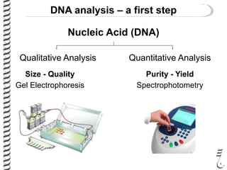
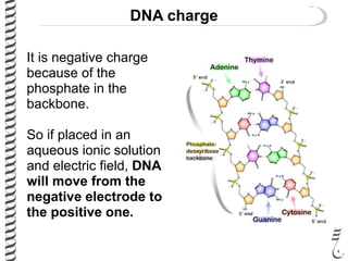
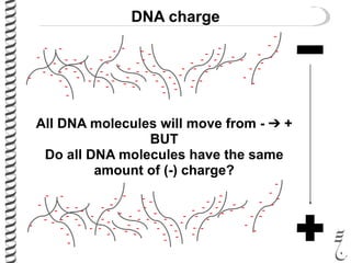
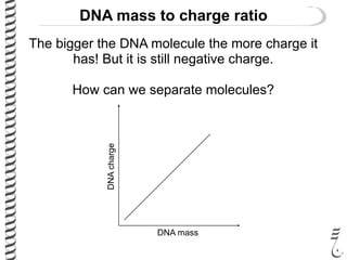
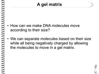
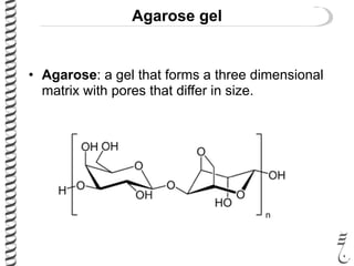
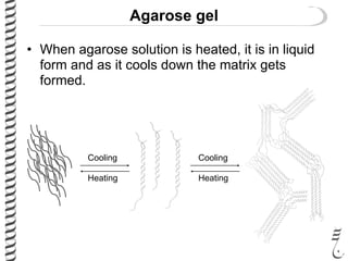
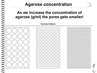
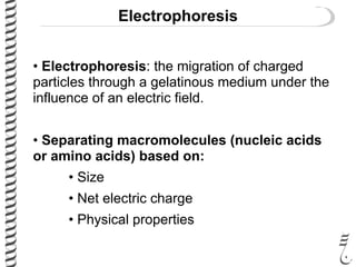
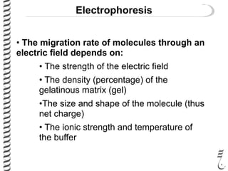
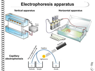
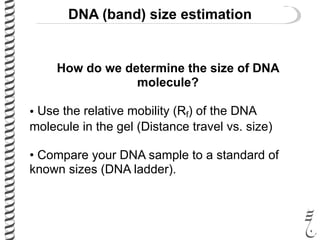
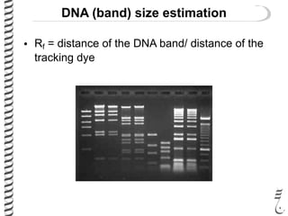
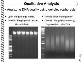
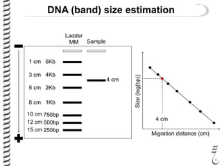
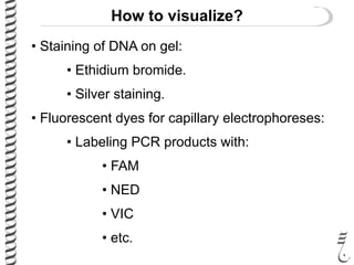
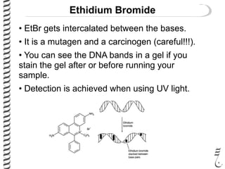
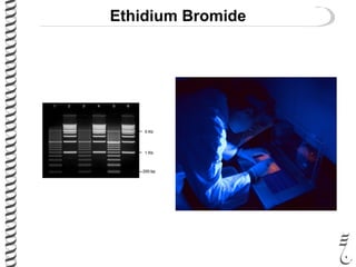
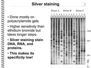
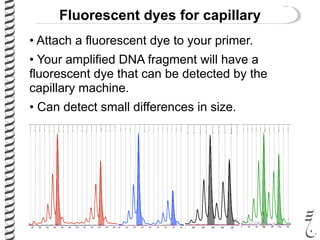
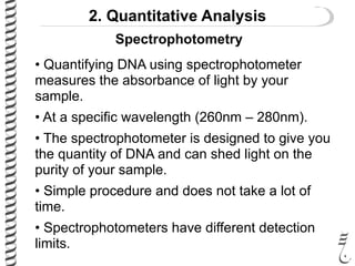
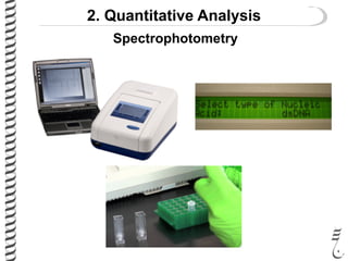
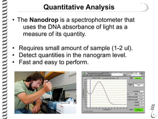
Lecture 30:
Molecular techniques
II. DNA quality and quantity
Readings (chapter 10)
Course 281
Lessons for life
AIMS
• Learn the fundamentals of analyzing the
quality and quantity of DNA.
• Understand why one needs to know:
• The quality of DNA
• The quantity of DNA
• Learn analyzing DNA qualitatively using gel
electrophoresis.
AIMS
• Learn the types of electrophoresis methods.
• Learn how using charge of DNA to separate
the sizes.
• Learn what DNA staining method you should
use and when.
• Learn the standard quantitative methods.
DNA analysis – a first step
Nucleic Acid (DNA)
Quantitative AnalysisQualitative Analysis
Purity - YieldSize - Quality
SpectrophotometryGel Electrophoresis
1. Qualitative Analysis
• One standard method to test the quality of a
DNA sample is to run the sample on a gel.
•This method allows you to visualize the quality of
DNA.
• Large amount?
• Small amount?
• Big size (genomic)?
• Small size (degraded)?
• The intensity of the band may give you a hint
about the quantity but this is not precise.
DNA charge
What is the net charge of DNA?
DNA charge
It is negative charge
because of the
phosphate in the
backbone.
So if placed in an
aqueous ionic solution
and electric field, DNA
will move from the
negative electrode to
the positive one.
DNA charge
-
-
-
-
- -
-
-
-
-
- - -
-
-
-
-
-
- -
-
-
-
--
- - -
-
-
-
-
-
-
-
- -
-
-
--
- -
-
-
-
-
-
-
-
-
-
-
- -
-
-
-
-
-
-
-
-
-
-
- --
-
-
- -
-
- -
-
-
-
-
-
-
-
- -
-
-
-
-
- - -
-
-
-
-
-
- -
-
-
-
--
- - -
-
-
-
-
-
-
-
- -
-
-
--
- -
-
-
-
-
-
-
-
-
-
-
- -
-
-
-
-
-
-
-
-
-
-
- --
-
-
- -
-
- -
-
-
-
All DNA molecules will move from - ➔ +
BUT
Do all DNA molecules have the same
amount of (-) charge?
DNA mass to charge ratio
The bigger the DNA molecule the more charge it
has! But it is still negative charge.
How can we separate molecules?
DNA mass
DNAcharge
A gel matrix
• How can we make DNA molecules move
according to their size?
• We can separate molecules based on their size
while all being negatively charged by allowing
the molecules to move in a gel matrix.
A gel matrix
• Types of gelatinous
mediums:
• Agarose
• Polyacrylamide
• Cellulose acetate and
starch
Sea weed and Agarose
Agarose gel
• Agarose: a gel that forms a three dimensional
matrix with pores that differ in size.
Agarose gel
• When agarose solution is heated, it is in liquid
form and as it cools down the matrix gets
formed.
Cooling
Heating
Cooling
Heating
Agarose concentration
As we increase the concentration of
agarose (g/ml) the pores gets smaller!
Concentration
Agarose concentration
Low concentration gel separates large DNA
molecules more clearly.
Why?
Agarose concentration
Intermediate concentration gel separates DNA
molecules almost uniformly.
Why?
Agarose concentration
High concentration gel separates small DNA
molecules more clearly.
Why?
Electrophoresis
• Electrophoresis: the migration of charged
particles through a gelatinous medium under the
influence of an electric field.
• Separating macromolecules (nucleic acids
or amino acids) based on:
• Size
• Net electric charge
• Physical properties
Electrophoresis
• The migration rate of molecules through an
electric field depends on:
• The strength of the electric field
• The density (percentage) of the
gelatinous matrix (gel)
•The size and shape of the molecule (thus
net charge)
• The ionic strength and temperature of
the buffer
Electrophoresis apparatus
Vertical apparatus Horizontal apparatus
Capillary
electrophoresis
DNA (band) size estimation
How do we determine the size of DNA
molecule?
• Use the relative mobility (Rf) of the DNA
molecule in the gel (Distance travel vs. size)
• Compare your DNA sample to a standard of
known sizes (DNA ladder).
DNA (band) size estimation
• Rf = distance of the DNA band/ distance of the
tracking dye
Qualitative Analysis
• Analyzing DNA quality using gel electrophoresis.
Genomic DNA Degraded low quality DNA
• Up in the gel (large in size)
• Down in the gel (small in size)
• Intense color (high quantity)
• Down in the gel (low quantity)
DNA (band) size estimation
Ladder
MM Sample
6Kb1 cm
4Kb3 cm
2Kb5 cm
8 cm 1Kb
750bp10 cm
500bp12 cm
250bp15 cm
4 cm
Migration distance (cm)
Size(log(bp))
4 cm
How to visualize?
• Staining of DNA on gel:
• Ethidium bromide.
• Silver staining.
• Fluorescent dyes for capillary electrophoreses:
• Labeling PCR products with:
• FAM
• NED
• VIC
• etc.
Ethidium Bromide
• EtBr gets intercalated between the bases.
• It is a mutagen and a carcinogen (careful!!!).
• You can see the DNA bands in a gel if you
stain the gel after or before running your
sample.
• Detection is achieved when using UV light.
Ethidium Bromide
Silver staining
• Done mostly on
polyacrylamide gels.
• Higher sensitivity than
ethidium bromide but
takes longer steps.
• Silver staining stain
DNA, RNA, and
proteins.
• This makes its
specificity low!
Fluorescent dyes for capillary
• Attach a fluorescent dye to your primer.
• Your amplified DNA fragment will have a
fluorescent dye that can be detected by the
capillary machine.
• Can detect small differences in size.Microsatellite Genotyping
2. Quantitative Analysis
• Quantifying DNA using spectrophotometer
measures the absorbance of light by your
sample.
• At a specific wavelength (260nm – 280nm).
• The spectrophotometer is designed to give you
the quantity of DNA and can shed light on the
purity of your sample.
• Simple procedure and does not take a lot of
time.
• Spectrophotometers have different detection
limits.
Spectrophotometry
2. Quantitative Analysis
Spectrophotometry
Quantitative Analysis
• The Nanodrop is a spectrophotometer that
uses the DNA absorbance of light as a
measure of its quantity.
• Requires small amount of sample (1-2 ul).
• Detect quantities in the nanogram level.
• Fast and easy to perform.
To study
agarose
Relative mobility
Relative mobility
Polyacrylamide
DNA charge
Ethidium bromide Silver staining
Fluorescent dyesGel electrophoresis
Capillary electrophoresis
UV light
NED
FAM VIC
Mutagen
Carcinogen
Spectrophotometer
Nanodrop
Expectations
• You know why DNA moves in an electric field.
• You know why to use a gel matrix for DNA
separation.
• You know why you need to stain DNA on a gel
and how.
• You know capillary electrophoresis and why
you use fluorescent labeled primers.
Expectations
• You know the qualitative methods you can run
to check the size and quality of your DNA
sample.
• You know some of the quantitative methods
you can use to assess the yield and purity of
your DNA sample.
• You know when to use a specific method of
DNA quantification and why.
For a smile

281 lec30 mol_tech2

  • 1.
    Lecture 30: Molecular techniques II.DNA quality and quantity Readings (chapter 10) Course 281
  • 2.
  • 3.
    AIMS • Learn thefundamentals of analyzing the quality and quantity of DNA. • Understand why one needs to know: • The quality of DNA • The quantity of DNA • Learn analyzing DNA qualitatively using gel electrophoresis.
  • 4.
    AIMS • Learn thetypes of electrophoresis methods. • Learn how using charge of DNA to separate the sizes. • Learn what DNA staining method you should use and when. • Learn the standard quantitative methods.
  • 5.
    DNA analysis –a first step Nucleic Acid (DNA) Quantitative AnalysisQualitative Analysis Purity - YieldSize - Quality SpectrophotometryGel Electrophoresis
  • 6.
    1. Qualitative Analysis •One standard method to test the quality of a DNA sample is to run the sample on a gel. •This method allows you to visualize the quality of DNA. • Large amount? • Small amount? • Big size (genomic)? • Small size (degraded)? • The intensity of the band may give you a hint about the quantity but this is not precise.
  • 7.
    DNA charge What isthe net charge of DNA?
  • 8.
    DNA charge It isnegative charge because of the phosphate in the backbone. So if placed in an aqueous ionic solution and electric field, DNA will move from the negative electrode to the positive one.
  • 9.
    DNA charge - - - - - - - - - - -- - - - - - - - - - - - -- - - - - - - - - - - - - - - -- - - - - - - - - - - - - - - - - - - - - - - - - - -- - - - - - - - - - - - - - - - - - - - - - - - - - - - - - - - - - -- - - - - - - - - - - - - - - -- - - - - - - - - - - - - - - - - - - - - - - - - - -- - - - - - - - - - - All DNA molecules will move from - ➔ + BUT Do all DNA molecules have the same amount of (-) charge?
  • 10.
    DNA mass tocharge ratio The bigger the DNA molecule the more charge it has! But it is still negative charge. How can we separate molecules? DNA mass DNAcharge
  • 11.
    A gel matrix •How can we make DNA molecules move according to their size? • We can separate molecules based on their size while all being negatively charged by allowing the molecules to move in a gel matrix.
  • 12.
    A gel matrix •Types of gelatinous mediums: • Agarose • Polyacrylamide • Cellulose acetate and starch Sea weed and Agarose
  • 13.
    Agarose gel • Agarose:a gel that forms a three dimensional matrix with pores that differ in size.
  • 14.
    Agarose gel • Whenagarose solution is heated, it is in liquid form and as it cools down the matrix gets formed. Cooling Heating Cooling Heating
  • 15.
    Agarose concentration As weincrease the concentration of agarose (g/ml) the pores gets smaller! Concentration
  • 16.
    Agarose concentration Low concentrationgel separates large DNA molecules more clearly. Why?
  • 17.
    Agarose concentration Intermediate concentrationgel separates DNA molecules almost uniformly. Why?
  • 18.
    Agarose concentration High concentrationgel separates small DNA molecules more clearly. Why?
  • 19.
    Electrophoresis • Electrophoresis: themigration of charged particles through a gelatinous medium under the influence of an electric field. • Separating macromolecules (nucleic acids or amino acids) based on: • Size • Net electric charge • Physical properties
  • 20.
    Electrophoresis • The migrationrate of molecules through an electric field depends on: • The strength of the electric field • The density (percentage) of the gelatinous matrix (gel) •The size and shape of the molecule (thus net charge) • The ionic strength and temperature of the buffer
  • 21.
    Electrophoresis apparatus Vertical apparatusHorizontal apparatus Capillary electrophoresis
  • 22.
    DNA (band) sizeestimation How do we determine the size of DNA molecule? • Use the relative mobility (Rf) of the DNA molecule in the gel (Distance travel vs. size) • Compare your DNA sample to a standard of known sizes (DNA ladder).
  • 23.
    DNA (band) sizeestimation • Rf = distance of the DNA band/ distance of the tracking dye
  • 24.
    Qualitative Analysis • AnalyzingDNA quality using gel electrophoresis. Genomic DNA Degraded low quality DNA • Up in the gel (large in size) • Down in the gel (small in size) • Intense color (high quantity) • Down in the gel (low quantity)
  • 25.
    DNA (band) sizeestimation Ladder MM Sample 6Kb1 cm 4Kb3 cm 2Kb5 cm 8 cm 1Kb 750bp10 cm 500bp12 cm 250bp15 cm 4 cm Migration distance (cm) Size(log(bp)) 4 cm
  • 26.
    How to visualize? •Staining of DNA on gel: • Ethidium bromide. • Silver staining. • Fluorescent dyes for capillary electrophoreses: • Labeling PCR products with: • FAM • NED • VIC • etc.
  • 27.
    Ethidium Bromide • EtBrgets intercalated between the bases. • It is a mutagen and a carcinogen (careful!!!). • You can see the DNA bands in a gel if you stain the gel after or before running your sample. • Detection is achieved when using UV light.
  • 28.
  • 29.
    Silver staining • Donemostly on polyacrylamide gels. • Higher sensitivity than ethidium bromide but takes longer steps. • Silver staining stain DNA, RNA, and proteins. • This makes its specificity low!
  • 30.
    Fluorescent dyes forcapillary • Attach a fluorescent dye to your primer. • Your amplified DNA fragment will have a fluorescent dye that can be detected by the capillary machine. • Can detect small differences in size.Microsatellite Genotyping
  • 31.
    2. Quantitative Analysis •Quantifying DNA using spectrophotometer measures the absorbance of light by your sample. • At a specific wavelength (260nm – 280nm). • The spectrophotometer is designed to give you the quantity of DNA and can shed light on the purity of your sample. • Simple procedure and does not take a lot of time. • Spectrophotometers have different detection limits. Spectrophotometry
  • 32.
  • 33.
    Quantitative Analysis • TheNanodrop is a spectrophotometer that uses the DNA absorbance of light as a measure of its quantity. • Requires small amount of sample (1-2 ul). • Detect quantities in the nanogram level. • Fast and easy to perform.
  • 34.
    To study agarose Relative mobility Relativemobility Polyacrylamide DNA charge Ethidium bromide Silver staining Fluorescent dyesGel electrophoresis Capillary electrophoresis UV light NED FAM VIC Mutagen Carcinogen Spectrophotometer Nanodrop
  • 35.
    Expectations • You knowwhy DNA moves in an electric field. • You know why to use a gel matrix for DNA separation. • You know why you need to stain DNA on a gel and how. • You know capillary electrophoresis and why you use fluorescent labeled primers.
  • 36.
    Expectations • You knowthe qualitative methods you can run to check the size and quality of your DNA sample. • You know some of the quantitative methods you can use to assess the yield and purity of your DNA sample. • You know when to use a specific method of DNA quantification and why.
  • 37.