BLOTTING TECHNIQUES 
Present by… 
CHOVATIYA MANISH M. 
M.SC BIOTECHNOLOGY
 Blots are techniques for transferring DNA , RNA 
and proteins onto a carrier so they can be 
separated, and follows the use of a gel 
electrophoresis. The Southern blot is used for 
transferring DNA, the Northern blot for RNA and 
the western blot for PROTEIN.
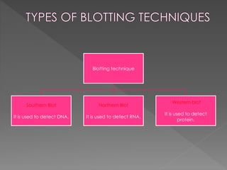
Blotting technique 
Southern Blot 
It is used to detect DNA. 
Northern Blot 
It is used to detect RNA. 
Western blot 
It is used to detect 
protein.
 Professor Sir Edwin Southern, 
Professor of Biochemistry and 
Fellow of Trinity developed this 
method in 1975. 
 Southern won the Lasker 
Award for Clinical Medical 
Research prize for the method 
of finding specific DNA 
sequences he developed this 
procedure at Edinburgh 
University more than 30 years 
ago. The technique is known 
as DNA transfer or 'Southern 
blotting' 
Professor Sir Edwin Southern
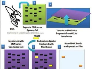
 This method Involves separation, transfer and 
hybridization. 
 It is a method routinely used in molecular biology for 
detection of a specific DNA sequence in DNA samples. 
 The DNA detected can be a single gene, or it can be part of 
a larger piece of DNA such as a viral genome.
 Southern blotting combines agarose gel electrophoresis for 
size separation of DNA with methods to transfer the size 
separated DNA to a filter membrane for probe 
hybridization. 
 The key to this method is Hybridization. 
 Hybridization - Process of forming a double-stranded 
DNA molecule between a single-stranded DNA probe and a 
single-stranded target patient DNA.
1. The mixture of molecules is separated. 
2. The molecules are immobilized on a MEMBRANE. 
3. The probe is added to the membrane to bind to 
the molecules. 
4. Any unbound probes are then removed. 
5. The place where the probe is connected corresponds to 
the location of the immobilized target molecule.
Cellulose nitrate (nitrocellulose) 
 It is produced by treating cellulose with 
nitric acid. 
 Each glucose unit in the cellulose polymer is 
esterified with three nitrate groups, and 
these nitrate groups are responsible for both 
the negative charge of nitrocellulose at 
neutral pH and the unusual flammability of 
dry nitrocellulose.
 The major improvement has been the introduction of nylon membranes, 
which have three advantages over their nitrocellulose counterparts. 
 First,nylon membranes are less fragile than nitrocellulose sheets,the latter 
tending to crack if handled roughly during Southern blotting,and usually 
disintegrating if attempts are made to carry out more than two or three 
hybridization analyses with the same blot. 
 Nylon membranes cannot be damaged by handling and a single blot 
can be rehybridized up to ten times,this limit being due not to eventual 
breakage of the membrane but to the gradual loss of the blotted DNA 
during repeated hybridizations. 
 The second advantage of nylon membranes is that under certain 
conditions (a positively charged membrane and an alkaline transfer 
buffer) the transferred DNA becomes covalently bound to the membrane 
during the transfer process. This is not the case with a nitrocellulose 
membrane,which initially binds DNA in a semipermanent 
manner,immobilization occurring only when the membrane is baked at 
80C.
 Transfer onto a positively charged nylon membrane 
can therefore reduce the possible loss of DNA that 
might occur by leaching through the membrane 
during the blotting process; 
 it is also quicker,the transfer time being reduced from 
18 h to 2 h. 
 Finally,nylon membranes efficiently bind DNA 
fragments down to 50 bp in length,whereas 
nitrocellulose membranes are effective only with 
molecules longer than 500 bp. 
 Nitrocellulose has not,however,been completely 
superseded because it has one significant 
advantage compared with nylon membranes: a 
reduced amount of background 
hybridization,especially with probes that have been 
labelled with nonradioactive markers.
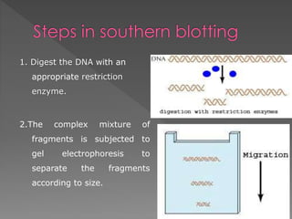
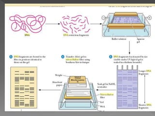
1. Digest the DNA with an 
appropriate restriction 
enzyme. 
2.The complex mixture of 
fragments is subjected to 
gel electrophoresis to 
separate the fragments 
according to size.
The restriction fragments present in the gel are denatured with alkali 
TO GET ss DNA (break Hydrogen bond). 
To break the DNA molecules in individual bands within the gel into smaller 
fragments, because smaller fragments transfer more quickly than larger 
ones. This is achieved by soaking the gel in 0.25 molL21 HCl for 30 
min,which results in a small amount of depurination – cleavage of the 
b-N-glycosidic bond between purine bases (adenine or guanine) and 
the sugar component of their nucleotides – which is followed by 
decomposition of the sugar structure and breakage of the 
polynucleotide chain.
 If a nitrocellulose membrane is being 
used then the alkali pretreatment is 
followed by neutralization of the gel by 
soaking in a Tris-salt buffer,this step being 
essential because DNA does not bind to 
nitrocellulose at a pH of greater than 9.0.
 Nitrocellulose membrane soaked in salt solution 
is placed over gel. Nucleic acid move from gel 
to membrane . a high concentration of salt 
(NaCl THAT IS the positive Na+ ions shield the 
negative charges on the phosphodiester 
backbone AND LOWERS –ve DNA = -ve 
membrane repelling each other 
 BAKING Leads to fixing of DNA strongly to the 
nitocellulose membrane. UV irradiation,which 
results in covalent attachment of DNA to a 
nylon membrane.
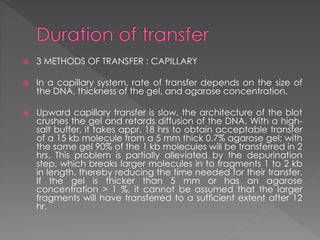
 3 METHODS OF TRANSFER : CAPILLARY 
 In a capillary system, rate of transfer depends on the size of 
the DNA, thickness of the gel, and agarose concentration. 
 Upward capillary transfer is slow, the architecture of the blot 
crushes the gel and retards diffusion of the DNA. With a high-salt 
buffer, it takes appr. 18 hrs to obtain acceptable transfer 
of a 15 kb molecule from a 5 mm thick 0.7% agarose gel; with 
the same gel 90% of the 1 kb molecules will be transferred in 2 
hrs. This problem is partially alleviated by the depurination 
step, which breaks larger molecules in to fragments 1 to 2 kb 
in length, thereby reducing the time needed for their transfer. 
If the gel is thicker than 5 mm or has an agarose 
concentration > 1 %, it cannot be assumed that the larger 
fragments will have transferred to a sufficient extent after 12 
hr.
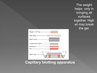
The weight 
helps only in 
bringing all 
Capillary blotting apparatus 
surfaces 
together. High 
wt may break 
the gel.
First,the membrane is prehybridized in a solution designed to 
block the unused DNA binding sites on the membrane 
surface. If this step is omitted then the probe will bind 
nonspecifically to the surface of the membrane and the 
signal resulting from hybridization to the specific restriction 
fragment will be difficult if not impossible to identify. 
The prehybridization solution therefore contains nonbiological 
polymeric compounds such as polyvinylpyrrolidone and/or 
biological polymers such as Ficoll (a carbohydrate-based 
compound),bovine serum albumin or dried milk. DNA from 
an organism unrelated to the one whose DNA has been 
blotted can also be used (salmon sperm DNA is a popular 
choice). 
Prehybridization takes between 15 min and 3 h at 
68C,depending on the type of membrane
 enough labeled probe DNA is added to hybridize to the 
target restriction fragment to produce a clear signal that 
can be discerned by the detection system appropriate for 
the label carried by the probe. 
The second critical factor that must be considered during the 
hybridization step is the specificity of the reaction. If the 
probe DNA has been carefully chosen then it will contain a 
region that is completely complementary to all or a part of 
the blotted restriction fragment that is being sought. If this 
hybridizing region in the probe is not completely 
complementary to the target,then it will at least have a 
region of strong similarity so that a stable hybrid can form. 
The problem is that the probe also has the potential to 
hybridize to any other blotted DNA fragments with which it 
has partial complementarity.
 Temperature is relevant because the melting 
temperature (Tm,the highest temperature at 
which the hybrid is stable) of a fully base-paired 
hybrid is higher than that for one in 
which some base pairs have not formed 
because the probe and target DNAs are not 
fully complementary. Hybridization Maximum 
rate occurs at 20-25°C below the Tm for DNA-DNA 
hybrids, 10-15°C below Tm for DNA-RNA 
hybrids 
 Formation of the desired hybrid,and 
destabilization of nonspecific hybrids,can 
therefore be achieved by utilizing an 
appropriate combination of buffer composition 
and hybridization temperature.
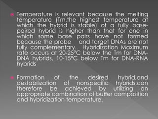
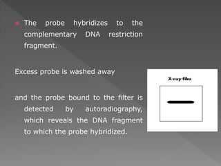
 The probe hybridizes to the 
complementary DNA restriction 
fragment. 
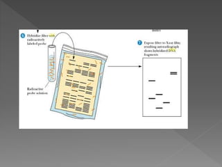
Excess probe is washed away 
and the probe bound to the filter is 
detected by autoradiography, 
which reveals the DNA fragment 
to which the probe hybridized.
 Southern blots are used in gene discovery , mapping, 
evolution and development studies, diagnostics and 
forensics (It is used for DNA fingerprinting, preparation of 
RFLP maps) 
 identification of the transferred genes in transgenic 
individuals, etc.
 Southern blots allow investigators to determine the 
molecular weight of a restriction fragment and to measure 
relative amounts in different samples. 
 Southern blot is used to detect the presence of a particular 
bit of DNA in a sample 
 analyze the genetic patterns which appear in a person's 
DNA.
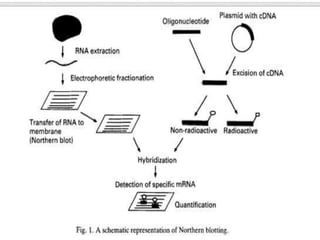
Northern blotting is a technique for detection of specific 
RNA sequences. 
Northern blotting was developed by James Alwine and 
George Stark at Stanford University (1979) and was 
named such by analogy to Southern blotting
 The first is to determine which tissues 
express a particular gene, and this can 
give some indication of the physiological 
function of the encoded protein. 
 The second principal reason for measuring 
an mRNA is to determine the factors which 
regulate the expression of a given gene, 
be they nutritional, hormonal, or 
environmental.
 Northern blotting. 
 A second method is the RNase protection 
assay, which is generally considered to offer 
improved sensitivity. 
 The third method utilizes the reverse 
transcriptase polymerase chain reaction; this 
provides considerable increases in sensitivity 
over Northern blotting and the RNase 
protection assay, and may be useful for 
measuring very low levels (a few copies) of an 
mRNA in a tissue.
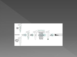
 Then, in a single step reaction, the 
nucleases are inactivated and the 
remaining probe:target hybrids are 
precipitated. These products are 
separated on a denaturing 
polyacrylamide gel and are visualized by 
autoradiography.
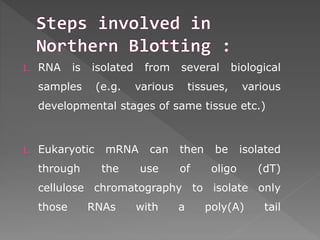
1. RNA is isolated from several biological 
samples (e.g. various tissues, various 
developmental stages of same tissue etc.) 
1. Eukaryotic mRNA can then be isolated 
through the use of oligo (dT) 
cellulose chromatography to isolate only 
those RNAs with a poly(A) tail
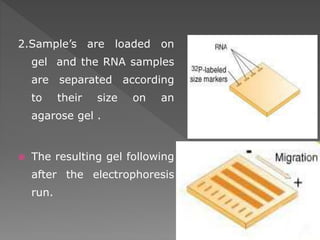
2.Sample’s are loaded on 
gel and the RNA samples 
are separated according 
to their size on an 
agarose gel . 
 The resulting gel following 
after the electrophoresis 
run.
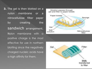
3. The gel is then blotted on a 
nylon membrane or a 
nitrocellulose filter paper 
by creating the 
sandwich arrangement. 
Nylon membrane with a 
positive charge is the most 
effective for use in northern 
blotting since the negatively 
charged nucleic acids have 
a high affinity for them.
 Once the RNA has been transferred to 
the membrane, it is immobilized 
through covalent linkage to the 
membrane by UV light or heat.
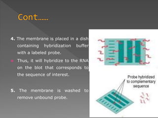
4. The membrane is placed in a dish 
containing hybridization buffer 
with a labeled probe. 
 Thus, it will hybridize to the RNA 
on the blot that corresponds to 
the sequence of interest. 
5. The membrane is washed to 
remove unbound probe.
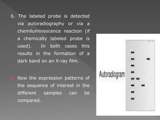
6. The labeled probe is detected 
via autoradiography or via a 
chemiluminescence reaction (if 
a chemically labeled probe is 
used). In both cases this 
results in the formation of a 
dark band on an X-ray film. 
 Now the expression patterns of 
the sequence of interest in the 
different samples can be 
compared.
 The RNA samples are most commonly 
separated on agarose gels 
containing formaldehyde as a 
denaturing agent for the RNA to limit 
secondary structure. 
 Polyacrylamide gel electrophoeresis 
with urea can also be used in RNA 
separation but it is most commonly used 
for fragmented RNA or microRNAs
 A standard for the study of gene expression at the level of 
mRNA (messenger RNA transcripts) 
 Detection of mRNA transcript size 
 Study RNA degradation 
 Study RNA splicing 
 Study RNA half-life 
 Often used to confirm and check transgenic / knockout 
mice (animals)
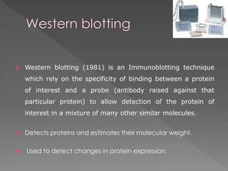
 Western blotting (1981) is an Immunoblotting technique 
which rely on the specificity of binding between a protein 
of interest and a probe (antibody raised against that 
particular protein) to allow detection of the protein of 
interest in a mixture of many other similar molecules. 
 Detects proteins and estimates their molecular weight. 
 Used to detect changes in protein expression.
 Tissue preparation 
 Samples can be taken from whole tissue or from cell culture. Solid tissues 
are first broken down mechanically using a blender (for larger sample 
volumes), using a homogenizer (smaller volumes), or by sonication. Cells 
may also be broken open by one of the above mechanical methods. 
However, virus or environmental samples can be the source of protein 
and thus western blotting is not restricted to cellular studies only. 
 Assorted detergents, salts, and buffers may be employed to 
encourage lysis of cells and to solubilize proteins. Protease and 
phosphatase inhibitors are often added to prevent the digestion of the 
sample by its own enzymes. Tissue preparation is often done at cold 
temperatures to avoid protein denaturing and degradation. 
 A combination of biochemical and mechanical techniques – 
comprising various types of filtration and centrifugation – can be used to 
separate different cell compartments and organelles.
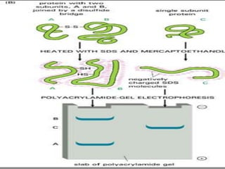
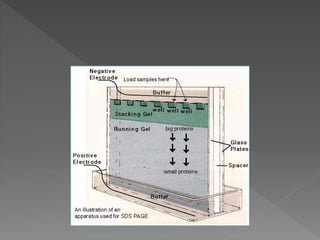
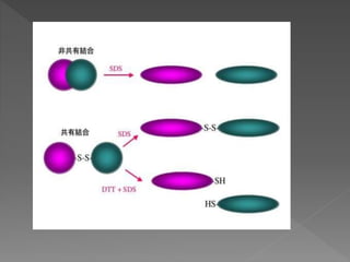
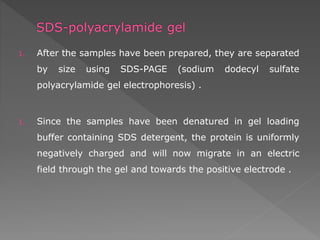
1. After the samples have been prepared, they are separated 
by size using SDS-PAGE (sodium dodecyl sulfate 
polyacrylamide gel electrophoresis) . 
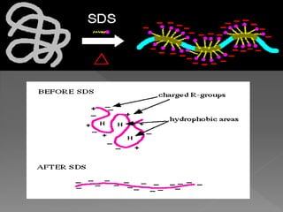
1. Since the samples have been denatured in gel loading 
buffer containing SDS detergent, the protein is uniformly 
negatively charged and will now migrate in an electric 
field through the gel and towards the positive electrode .
 SDS-PAGE ( sodium dodecylsulphate-polyacrylamide 
gel electrophoresis) 
 The purpose of this method is to separate 
proteins according to their size, and no 
other physical feature.
 Stacking Gel
 Transfer of the proteins fractionated by SDS-PAGE 
to a solid support membrane (Western blotting) 
can be accomplished by electroblotting
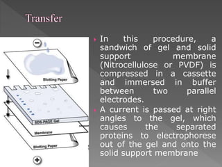
 In this procedure, a 
sandwich of gel and solid 
support membrane 
(Nitrocellulose or PVDF) is 
compressed in a cassette 
and immersed in buffer 
between two parallel 
electrodes. 
 A current is passed at right 
angles to the gel, which 
causes the separated 
proteins to electrophorese 
out of the gel and onto the 
solid support membrane
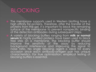
 The membrane supports used in Western blotting have a 
high affinity for proteins. Therefore, after the transfer of the 
proteins from the gel, it is important to block the remaining 
surface of the membrane to prevent nonspecific binding 
of the detection antibodies during subsequent steps. 
 A variety of blocking buffers ranging from milk or normal 
serum to highly purified proteins have been used to block 
free sites on a membrane. The blocking buffer should 
improve the sensitivity of the assay by reducing 
background interference and improving the signal to 
noise ratio. No single blocking agent is ideal for every 
occasion since each antibody-antigen pair has unique 
characteristics. For true optimization, empirical testing of 
blocking buffers is essential.
 The balance of SDS in the transfer buffer, 
protein size, and gel percentage are the 
main factors that affect transfer efficiency. 
 About the current and transfer time for 
western blot, it's critical to choose the 
appropriate current and transfer time for a 
successful western blotting. 
 Too low current or/and transfer time will 
lead to incomplete transfer; if the current 
or/and transfer time is too high, on the 
contrary, the proteins may migrate 
through the membrane too fast without 
being absorbed.
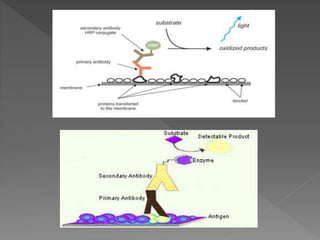
 One of the critical features of any 
successful Western blot is the highly 
 specific interaction between an antibody 
and an antigen. The antigen, 
 usually a protein or peptide, is the target of 
the antibody. The precise point 
 of interaction is between a small region of 
the antigen, an epitope, 
 and the recognition sites found on the arms 
of the antibody molecule.
1.The confirmatory HIV test 
2.Western blot is also used as the definitive test for 
Bovine spongiform encephalopathy (BSE( 
3.Some forms of Lyme disease testing employ Western 
blotting .
Blotting techniques (manish)