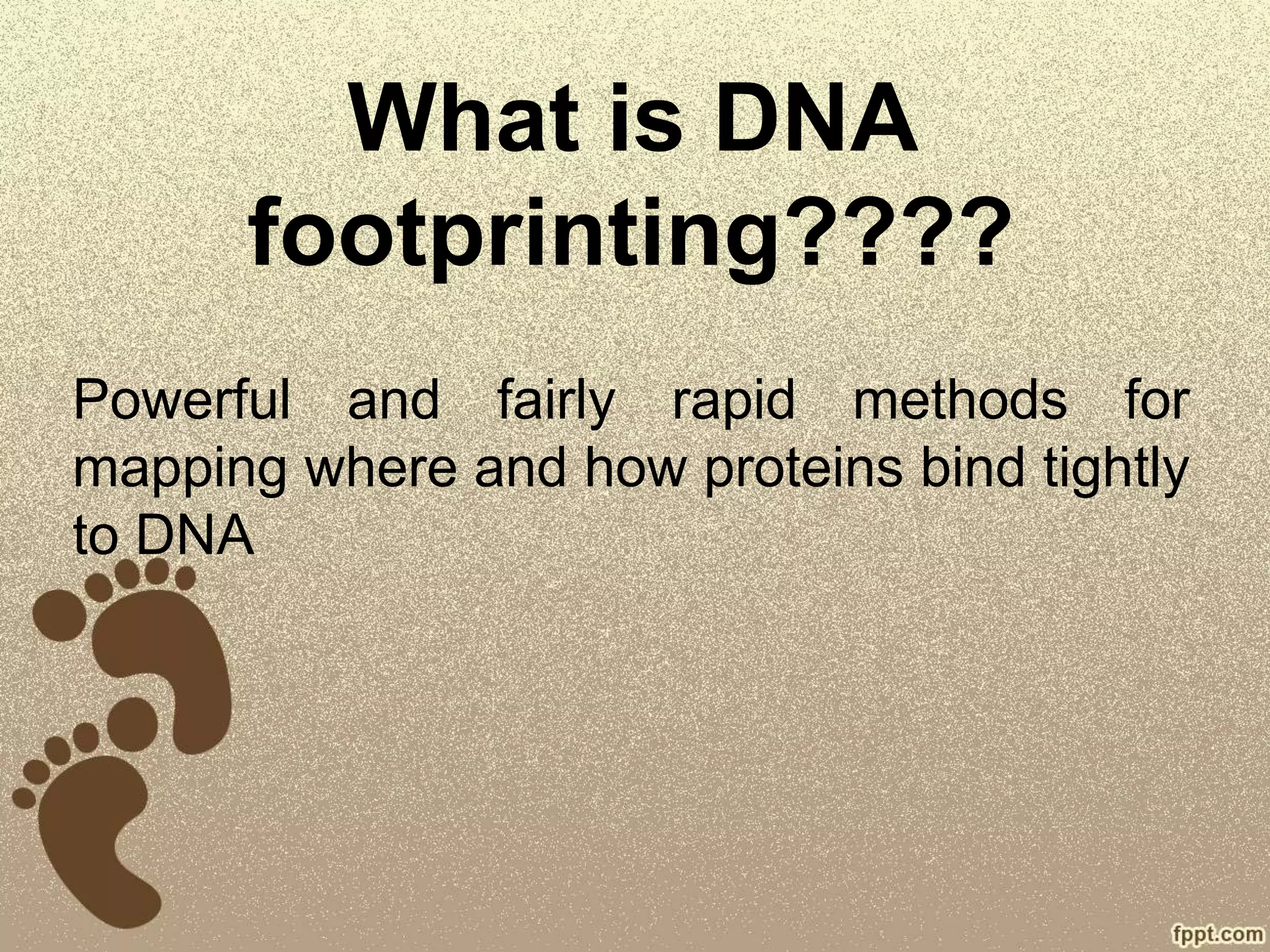
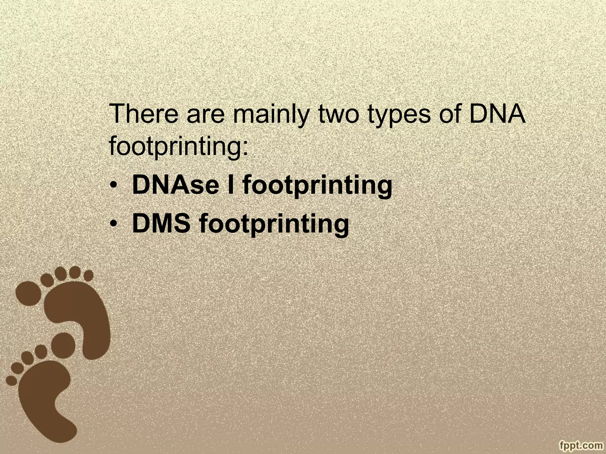
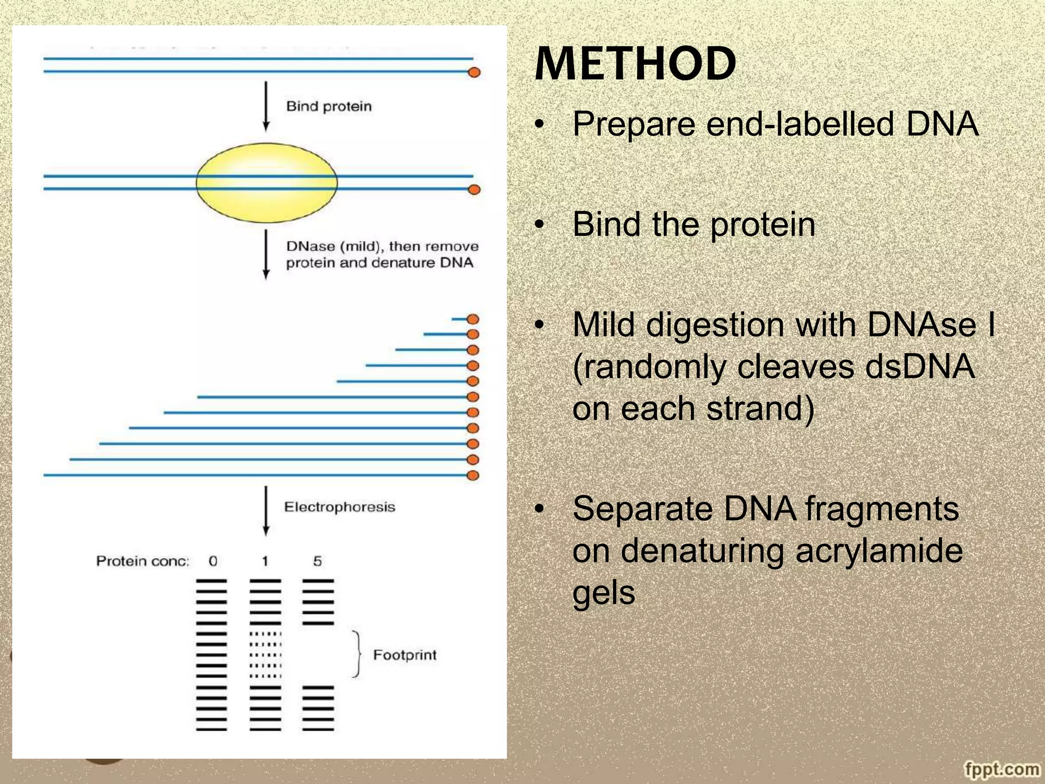
DNA
Footprinting
Adarsh P P
M.Sc. Bioinformatics
Union Christian College, Aluva
What is DNA
footprinting????
Powerful and fairly rapid methods for
mapping where and how proteins bind tightly
to DNA
HISTORY
• Developed by David Galas and
Albert Schmilz in 1978
• Used to study the binding of lac
repressor
• Modification of Maxam-Gilbert
chemical sequencing technique
PRINCIPLE
• Assay used to identify the site of protein-DNA
interaction.
• It has been found that a protein bound to the
DNA protect it from the action of degradative
enzymes.
• The main application of this method is to find
the binding regions of DNA transcription
factors.
PROCEDURE
• The DNA of interest is first amplified using PCR
• The double stranded amplified DNA is labelled
at one end of each strand.
• Next, the protein of interest is added.
• The DNA is then cleaved by chemical or
enzymatic cleavage agent.
• The fragments are then run on a denaturing
polyacrylamide gel.
There are mainly two types of DNA
footprinting:
• DNAse I footprinting
• DMS footprinting
DNAse I Footprinting
• Here the DNAse I enzyme is used
• It not only helps to find the target protein that
binds to specific DNA, but also identify which
sequence to which the protein is bound.
• The protein binding to the DNA protect it from
DNAse I
• The fragments are then left after cleavage, so it
can be sequenced
METHOD
• Prepare end-labelled DNA
• Bind the protein
• Mild digestion with DNAse I
(randomly cleaves dsDNA
on each strand)
• Separate DNA fragments
on denaturing acrylamide
gels
• Samples in lane 2-4 had
increasing concentration of
DNA-binding protein.
DIMETHYLSULFAT
E FOOTPRINTING
• DMS induces methylation of guanine residues.
• Similar to DNAse footprinting
• Mildly treat the DNA with DMS (after the addition of
protein) so that on an average only one methylation
occurs per DNA.
• The DNA is then treated with a reagent that removes the
methylated purins.
• The apurinic sites thus are removed with the help of
cleaving agents.
• Each band end next to a nucleotide that was methylated
and thus unprotected by the protein.
METHOD
• End-labelled DNA fragment
• Bind protein
• Treat with DMS, methylates
purines
• Partially cleave DNA at
methylated bases.
• Separate fragments on the
gel
• Lane 1 and 4 have no protein
• Lane 2 and 3 have different
amount of protein.
Disadvantages
• Protein binding protects some
purines from modification by
DMS, it but can stimulate
modification of others (helix
distorted or partially melted)
• Cannot be used to detect the
proteins binding to the AT rich
sequences.
APPLICATIONS
• In vivo Foot printing
• Quantitative Footprinting
• Detection by capillary
electrophoresis.
In vivo Foot printing
• Used to analyse protein-DNA interaction occurring at a
given time in a cell.
• The cell membrane is first permeabilized using UV rays.
• Then the DNAse I is used.
• After the cleavage, the single stranded DNA is isolated
and purified, a linker DNA is added into the break
points.
• The region is then amplified and run on gel.
• This method can be coupled with immunoprecipitation
The DNA binding to the protein of interest can be
precipitated with an antibody to the protein, and then
specific region of protein binding can be assessed.
Quantitative Footprinting
• Modification of normal footprinting
• Varying concentration of protein is used
• Protein binding affinity can be estimated,
because the formation of footprinting can be
observed with increasing concentration of
protein
Detection by Capillary
Electrophoresis
• Capillary electrophoresis device is used.
• The PCR primers are coupled with
carboxyflourescein, thus the fragments
produced by digestion will contain
carboxyflourescein
• This fluorescence can be detected using
capillary electrophoresis device
• Transcription factor binding sites can be
effectively identified.
THANK YOU

Dna Footprinting

  • 1.
    DNA Footprinting Adarsh P P M.Sc.Bioinformatics Union Christian College, Aluva
  • 2.
    What is DNA footprinting???? Powerfuland fairly rapid methods for mapping where and how proteins bind tightly to DNA
  • 3.
    HISTORY • Developed byDavid Galas and Albert Schmilz in 1978 • Used to study the binding of lac repressor • Modification of Maxam-Gilbert chemical sequencing technique
  • 4.
    PRINCIPLE • Assay usedto identify the site of protein-DNA interaction. • It has been found that a protein bound to the DNA protect it from the action of degradative enzymes. • The main application of this method is to find the binding regions of DNA transcription factors.
  • 5.
    PROCEDURE • The DNAof interest is first amplified using PCR • The double stranded amplified DNA is labelled at one end of each strand. • Next, the protein of interest is added. • The DNA is then cleaved by chemical or enzymatic cleavage agent. • The fragments are then run on a denaturing polyacrylamide gel.
  • 7.
    There are mainlytwo types of DNA footprinting: • DNAse I footprinting • DMS footprinting
  • 8.
    DNAse I Footprinting •Here the DNAse I enzyme is used • It not only helps to find the target protein that binds to specific DNA, but also identify which sequence to which the protein is bound. • The protein binding to the DNA protect it from DNAse I • The fragments are then left after cleavage, so it can be sequenced
  • 9.
    METHOD • Prepare end-labelledDNA • Bind the protein • Mild digestion with DNAse I (randomly cleaves dsDNA on each strand) • Separate DNA fragments on denaturing acrylamide gels
  • 10.
    • Samples inlane 2-4 had increasing concentration of DNA-binding protein.
  • 11.
    DIMETHYLSULFAT E FOOTPRINTING • DMSinduces methylation of guanine residues. • Similar to DNAse footprinting • Mildly treat the DNA with DMS (after the addition of protein) so that on an average only one methylation occurs per DNA. • The DNA is then treated with a reagent that removes the methylated purins. • The apurinic sites thus are removed with the help of cleaving agents. • Each band end next to a nucleotide that was methylated and thus unprotected by the protein.
  • 12.
    METHOD • End-labelled DNAfragment • Bind protein • Treat with DMS, methylates purines • Partially cleave DNA at methylated bases. • Separate fragments on the gel
  • 13.
    • Lane 1and 4 have no protein • Lane 2 and 3 have different amount of protein. Disadvantages • Protein binding protects some purines from modification by DMS, it but can stimulate modification of others (helix distorted or partially melted) • Cannot be used to detect the proteins binding to the AT rich sequences.
  • 14.
    APPLICATIONS • In vivoFoot printing • Quantitative Footprinting • Detection by capillary electrophoresis.
  • 15.
    In vivo Footprinting • Used to analyse protein-DNA interaction occurring at a given time in a cell. • The cell membrane is first permeabilized using UV rays. • Then the DNAse I is used. • After the cleavage, the single stranded DNA is isolated and purified, a linker DNA is added into the break points. • The region is then amplified and run on gel. • This method can be coupled with immunoprecipitation The DNA binding to the protein of interest can be precipitated with an antibody to the protein, and then specific region of protein binding can be assessed.
  • 16.
    Quantitative Footprinting • Modificationof normal footprinting • Varying concentration of protein is used • Protein binding affinity can be estimated, because the formation of footprinting can be observed with increasing concentration of protein
  • 17.
    Detection by Capillary Electrophoresis •Capillary electrophoresis device is used. • The PCR primers are coupled with carboxyflourescein, thus the fragments produced by digestion will contain carboxyflourescein • This fluorescence can be detected using capillary electrophoresis device • Transcription factor binding sites can be effectively identified.
  • 18.