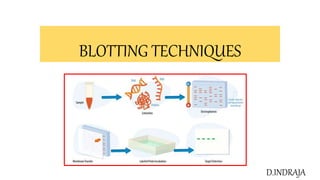
This document summarizes several blotting techniques used in molecular biology to detect biomolecules like DNA, RNA, and proteins. Southern blotting detects specific DNA sequences, Northern blotting detects RNA, and Western blotting detects proteins. Eastern blotting detects post-translational modifications to proteins. Dot blotting directly applies biomolecule samples to a membrane as dots to detect presence or absence without separation by size. These techniques involve transferring molecules to a membrane, probing with a labeled probe, washing, and detecting hybridized probes.
To modifying the structure of a specific gene.
Gene targeting vector introduced into the cell.
Vector modifies the normal chromosomal gene through homologous recombination.
Useful in treating some human genetic disorders – Hemophilia, Duchenne Muscular Dystrophy.
Treating human diseases by genetic approaches – Gene Therapy.
Gene Therapy – Replacing the defective gene by normal copy of the gene.
Expressed sequence tag/EST is a short partial sequence, typically 200-400 bp long, of a complimentary DNA/Cdna.
EST is a short sub-sequence of a cDNA sequence.
Used to identify gene transcripts, and are instrumental in gene discovery and in gene-sequence determination.
Approximately 74.2 million ESTs are available in public databases.
EST results from one-short sequencing of a cloned cDNA.
Low-quality fragments.
Length is approximately 500 to 800 nucleotides.
MBB 501 PLANT BIOTECHNOLOGY
INFORMATION ABOUT DIFFERENT DNA MODIFYING ENZYMES
WHAT IS AN ENZYME?
Alkaline Phosphatase
Polynucleotide kinase
Terminal deoxyneucleotidyl transferase
Nucleases
Exonuclease
Bal31 Exonuclease III
Endonuclease
S1 endonulease
Deoxyribonuclease 1 (Dnase 1)
RNase A
RNase H
Restriction Endonuclease
PvuI
PvuII
Different types of endonuclease enzymes
The recognition sequences for some of the most frequently used restriction endonucleases.
Categorization of enzymes
Isoschizomers
Neoschizomers
Isocaudomers
Creation of a cDNA library starts with mRNA instead of DNA. Messenger RNA carries encoded information from DNA to ribosomes for translation into protein. To create a cDNA library, these mRNA molecules are treated with the enzyme reverse transcriptase, which is used to make a DNA copy of an mRNA (i.e., cDNA). A cDNA library represents a sampling of the transcribed genes, but a genomic library includes untranscribed regions.
Blotting technique including Southern , Northern and Western blotting Rohit Mondal
he given ppt contains all the blotting techniques which is being studied by students in Biotechnology related subject and this PPT contais all blotting techniques in a very elaborative concise manner includes procedure principle application etc so which itwould help any bio student to take proper knowledge in this topic. I hope you will enjoy the content of the topic and would be able to grasp the topic properly
Concept: reannealing nucleic acids to identify sequence of interest.
Separates DNA/RNA in an agarose gel, then detects specific bands using probe and hybridization.
Hybridization takes advantage of the ability of a single stranded DNA or RNA molecule to find its complement, even in the presence of large amounts of unrelated DNA.
Allows detection of specific bands (DNA fragments or RNA molecules) that have complementary sequence to the probe.
Size bands and quantify abundance of molecule.
The methods used for DNA finger printing are the same Molecular markers...so for detailed note on the steps which is explained in DNA typing can be used to study the performance pf markers too...
Southern blotting is a laboratory technique used to detect specific DNA sequences in DNA samples. It involves several steps:
Restriction Digestion: DNA from a biological sample (such as blood or tissue) is broken into smaller fragments using restriction enzymes, which cut the DNA at specific sequences.
Electrophoresis: The DNA fragments are separated based on their molecular weights using gel electrophoresis. This process allows smaller fragments to move faster than larger fragments.
Transfer to Membrane: The DNA fragments are transferred from the gel onto a solid membrane, typically a positively charged nylon membrane, using capillary action.
Hybridization: The membrane is then exposed to a DNA probe labeled with a radioactive, fluorescent, or chemical tag. The probe is designed to be complementary to the target DNA sequence, allowing it to bind to the specific DNA fragment on the membrane.
Detection: The bound probe is detected using methods such as X-ray film, phosphorimaging, or chemiluminescent substrates, depending on the type of label used.
Southern blotting is used in various applications, including:
Identifying specific DNA sequences in DNA samples
Studying gene rearrangements and mutations
Analyzing viral and bacterial infections
Forensic analysis and personal identification
Gene mapping and restriction enzyme mapping
Identifying methylated sites in genes.
This technique is named after its inventor, Dr. Edwin Southern, who first published it in 1975.
This is all about southern blotting
A detailed explanation of cloning strategies which involves isolation of DNA fragments from the sample and introduction in to a vector with restriction enzymes and introduced in to host by different methods and finally screening of the host cells with the recombinants based on protein,nucleicacid and antibiotic assays
control of gene expression by sigma factor and post transcriptional controlIndrajaDoradla
explanation of control of gene expression by sigma factor and decription of sigma factor and detailed explation of post transcriptional control by antisense technology and rna i
description of transgenic animals and production with desired traits using different methods and their applications and their advantages and disadvantages
A brief information about the SCOP protein database used in bioinformatics.
The Structural Classification of Proteins (SCOP) database is a comprehensive and authoritative resource for the structural and evolutionary relationships of proteins. It provides a detailed and curated classification of protein structures, grouping them into families, superfamilies, and folds based on their structural and sequence similarities.
Earliest Galaxies in the JADES Origins Field: Luminosity Function and Cosmic ...Sérgio Sacani
We characterize the earliest galaxy population in the JADES Origins Field (JOF), the deepest
imaging field observed with JWST. We make use of the ancillary Hubble optical images (5 filters
spanning 0.4−0.9µm) and novel JWST images with 14 filters spanning 0.8−5µm, including 7 mediumband filters, and reaching total exposure times of up to 46 hours per filter. We combine all our data
at > 2.3µm to construct an ultradeep image, reaching as deep as ≈ 31.4 AB mag in the stack and
30.3-31.0 AB mag (5σ, r = 0.1” circular aperture) in individual filters. We measure photometric
redshifts and use robust selection criteria to identify a sample of eight galaxy candidates at redshifts
z = 11.5 − 15. These objects show compact half-light radii of R1/2 ∼ 50 − 200pc, stellar masses of
M⋆ ∼ 107−108M⊙, and star-formation rates of SFR ∼ 0.1−1 M⊙ yr−1
. Our search finds no candidates
at 15 < z < 20, placing upper limits at these redshifts. We develop a forward modeling approach to
infer the properties of the evolving luminosity function without binning in redshift or luminosity that
marginalizes over the photometric redshift uncertainty of our candidate galaxies and incorporates the
impact of non-detections. We find a z = 12 luminosity function in good agreement with prior results,
and that the luminosity function normalization and UV luminosity density decline by a factor of ∼ 2.5
from z = 12 to z = 14. We discuss the possible implications of our results in the context of theoretical
models for evolution of the dark matter halo mass function.
(May 29th, 2024) Advancements in Intravital Microscopy- Insights for Preclini...Scintica Instrumentation
Intravital microscopy (IVM) is a powerful tool utilized to study cellular behavior over time and space in vivo. Much of our understanding of cell biology has been accomplished using various in vitro and ex vivo methods; however, these studies do not necessarily reflect the natural dynamics of biological processes. Unlike traditional cell culture or fixed tissue imaging, IVM allows for the ultra-fast high-resolution imaging of cellular processes over time and space and were studied in its natural environment. Real-time visualization of biological processes in the context of an intact organism helps maintain physiological relevance and provide insights into the progression of disease, response to treatments or developmental processes.
In this webinar we give an overview of advanced applications of the IVM system in preclinical research. IVIM technology is a provider of all-in-one intravital microscopy systems and solutions optimized for in vivo imaging of live animal models at sub-micron resolution. The system’s unique features and user-friendly software enables researchers to probe fast dynamic biological processes such as immune cell tracking, cell-cell interaction as well as vascularization and tumor metastasis with exceptional detail. This webinar will also give an overview of IVM being utilized in drug development, offering a view into the intricate interaction between drugs/nanoparticles and tissues in vivo and allows for the evaluation of therapeutic intervention in a variety of tissues and organs. This interdisciplinary collaboration continues to drive the advancements of novel therapeutic strategies.
Richard's entangled aventures in wonderlandRichard Gill
Since the loophole-free Bell experiments of 2020 and the Nobel prizes in physics of 2022, critics of Bell's work have retreated to the fortress of super-determinism. Now, super-determinism is a derogatory word - it just means "determinism". Palmer, Hance and Hossenfelder argue that quantum mechanics and determinism are not incompatible, using a sophisticated mathematical construction based on a subtle thinning of allowed states and measurements in quantum mechanics, such that what is left appears to make Bell's argument fail, without altering the empirical predictions of quantum mechanics. I think however that it is a smoke screen, and the slogan "lost in math" comes to my mind. I will discuss some other recent disproofs of Bell's theorem using the language of causality based on causal graphs. Causal thinking is also central to law and justice. I will mention surprising connections to my work on serial killer nurse cases, in particular the Dutch case of Lucia de Berk and the current UK case of Lucy Letby.
Slide 1: Title Slide
Extrachromosomal Inheritance
Slide 2: Introduction to Extrachromosomal Inheritance
Definition: Extrachromosomal inheritance refers to the transmission of genetic material that is not found within the nucleus.
Key Components: Involves genes located in mitochondria, chloroplasts, and plasmids.
Slide 3: Mitochondrial Inheritance
Mitochondria: Organelles responsible for energy production.
Mitochondrial DNA (mtDNA): Circular DNA molecule found in mitochondria.
Inheritance Pattern: Maternally inherited, meaning it is passed from mothers to all their offspring.
Diseases: Examples include Leber’s hereditary optic neuropathy (LHON) and mitochondrial myopathy.
Slide 4: Chloroplast Inheritance
Chloroplasts: Organelles responsible for photosynthesis in plants.
Chloroplast DNA (cpDNA): Circular DNA molecule found in chloroplasts.
Inheritance Pattern: Often maternally inherited in most plants, but can vary in some species.
Examples: Variegation in plants, where leaf color patterns are determined by chloroplast DNA.
Slide 5: Plasmid Inheritance
Plasmids: Small, circular DNA molecules found in bacteria and some eukaryotes.
Features: Can carry antibiotic resistance genes and can be transferred between cells through processes like conjugation.
Significance: Important in biotechnology for gene cloning and genetic engineering.
Slide 6: Mechanisms of Extrachromosomal Inheritance
Non-Mendelian Patterns: Do not follow Mendel’s laws of inheritance.
Cytoplasmic Segregation: During cell division, organelles like mitochondria and chloroplasts are randomly distributed to daughter cells.
Heteroplasmy: Presence of more than one type of organellar genome within a cell, leading to variation in expression.
Slide 7: Examples of Extrachromosomal Inheritance
Four O’clock Plant (Mirabilis jalapa): Shows variegated leaves due to different cpDNA in leaf cells.
Petite Mutants in Yeast: Result from mutations in mitochondrial DNA affecting respiration.
Slide 8: Importance of Extrachromosomal Inheritance
Evolution: Provides insight into the evolution of eukaryotic cells.
Medicine: Understanding mitochondrial inheritance helps in diagnosing and treating mitochondrial diseases.
Agriculture: Chloroplast inheritance can be used in plant breeding and genetic modification.
Slide 9: Recent Research and Advances
Gene Editing: Techniques like CRISPR-Cas9 are being used to edit mitochondrial and chloroplast DNA.
Therapies: Development of mitochondrial replacement therapy (MRT) for preventing mitochondrial diseases.
Slide 10: Conclusion
Summary: Extrachromosomal inheritance involves the transmission of genetic material outside the nucleus and plays a crucial role in genetics, medicine, and biotechnology.
Future Directions: Continued research and technological advancements hold promise for new treatments and applications.
Slide 11: Questions and Discussion
Invite Audience: Open the floor for any questions or further discussion on the topic.
Nutraceutical market, scope and growth: Herbal drug technologyLokesh Patil
As consumer awareness of health and wellness rises, the nutraceutical market—which includes goods like functional meals, drinks, and dietary supplements that provide health advantages beyond basic nutrition—is growing significantly. As healthcare expenses rise, the population ages, and people want natural and preventative health solutions more and more, this industry is increasing quickly. Further driving market expansion are product formulation innovations and the use of cutting-edge technology for customized nutrition. With its worldwide reach, the nutraceutical industry is expected to keep growing and provide significant chances for research and investment in a number of categories, including vitamins, minerals, probiotics, and herbal supplements.
2. • Blotting is used in molecular biology for the identification of proteins and nucleic acids and is widely
used for diagnostic purposes.
• This technique immobilizes the molecule of interest on a support, which is a nitrocellulosic membrane or
nylon.
• It uses hybridization techniques for the identification of the specific nucleic acids and genes.
• The blotting technique is a tool used in the identification of biomolecules such ad DNA, mRNA and
protein during different stages of gene expression
• Southern blotting was introduced by Edwin Southern in 1975 as a method to detect specific sequences of
DNA in DNA samples. The other blotting techniques emerged from this method have been termed as
Northern (for RNA), Western (for proteins), Eastern (for post-translational protein modifications)
• TYPES OF BLOTTING TECHNIQUES
• Southern blotting(DNA)
• Northern blotting(RNA)
• Western blotting(proteins), Far western blott(protein-protein interaction)
• Eastern blotting, Far eastern blott (lipds)
• Dotblot (bio molecules)
3. Southern Blot
• Southern Blot is the analytical technique used in molecular biology, immunogenetics and other
molecular methods to detect or identify DNA of interest from a mixture of DNA sample or a specific
base sequence within a strand of DNA.
• The technique was developed by a molecular biologist E.M. Southern in 1975 for analysing the related
genes in a DNA restriction fragment and thus named as Southern blotting in his honour
Principle of Southern Blot
• The process involves the transfer of electrophoresis-separated DNA fragments to a carrier membrane
which is usually nitrocellulose and the subsequent detection of the target DNA fragment by probe
hybridization.
• Hybridization Hybridization refers to the process of forming a double-stranded DNA
molecule between a single-stranded DNA probe and a single-stranded target DNA. Since the probe and
target DNA are complementary to each other, the reaction is specific which aids in the detection of the
specific DNA fragment.
• Key point If some of the DNA fragments are larger than 15 kb, then prior to blotting, the gel
may be treated with an acid, such as dilute HCl. This depurinates the DNA fragments, breaking the DNA
into smaller pieces, thereby allowing more efficient transfer from the gel to membrane
4. Steps Involved in Southern Blot
1.Extraction and purification of DNA from cells
• DNA is first separated from target cells following standard methods of genomic
DNA extraction and then purified.
2.Restriction Digestion or DNA Fragmentation
• Restriction endonucleases are used to cut high-molecular-weight DNA strands
into smaller fragments. One or more restriction enzymes can be used to achieve
such fragments.
3.Separation by Electrophoresis
• The separation may be done by agarose gel electrophoresis in which the
negatively charged DNA fragments move towards the positively charged anode,
the distance moved depending upon its size.
5.Denaturation
• DNA is then denatured with a mild alkali such as an alkaline solution of NaOH.
This causes the double stranded DNA to become single-stranded, making them
suitable for hybridization. DNA is then neutralized with NaCl to prevent re-
hybridization before addition of the probe.
5. 6.Blotting
• The denatured fragments are then transferred onto a nylon or
nitrocellulose filter membrane which is done by placing the gel on top of a
buffer saturated filter paper, then laying nitrocellulose filter membrane on
the top of gel. Finally some dry filter papers are placed on top of the
membrane. Fragments are pulled towards the nitrocellulose filter
membrane by capillary action and result in the contact print of the gel.
7.Baking
• The nitrocellulose membrane is removed from the blotting stack, and the
membrane with single stranded DNA bands attached on to it is baked in a
vacuum or regular oven at 80 °C for 2-3 hours or exposed to ultraviolet
radiation to permanently attach the transferred DNA onto the membrane.
8.Hybridization
• The membrane is then exposed to a hybridization probe which is a single
DNA fragment with a specific sequence whose presence in the target DNA
is to be determined. The probe DNA is labeled so that it can be detected,
usually by incorporating radioactivity or tagging the molecule with a
fluorescent or chromogenic dye.
6. 9.Washing of unbound probes
• After hybridization, the membrane is thoroughly washed with a
buffer to remove the probe that is bound nonspecifically or any
unbound probes present.
10.Autoradiograph
• The hybridized regions are detected autoradiographically by
placing the nitrocellulose membrane in contact with a
photographic film which shows the hybridized DNA molecules.
The pattern of hybridization is visualized on X-ray film by
autoradiography in case of a radioactive or fluorescent probe is
used or by the development of color on the membrane if a
chromogenic detection method is used
Excess probe is
washed off
7.
8. Applications of Southern Blot
• Identifying specific DNA in a DNA sample.
• Preparation of RFLP (Restriction Fragment Length Polymorphism) maps
• Detection of mutations, deletions or gene rearrangements in DNA
• For criminal identification and DNA fingerprinting (VNTR)
• Detection and identification of trans gene in transgenic individual
• Mapping of restriction sites
• For diagnosis of infectious diseases
• Prognosis of cancer and prenatal diagnosis of genetic diseases
• Determination of the molecular weight of a restriction fragment and to measure relative amounts in
different samples.
9. Northern blotting
• The technique that is used in molecular biology research to study gene expression by detection of RNA
or isolated mRNA in a sample is called northern blotting (RNA blotting).
• It is a classical method for analysis of the size and steady state level of a specific RNA in a complex
sample.
• Northern blotting is a technique for detection of specific RNA sequences. Northern blotting was
developed by James Alwine and George Stark at Stanford University (1979) and was named such by
analogy to Southern blotting
• The Northern blot involves the size separation of RNA in gels like that of DNA. Because we wish to
determine the native size of the RNA transcript (and because RNA is single stranded) no restriction
enzymes are ever used.
• Key point Because most RNA is single stranded and can fold into various conformations through
intra-molecular base pairing, the electrophoresis separation is more haphazard and the bands are often
less sharp, compared to that of double stranded DNA.
10. • Steps Involved in Northern Blot
1. RNA isloation
• RNA is isolated from several biological samples (e.g. various tissues,
various developmental stages of same tissue etc.) by standard methods of
islolation
• The tissue or culture sample collected is first homogenized. The samples
may be representative of different types of culture for comparison or it can
be for the study of different stages of growth inside the culture.
2. Separation by Electrophoresis
• Sample’s are loaded on gel and the RNA samples are separated according
to their size on an agarose gel
• The RNA samples are most commonly separated on agarose gels
containing formaldehyde as a denaturing agent for the RNA to limit
secondary structure
• Denaturing agent (formaldehyde or glyoxal/DMSO) disrupts the secondary
structure.
• Separation of RNA is better in glyoxal/DMSO system as sharper band for
specific RNA is detected by hybridization.
11. 3. Transfer of RNA to a membrane
• Nitrocellulose and nylons are commonly used membranes. (Nylon
membrane are more durable)
• RNA are transferred via capillary or vacuum transfer. Vacuum transfer
are more efficient but require special transfer apparatus.
• Electrophoretic transfer method is available only for nylon membrane
due to low concentration of salts required to bind the RNA.
• Transferred RNA are then immobilized to membrane by baking at 80 °C
or through UV crosslinking in case of nylon membrane.
4. Hybridization and Washing
• Hybridization is performed using radio or fluorescently labelled probe to
identify specific RNA immobilization.
• Pre-hybridization blocks single stranded probe from binding on non-
specific sites on the membrane.
• Hybridization solution should contain 50% formamide to ensure
hybridization at lower temperature and minimize RNA degradation.
• The membrane is washed in the buffer containing lower concentration of
salt to remove excess probes
12. 5. Visualization
• Detection of specific transcript through autoradiography.
• Membranes are place over X-ray film.
• The X-ray film darkens where fragments are corresponding to the radioactive
probes.
13. APPLICATIONS
• A standard for the study of gene expression at the level of mRNA (messenger RNA transcripts)
• Detection of mRNA transcript size
• Study RNA degradation
• Study RNA splicing
• Study RNA half-life
• Often used to confirm and check transgenic / knockout mice (animals)
Disadvantages
• Time consuming (Only one gene can be analyzed at a time).
• RNA degradation risks because of RNases contamination in work environment.
• Relatively expensive for large scale analysis as huge amount of RNA and reagents are required.
14. Western blotting
• Western blot is the analytical technique used in molecular biology, immunogenetics and other molecular
biology to detect specific proteins in a sample of tissue homogenate or extract.
• Western blotting is called so as the procedure is similar to Southern blotting.
• While Southern blotting is done to detect DNA, Western blotting is done for the detection of proteins.
• Western blotting is also called protein immunoblotting because an antibody is used to specifically detect
its antigen
• Western blotting was first described by G.stark in 1979. It was in 1981 when W. Neal Burnette
developed an improved version of the method and gave the name “western blotting”
• Principle
• Western blotting (protein blotting or immunoblotting) is a rapid and sensitive assay for detection and
characterization of proteins. It is based on the principle of immunochromatography where proteins are
separated into polyacrylamide gel according to their molecular weight.
• The protein thus separated are then transferred or electrotransferred onto nitrocellulose membrane and
are detected using specific primary antibody and secondary enzyme labeled antibody and substrate.
15. • Steps Involved in Western Blot
1. Extraction of Protein
• Cell lysate is most common sample for western blotting.
• Protein is extracted from cell by mechanical or chemical lysis of cell. This
step is also known as tissue preparation.
• To prevent denaturing of protein protease inhibitor is used.
• The concentration of protein is determined by spectroscopy.
• When sufficient amount of protein sample is obtained, it is diluted in loading
buffer containing glycerol which helps to sink the sample in well.
• Tracking dye (bromothymol blue) is also added in sample to monitor the
movement of proteins.
2. Gel electrophoresis
• The sample is loaded in well of SDS-PAGE Sodium dodecyl sulfate- poly-
acrylamide gel electrophoresis.
• The proteins are separated on the basis of electric charge, isoelectric point,
molecular weight, or combination of these all.
• The small size protein moves faster than large size protein.
• Protein are negatively charged, so they move toward positive (anode) pole as
electric current is applied.
16. 3. Blotting
• The nitrocellulose membrane is placed on the gel. The separated protein from gel get
transferred to nitrocellulose paper by capillary action. This type of blotting is time
consuming and may take 1-2 days
• For fast and more efficient transfer of desired protein from the gel to nitrocellulose
paper electro-blotting can be used.
• In electro-blotting nitrocellulose membrane is sandwich between gel and cassette of
filter paper and then electric current is passed through the gel causing transfer of
protein to the membrane.
4. Blocking
• Blocking is very important step in western blotting.
• Antibodies are also protein so they are likely to bind the nitrocellulose paper. So
before adding the primary antibody the membrane is non-specifically saturated or
masked by using casein or Bovine serum albumin (BSA)
5. Treatment with Primary Antibody
• The primary antibody (1° Ab) is specific to desired protein so it form Ag-Ab complex
6.Treatment with secondary antibody
• The secondary antibody is enzyme labelled. For eg. alkaline phosphatase or
Horseradish peroxidase (HRP) is labelled with secondary antibody.
• Secondary antibody (2° Ab) is antibody against primary antibody (anti-antibody) so it
can bind with Ag-Ab complex.
17. 7. Treatment with suitable substrate
• To visualize the enzyme action, the reaction mixture is incubated with
specific substrate.
• The enzyme convert the substrate to give visible colored product, so
band of color can be visualized in the membrane.
• Western blotting is also a quantitative test to determine the amount of
protein in sample
Detection can be done by other methods such as:
•Colorimetric detection
•Radioactive detection
•Chemiluminescent detection
•Fluorescent detection
18.
19. Applications:
• To determine the size and amount of protein in given sample.
• Disease diagnosis: detects antibody against virus or bacteria in serum.
• Western blotting technique is the confirmatory test for HIV. It detects anti HIV antibody inpatient's
serum.
• Useful to detect defective proteins. For eg Prions disease.
• Definitive test for Creutzfeldt-Jacob disease Lyme disease Hepatitis B and Herpes
20. Eastern Blotting
• Developed by Towbin in 1979
• The eastern blot is a biochemical technique used to analyze protein post translational modifications
(PTM) such as lipids, phosphomoieties and glycoconjugates.
• It is most often used to detect carbohydrate epitopes
• Thus, eastern blotting can be considered an extension of the biochemical technique of western blotting.
• Multiple techniques have been described by the term eastern blotting, most use proteins blotted from
SDS-PAGE gel on to a nitrocellulose membrane
• Transferred proteins are analyzed for post-translational modifications using probes that may detect lipids,
carbohydrate, phosphorylation or any other protein modification
• Eastern blotting should be used to refer to methods that detect their targets through specific interaction of
the PTM and the probe, distinguishing them from a standard far-western blot.
• In principle, eastern blotting is similar to lectin blotting (i.e. detection of carbohydrate epitopes on
proteins or lipids).
21. DOT BLOT TECHNIQUE
• This technique is used to detect the presence of a given sequence of DNA/RNA in the non-
fractionated(not subjected to electrophoresis) DNA sample
• DNA from many samples can be tested in a single test.
• A Dot blot (or Slot blot) is a technique used to detect biomolecules.
• It represents a simplification of the northern blot, Southern blot, or western blot methods. In a dot blot
the biomolecules to be detected are not first separated
• Instead, a mixture containing the molecule to be detected is applied directly on a membrane as a dot.
• Then is spotted through circular templates directly onto the membrane or paper substrate.
• Then followed by detection by either nucleotide probes (for a northern blot and Southern blot) or
antibodies (for a western blot).
• It offers no information on the size of the target biomolecule. Furthermore, if two molecules of different
sizes are detected, they will still appear as a single dot.
• Can only confirm the presence or absence of a biomolecule.