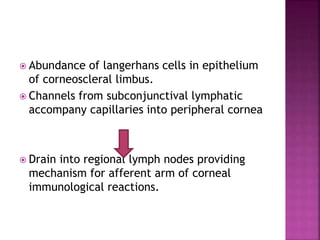
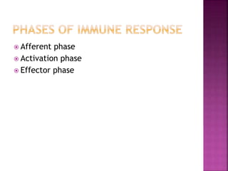
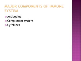
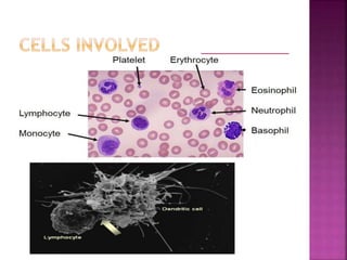
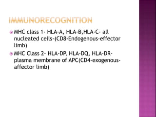
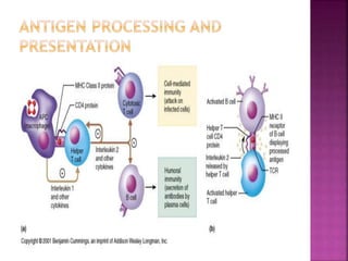
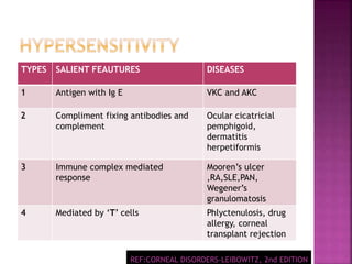
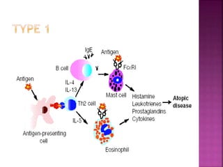
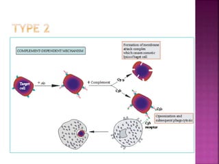
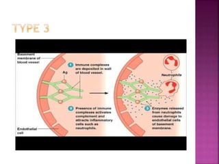
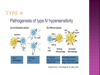
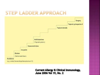
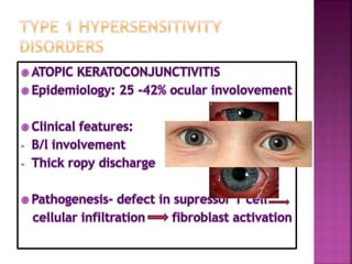
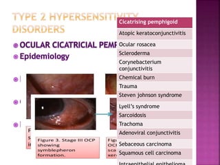
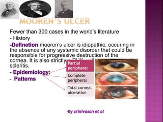
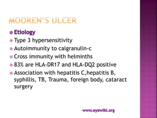
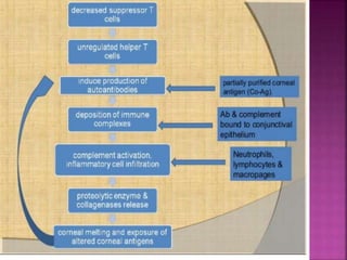
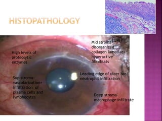
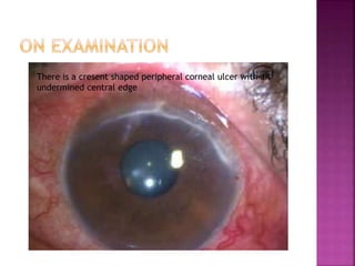
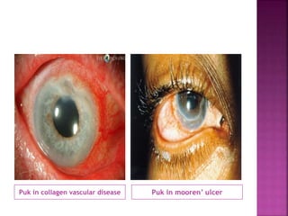
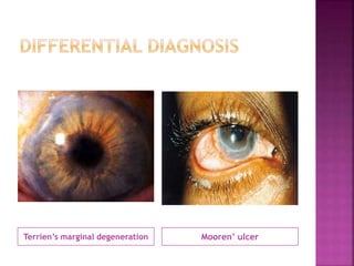
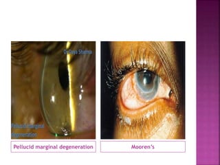
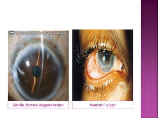
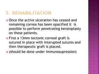
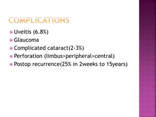
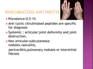
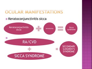
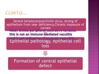
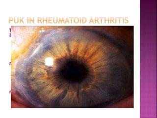
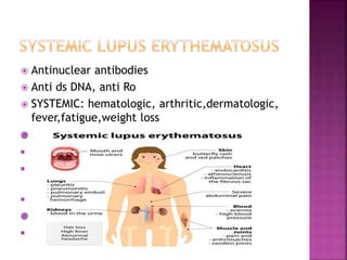
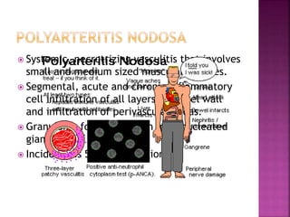
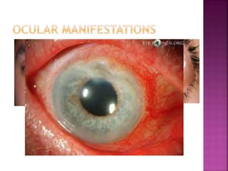
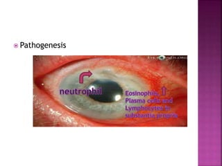
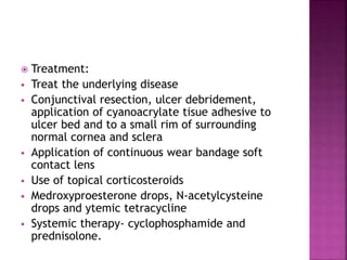
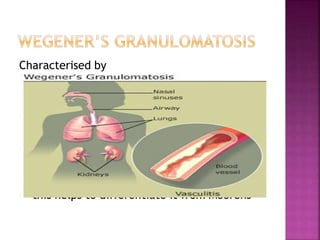
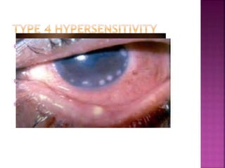
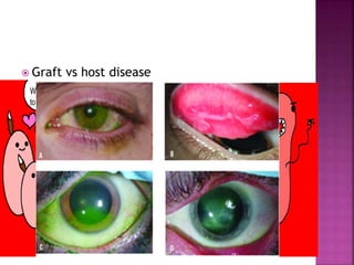
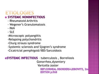
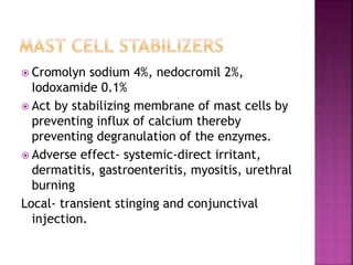
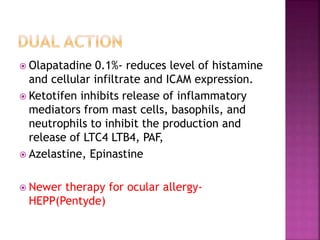
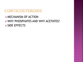
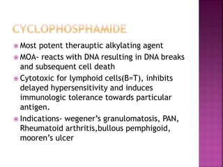
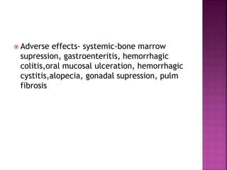
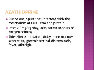
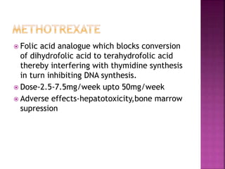
this slide share admixed with pictures and animations will give an overall idea of immunological disorders of cornea. it covers anatomy immunology, and pharmacology as well
4. Abundance of langerhans cells in epithelium
of corneoscleral limbus.
Channels from subconjunctival lymphatic
accompany capillaries into peripheral cornea
Drain into regional lymph nodes providing
mechanism for afferent arm of corneal
immunological reactions.
8. Neutrophil
Eosinophil
Lymphocytes
Dendritic cells
T lymphocytes
B lymphocyte
Helper T cell
Cytotoxic T cell
Supressor T cell
Memory T cell
Interdigitating follicular
9. MHC class 1- HLA-A, HLA-B,HLA-C- all
nucleated cells-(CD8-Endogenous-effector
limb)
MHC Class 2- HLA-DP, HLA-DQ, HLA-DR-
plasma membrane of APC(CD4-exogenous-
affector limb)
10.
11. TYPES SALIENT FEAUTURES DISEASES
1 Antigen with Ig E VKC and AKC
2 Compliment fixing antibodies and
complement
Ocular cicatricial
pemphigoid,
dermatitis
herpetiformis
3 Immune complex mediated
response
Mooren’s ulcer
,RA,SLE,PAN,
Wegener’s
granulomatosis
4 Mediated by ‘T’ cells Phlyctenulosis, drug
allergy, corneal
transplant rejection
REF:CORNEAL DISORDERS-LEIBOWITZ, 2nd EDITION
12.
13.
14.
15.
16. •The absence of an identifiable lymphatic drainage pathway led to the
conclusion that antigenic material was sequestered in the
immunologically priviledged site and was probably ignored by the
immune system.
•However it was noted that cell mediated immunity is supressed and
humoral immunity is promoted.
•ACAID depends on unique features of both spleen and eye for its
initiation.
•Cells from iris and ciliary body are able to downregulate the earliest
events of antigen presentation and lymphocyte activation, thereby
initiating a selective impairment of delayed hypersensitivity.
- Leibowitz 2nd edition
17. Clinical features:
Pathogenesis:
Treatment:
a) Control the symptoms
b) Limit the sight threatening complications
18. mild moderate severe Blinding
Bulbar
conj
congestion congestion Thickenin
g H.T
spots
granuloma
Tarsal/pal micropapillae macropapillae Giant
papillae
Mega
cobblestone
s
Cornea -- microerosions macroersi
os
Shield ulcer
limbus -- Focal infl Diffuse inf Limbal def
19. Grade Findings Treatment
0(quiescent) Absence of symptoms No treatment
1(mild) Symptoms but no corneal
inv
Antiallergic e/d
occassionally
2(moderate) Symptoms with
photophobia but no
corneal inv
Antiallergic e/d daily
3(severe) Symptoms, photophobia
mild-mod spk
Antiallergic e/d daily
with pulsed low dose
topical steroid
Symptoms,photophobia,
diffuse spk or corneal
ulcer
Pulsed high dose
steroid eventually
asso with surgical
removal of cornea
.
20.
21.
22. Cicatrising pemphigoid
Atopic keratoconjunctivitis
Ocular rosacea
Scleroderma
Corynebacterium
conjunctivitis
Chemical burn
Trauma
Steven johnson syndrome
Lyell’s syndrome
Sarcoidosis
Trachoma
Adenoviral conjunctivitis
Sebaceous carcinoma
Squamous cell carcinoma
24. Epilation
Topical retinoids
Lid margin mucous membrane grafting
Punctal occlusion with lubricants
Tarsorrhaphy
If good lid function; then penetrating
keratoplasty.
25. Fewer than 300 cases in the world’s literature
- History
:mooren’s ulcer is idiopathic, occuring in
the absence of any systemic disorder that could be
responsible for progressive destruction of the
cornea. It is also strictly a PUK, with no associated
scleritis.
-
Partial
peripheral
Complete
peripheral
Total corneal
ulceration
-
26. Type 3 hypersensitivity
Autoimmunity to calgranulin-c
Cross immunity with helminths
83% are HLA-DR17 and HLA-DQ2 positive
Association with hepatitis C,hepatitis B,
syphillis, TB, Trauma, foreign body, cataract
surgery
-
28. High levels of
proteolytic
enzymes
Sup stroma-
vascularization+
infiltration of
plasma cells and
lymphocytes
Mid stroma-
disorganized
collagen lamellae+
hyperactive
fibroblats
Deep stroma-
macrophage infiltrate
Leading edge of ulcer has
neutrophil infiltration
29. : excesive painful
Seen in elderly
Affected eyes are red and congested but
inflammation doesnot extend 3mm beyond limbus.
Vascularization of ulcer bed is seen alomg with
leakage at tips of new vessels
Ulceration extends around the globe and leaves thick
opaque central cornea
Fluroscein angio: venular occlusion of local episcleral
and conjunctival blood vessels along with disruption
of limbal arcade and vascular leakage from deep
vessels at limbus and base of ulcer, vasoobliteration
of sup vascular arcades is characteristic
--
30. Type 2- bilateral, occurs in young
Pain is less severe , pain in one eye and
congestion in other eventually progress to
develop grey patches within corneal stroma
and 2mm from edge of limbus.
FA : shows vascular leakage and new vessel
formation that reaches base of ulcer, also
shows change in architecture of episcleral
vessels and blockage in addition to break up
of limbal arcade
31. Type 3- indolent, affects mid age
Corneal guttering in both eyes with little
inflammation.
Disease more severe in one eye and patient
complain of discomfort.
Most cases are progressive but some heal
spontaneously. Few may be quiscient.
FA- vascular architecture is normal except
new vessel may extend upto base of ulcer.
32. There is a cresent shaped peripheral corneal ulcer with an
undermined central edge
38. Historically
Subconj dichloride of mercury, carbonic and
nitric acid
Formalin, tincture iodine, subconj heparin
Galvanocautery, vit B1 injections, tuberculin
injection, irradiation, paracentesis
39. 1. Topical steroids
2. Conjuctival resection
3.Sytemic immunosupressives
4. Additional surgical procedures
5. Visual rehabilitation
Goals of therapy- halt the destructive
process
Promote healing and re epitheliasation of
cornea
DOS Times - Vol. 11, No. 7
40. Prednisolone acetate/phophate 1% hourly
+cycloplegic +prophylactic antibiotics
If no healing in 2-3days If heals
Continue steroids half
hourly
Taper the steroids over
several months
•Oral pulse therapy(oral prednisolone 60-100mg daily for 7-
10days)
•Topical teracycline/medroxyprogesterone drops,BCL
DOS Times - Vol. 11, No. 7
41. Conjunctival excision to bare sclera
extending atleast 2 clock hour to either side
of peripheral ulcer approx 4mm posterior to
limbus and parallel to ulcer
Overhanging lip of the cornea to be removed
Tissue adhesives and BCL
Keratoepithelioplasty- donor cornea
lenticules are sutured onto scleral bed after
conjunctival resection
42. Cyclophosphamide 2mg/kg/day
Methotrexate 7.5mg/week
Azathioprine 2mg/kg/day
Oral cyclosporine 10mg/kg/day (loading dose
of 8 mg/kg in two divided doses for 2 days,
thereafter being reduced to 3-4 mg/kg/d to
maintain serum level of 200-400 ng/ml).
43. Resection of the overhanging lip of the
ulcerating cornea and application of tissue
adhesive with bandage soft contact lens
application or amniotic membrane has been
described in a study healed 34 out of 40
eyes.
44. For an ulcer smaller than half circle of the
limbus and the central 7-8 mm of the cornea
uninvolved crescent shaped lamellar graft
can be used.
For an ulcer larger than 2/3 of a circle of the
limbus where the central 7-8 mm of cornea is
intact, a doughnut shaped lamellar graft is
recommended.
A full lamellar graft is used to
maintain useful vision.
45. Double lamellar grafts (a fresh thin inner
graft with corneal endothelial cells is used to
repair the perforation, on which another
lamellar graft shaped in accordance with the
shape of the ulcer is placed) can be
used for perforations of the peripheral
cornea.
46. Once the active ulceration has ceased and
remaining cornea has been opacified it is
possible to perform penetrating keratoplasty
on these patients.
First a 13mm tectonic corneal graft is
sutured in place with interupted sutures and
then therapeutic graft is placed.
(should be done under immunosupression)
47. Uveitis (6.8%)
Glaucoma
Complicated cataract(2-3%)
Perforation (limbus>peripheral>central)
Postop recurrence(25% in 2weeks to 15years)
48. Prevalence-0.5-1%
Anti cyclic citrullinated peptides are specific
for diagnosis
Systemic : articular:joint deformity and joint
destruction,
Non articular:subcutaneous
nodules,vasculitis,
pericarditis,pulmonary nodules or interstitial
fibrosis
50. Severe keratoconjunctivitis sicca, drying of
epithelium from tear deficiency,Chronic exposure of
cornea
Epithelial pathology, epithelial cell
loss
Formation of central epithelial
defect
51.
52. After wide conjunctival resection, ulcer
debridement, and application of tissue
adhesive and soft contact lens adminiter
systemic immunosuppressive therapy.
Effective drug:
Cyclophosphamide 2mg/kg/day
Methotrexate 7.5mg/week a one dose
Daily azathioprine 2mg/kg/day
Cyclosporine 5mg/kg/day
53. Antinuclear antibodies
Anti ds DNA, anti Ro
SYSTEMIC: hematologic, arthritic,dermatologic,
fever,fatigue,weight loss
54. Systemic, necrotrizing vasculitis that involves
small and medium sized muscular arteries.
Segmental, acute and chronic inflammatory
cell infiltration of all layers of vessel wall
and infiltration of perivascular areas.
Granuloma formation wuth multinucleated
giant cells.
Incidence is 5-220 per million
57. Treatment:
Treat the underlying disease
Conjunctival resection, ulcer debridement,
application of cyanoacrylate tisue adhesive to
ulcer bed and to a small rim of surrounding
normal cornea and sclera
Application of continuous wear bandage soft
contact lens
Use of topical corticosteroids
Medroxyproesterone drops, N-acetylcysteine
drops and ytemic tetracycline
Systemic therapy- cyclophosphamide and
prednisolone.
58. Characterised by
Peripheral ulcerative keratitis is the initial
ocular manifestation (50-60%)
- Sometimes maybe preceded by conjunctivitis
or episcleritis
- Crescentic peripheral corneal ulcer that
resembles Mooren’s ulcer
- Scleral involvement is invariably present
- this helps to differentiate it from Moorens
59. HISTOPATHOLOGY – lymphocytes and plasma cells
predominate the substantia propria
- The sclera and episclera may show a
- Granulomatous reaction with epithelioid cells
and giant cells,fibrinoid necrosis
- Areas of active collagen degradation can be
seen
- ANCA testing is positive (sensitivity-96%n for
active generalised ds, 67%-local regional ds,32%-
pts in remission)
- MANAGEMENT – Systemic immunosuppression
with cyclophosphamide and steroids is highly
effective
60. Fibrosis of the skin and visceral organs
c/f: keratoconjunctivitis sicca, shortening of
fornices, mucosal subepithelial fibrosis,
vascular congestion, telangiectasia, and
varicosities.
61. Autoimmunity to type2 collagen(sclera)
Ocular involvement in 50% of cases and in
19% ocular involvemnt is initial presentation.
Necrotising scleritis with or without
peripheral ulcerative keratitis.
Treatment:
62. Mooren's ulcer although a distinct clinical entity,
remains a diagnosis of exclusion.
One should always look for associated scleritis,limbal
involvement, corneal sensation, associated
blepharitis and keratitis, lipid deposition, ulcerated
corneal epithelium and stroma,and so on, to rule out
other causes of peripheral ulcerative keratitis,
including infections, collagen vascular diseases
anddegenerative processes.
The precise pathophysiology of Mooren's ulcer
remains uncertain.
Advances have been made in its management, but
significant percentage of cases still remain
refractory to available therapies and result in severe
visual morbidity
68. Antistine,Levocabastine(0.5%),emadastine
0.05%
Act on H1 receptors in conjunctiva.
Adverse effects: systemic- nausea, vomiting,
diarrhoea,dryness of mucous
membranes,sedation
Local- precipitates narrow angle glaucoma,
ciliary muscle paresis
69. Cromolyn sodium 4%, nedocromil 2%,
Iodoxamide 0.1%
Act by stabilizing membrane of mast cells by
preventing influx of calcium thereby
preventing degranulation of the enzymes.
Adverse effect- systemic-direct irritant,
dermatitis, gastroenteritis, myositis, urethral
burning
Local- transient stinging and conjunctival
injection.
70. Olapatadine 0.1%- reduces level of histamine
and cellular infiltrate and ICAM expression.
Ketotifen inhibits release of inflammatory
mediators from mast cells, basophils, and
neutrophils to inhibit the production and
release of LTC4 LTB4, PAF,
Azelastine, Epinastine
Newer therapy for ocular allergy-
HEPP(Pentyde)
71. MECHANISM OF ACTION
WHY PHOSPHATES AND WHY ACETATES?
SIDE EFFECTS
72. Most potent therauptic alkylating agent
MOA- reacts with DNA resulting in DNA breaks
and subsequent cell death
Cytotoxic for lymphoid cells(B=T), inhibits
delayed hypersensitivity and induces
immunologic tolerance towards particular
antigen.
Indications- wegener’s granulomatosis, PAN,
Rheumatoid arthritis,bullous pemphigoid,
mooren’s ulcer
74. Purine analogues that interfere with the
metabolism of DNA, RNA and protein
Dose-2-3mg/kg/day, acts within 48hours of
antigen priming.
Side effects- hepatotoxicity, bone marrow
supression, gastrointestinal distress,rash,
fever, athralgia
75. Folic acid analogue which blocks conversion
of dihydrofolic acid to terahydrofolic acid
thereby interfering with thymidine synthesis
in turn inhibiting DNA synthesis.
Dose-2.5-7.5mg/week upto 50mg/week
Adverse effects-hepatotoxicity,bone marrow
supression
76. Is a fungal metabolite isolated from cultures
of tolypocladium inflatum and
cylindrocarpon lucidum
MOA-they block the helper t cell response to
IL-1 and block IL-2 release.
Adverse effect- systemic-B cell lymphomas,
interstitial pneumonitis, renal tubular
necrosis
77. Applied to the area of perforation after careful debridement. The
surface is dried with a sponge & a small drop of tissue adhesive
from the undersurface of a bent iris repositer or a hypodermic
needle is placed immediately over the perforation. Drying takes 5-
10 minutes after which anterior chamber reforms. Following this,
continuous wear soft contact lens may be applied.
Advantage: good tensile strength
Drawback: it may form a solid, impermeable mass in situ and may
require removal.
78. Blood derived product that is absorbable,
easy to use and can be kept at room
temperature or refrigerator.
Can be prepared at a blood bank, or from
patients own blood or commercially available
preparation.
Advantage: forms a smooth seal along the
perforation, less postop discomfort.
79. Technique of Glue Application
Part preparation (paint and drapes), application of topical anesthetics and speculum
Debridement of necrotic tissue from ulcer crater
As tissue adhesive glue adheres best to basement membrane, 1-2 mm of normal epithelium should be debride
to allow the glue to properly adhere
Dry the site by methyl cellulose spear
As application in small amount to seal the perforation
Tissue adhesive solidify in few minutes via polymerization
Send this material for culture and sensitivity
A large heaped up mound is not required
80. Check the evidence Check for anterior chamber Apply bandage
of leakage by Seidel’s test maintenance (Air bubble contact lens
can also be used)
Tissue adhesive remains in place for weeks to
months until the perforation seals, it can be
removed or dislodges of its own