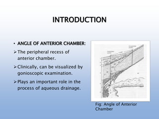
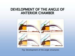
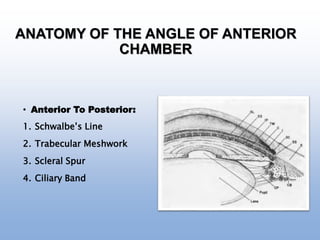
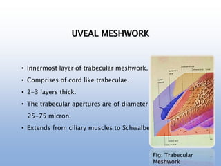
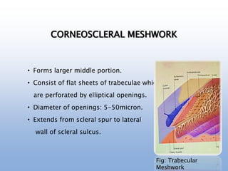
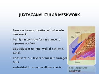
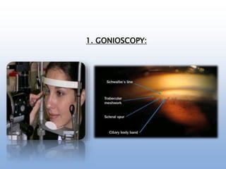
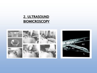
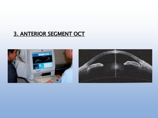
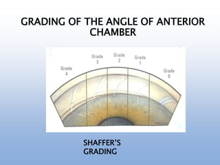
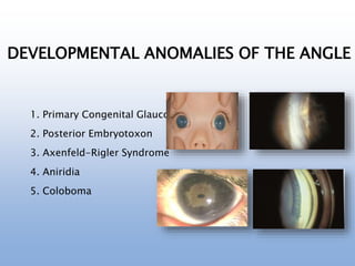
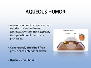
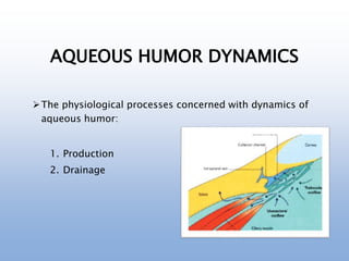
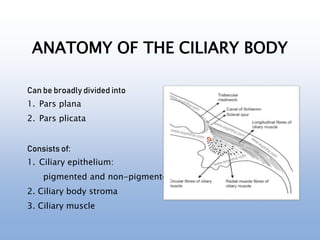
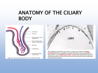
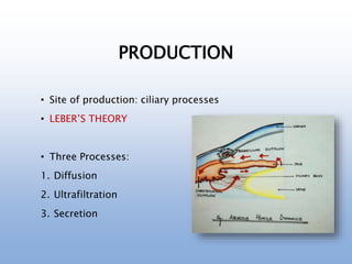
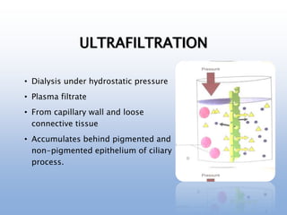
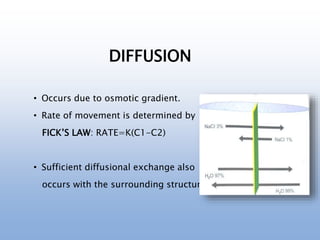
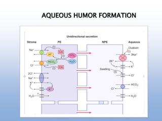
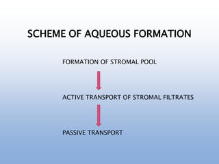
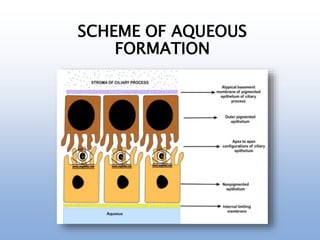
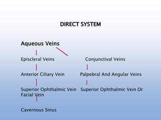
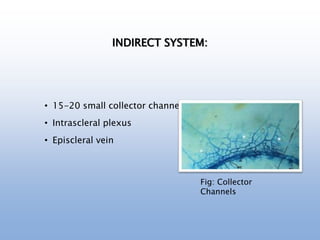
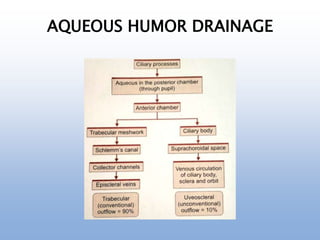
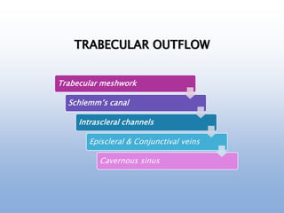
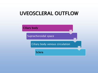
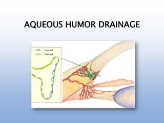
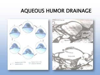
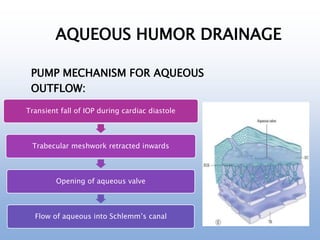
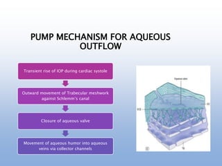
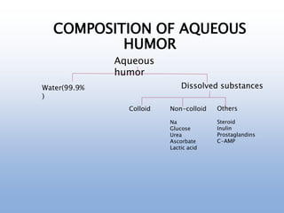
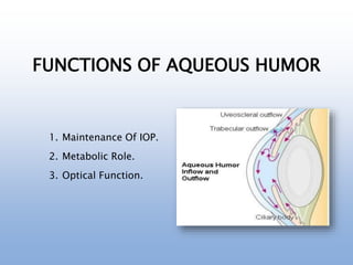
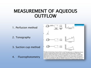
ANGLE OF ANTERIOR CHAMBER
AND AQUEOUS HUMOR DYNAMICS
Dr. GARIYASHEE LAHKAR
OUTLINE
1. Introduction.
2. Development of the angle of anterior chamber.
3. Anatomy of the angle of anterior chamber.
4. Diagnostic modalities for angle examination.
5. Grading of the angle of anterior chamber.
6. Developmental anomalies of the angle.
7. Aqueous humor dynamics.
8. Composition, physical properties and functions of aqueous
humor.
9. Measurement of aqueous outflow.
10. Summary.
INTRODUCTION
• ANGLE OF ANTERIOR CHAMBER:
The peripheral recess of
anterior chamber.
Clinically, can be visualized by
gonioscopic examination.
Plays an important role in the
process of aqueous drainage.
Fig: Angle of Anterior
Chamber
INTRODUCTION (Contd.)
• AQUEOUS HUMOR DYNAMICS:
The physiological processes concerned with dynamics of
aqueous humor:
1. Production
2. Drainage
Maintains structural integrity of eyeball.
Nourishes cornea and lens.
Provides optically clear media.
DEVELOPMENT OF THE ANGLE OF
ANTERIOR CHAMBER
• Formed by loosely arranged mesenchymal neural crest
cells.
• At 7th week cells accumulate near the angle.
• At 5 months, closed cavity of anterior chamber is formed.
• At the end of 3rd trimester, endothelial layer progressively
disappears.
• Development of trabecular spaces and intertrabecular
spaces.
• Normal anterior chamber is not formed uptil 1 year of age.
• Angle deepening continues even after birth.
DEVELOPMENT OF THE ANGLE OF
ANTERIOR CHAMBER
Fig: Development of the angle structures
ANATOMY OF THE ANGLE OF ANTERIOR
CHAMBER
• Anterior To Posterior:
1. Schwalbe’s Line
2. Trabecular Meshwork
3. Scleral Spur
4. Ciliary Band
1. SCHWALBE’S LINE:
• Anterior limit of the drainage angle.
• Fine scalloped border at the termination of Descemet’s
membrane.
• Lies in the plane of posterior corneal surface.
• Contains collagen fibres intermixed with elastic fibres.
• Also marks transition between corneal endothelium and
trabecular meshwork.
Fig: Schwalbe’s line
2. TRABECULAR MESHWORK:
• Spongework of connective tissue beams.
• Arranged as superimposed perforated sheets.
• Seen as a broad band just anterior to the scleral spur.
• Usually featureless in unpigmented eye.
• Extends from Scleral spur to Schwalbe’s line.
Fig: Trabecular
Meshwork
2. TRABECULAR MESHWORK:
• Microscopically, trabecular meshwork consists of 3
portions:
1. Uveal Meshwork
2. Corneoscleral Meshwork
3. Juxtacannalicular /Cribriform Meshwork
UVEAL MESHWORK
• Innermost layer of trabecular meshwork.
• Comprises of cord like trabeculae.
• 2-3 layers thick.
• The trabecular apertures are of diameter
25-75 micron.
• Extends from ciliary muscles to Schwalbe’s line.
Fig: Trabecular
Meshwork
CORNEOSCLERAL MESHWORK
• Forms larger middle portion.
• Consist of flat sheets of trabeculae which
are perforated by elliptical openings.
• Diameter of openings: 5-50micron.
• Extends from scleral spur to lateral
wall of scleral sulcus.
Fig: Trabecular
Meshwork
JUXTACANALICULAR MESHWORK
• Forms outermost portion of trabecular
meshwork.
• Mainly responsible for resistance to
aqueous outflow.
• Lies adjacent to inner wall of schlem’s
canal.
• Consist of 2-5 layers of loosely arranged
cells
embedded in an extracellular matrix. Fig: Trabecular
Meshwork
3.SCLERAL SPUR:
• Wedge shaped ridge.
• Posterior portion of scleral sulcus.
• Anterior: trabecular meshwork.
• Posterior: longitudinal fibres of ciliary body.
• Pale, transparent, narrow strip of scleral tissue.
• Composed of 75-85% collagen and 5% elastic tissue.
Fig: Scleral spur
4. CILIARY BAND:
• Anterior most part of the ciliary body.
• Lies between its attachment to scleral spur
and insertion of iris.
• Forms posterior most landmark of angle
recess.
• Width depends upon the level of iris
insertion.
• Consists of longitudinal fibres.
Fig: Ciliary band
1. GONIOSCOPY:
• Indirect Gonioscopy
commonly used.
• Assessment Of Angle.
DIAGNOSTIC MODALITIES FOR ANGLE EXAMINATI
1. GONIOSCOPY:
2. ULTRASOUND
BIOMICROSCOPY
3. ANTERIOR SEGMENT OCT
GRADING OF THE ANGLE OF ANTERIOR
CHAMBER
SHAFFER’S
GRADING
DEVELOPMENTAL ANOMALIES OF THE ANGLE
1. Primary Congenital Glaucoma
2. Posterior Embryotoxon
3. Axenfeld-Rigler Syndrome
4. Aniridia
5. Coloboma
AQUEOUS HUMOR DYNAMICS
AQUEOUS HUMOR
• Aqueous humor is a transparent ,
colorless solution formed
continuously from the plasma by
the epithelium of the ciliary
processes.
• Continuously circulated from
posterior to anterior chamber.
• Dynamic equilibrium.
AQUEOUS HUMOR DYNAMICS
The physiological processes concerned with dynamics of
aqueous humor:
1. Production
2. Drainage
ANATOMY OF THE CILIARY BODY
Can be broadly divided into
1. Pars plana
2. Pars plicata
Consists of:
1. Ciliary epithelium:
pigmented and non-pigmented
2. Ciliary body stroma
3. Ciliary muscle
ANATOMY OF THE CILIARY
BODY
PRODUCTION
• Site of production: ciliary processes
• LEBER’S THEORY
• Three Processes:
1. Diffusion
2. Ultrafiltration
3. Secretion
ULTRAFILTRATION
• Dialysis under hydrostatic pressure
• Plasma filtrate
• From capillary wall and loose
connective tissue
• Accumulates behind pigmented and
non-pigmented epithelium of ciliary
process.
SECRETION
• Active process.
• 80-90% of aqueous humor formation.
• Active transport of ions against
concentration gradient.
• Substances actively transported
to posterior chamber.
DIFFUSION
• Occurs due to osmotic gradient.
• Rate of movement is determined by
FICK’S LAW: RATE=K(C1-C2)
• Sufficient diffusional exchange also
occurs with the surrounding structures.
AQUEOUS HUMOR FORMATION
SCHEME OF AQUEOUS FORMATION
FORMATION OF STROMAL POOL
ACTIVE TRANSPORT OF STROMAL FILTRATES
PASSIVE TRANSPORT
SCHEME OF AQUEOUS
FORMATION
AQUEOUS HUMOR DRAINAGE
Mainly by two pathways:
1. Conventional/ Trabecular Pathway
2. Unconventional/Uveoscleral Pathway
ANATOMY OF THE OUTFLOW
SYSTEM
1. Trabecular Meshwork
2. Schlemn’s Canal
3. Collector Channels:
• Direct
• Indirect
2. SCHLEMM’S CANAL:
• Narrow circular tube.
• Endothelial lined oval channel.
• Present circumferentially in the
scleral sulcus.
• Conducts aqueous humor from
Trabecular meshwork to
Episcleral venous network.
3. COLLECTOR CHANNELS
• 2 Intrascleral System.
1. Direct : Large calibre vessels.
Short intrascleral course.
Drains directly into episcleral
system.
2. Indirect: Finer channels.
Forms intrascleral plexus
before
draining into episcleral veins.
Fig: Collector Channels
DIRECT SYSTEM
Aqueous Veins
Episcleral Veins Conjunctival Veins
Anterior Ciliary Vein Palpebral And Angular Veins
Superior Ophthalmic Vein Superior Ophthalmic Vein Or
Facial Vein
Cavernous Sinus
INDIRECT SYSTEM:
• 15-20 small collector channels
• Intrascleral plexus
• Episcleral vein
Fig: Collector
Channels
AQUEOUS HUMOR DRAINAGE
TRABECULAR OUTFLOW
Trabecular meshwork
Schlemm’s canal
Intrascleral channels
Episcleral & Conjunctival veins
Cavernous sinus
UVEOSCLERAL OUTFLOW
Ciliary body
Suprachoroidal space
Ciliary body venous circulation
Sclera
AQUEOUS HUMOR DRAINAGE
PUMP MECHANISM FOR AQUEOUS
OUTFLOW:
Transient fall of IOP during cardiac diastole
Trabecular meshwork retracted inwards
Opening of aqueous valve
Flow of aqueous into Schlemm’s canal
PUMP MECHANISM FOR AQUEOUS
OUTFLOW
Transient rise of IOP during cardiac systole
Outward movement of Trabecular meshwork
against Schlemm’s canal
Closure of aqueous valve
Movement of aqueous humor into aqueous
veins via collector channels
Aqueous
humor
Water(99.9%
)
Dissolved substances
Colloid Non-colloid
Na
Glucose
Urea
Ascorbate
Lactic acid
Others
Steroid
Inulin
Prostaglandins
C-AMP
COMPOSITION OF AQUEOUS
HUMOR
FACTORS AFFECTING
COMPOSITION
• Blood aqueous barrier.
• Hemodynamic factors affecting stromal pool.
• Diffusional exchange at iris.
• Metabolites.
• Rate of aqueous drainage.
PHYSICAL PROPERTIES
1. Volume: 0.31mL
2. Refractive index: 1.336
3. Viscosity: 1.025
4. Osmotic pressure: 3-5mosm/L
5. Ph: 7.2
6. Rate of formation: 2.3uL/min
FUNCTIONS OF AQUEOUS HUMOR
1. Maintenance Of IOP.
2. Metabolic Role.
3. Optical Function.
MEASUREMENT OF AQUEOUS
OUTFLOW
1. Perfusion method
2. Tonography
3. Suction cup method
4. Fluorophotometry
SUMMARY
• The angle of anterior chamber plays an important role in
the process of aqueous drainage.
• Gonioscopy is most reliable for assessment of angle
structures.
• A number of structural changes are observed in the angle
with advancing age that causes increased resistance in
ageing eyes.
• The aqueous humor fills the anterior and posterior
chamber of the eye.
• The aqueous humor remains in a state of dynamic
equilibrium by continuous process of production and
drainage.
REFERENCES
• Allingham, R. Rand. Shields Textbook of Glaucoma, 6th Ed.
2011
• Anthony J. Bron. Wolff’s Anatomy of the Orbit, 8th Ed.
1997, p 279-282
• Brad Bowling. Kanski’s Clinical Ophthalmology, 8th Ed.
2016, p 306-316
• Myron Yanoff, Jay S Duker. Yanoff Duker Ophthalmology,
5th Ed. 2019
• Leonard A. Levin, Siv F. E. Nilson, James Ver Hoeve, Samuel
M Wu. Adler’s Physiology of the Eye, 11th Ed. 2003, p 274-
307
Aqueous Humor Dynamics.pptx

Aqueous Humor Dynamics.pptx

  • 1.
    ANGLE OF ANTERIORCHAMBER AND AQUEOUS HUMOR DYNAMICS Dr. GARIYASHEE LAHKAR
  • 2.
    OUTLINE 1. Introduction. 2. Developmentof the angle of anterior chamber. 3. Anatomy of the angle of anterior chamber. 4. Diagnostic modalities for angle examination. 5. Grading of the angle of anterior chamber. 6. Developmental anomalies of the angle. 7. Aqueous humor dynamics. 8. Composition, physical properties and functions of aqueous humor. 9. Measurement of aqueous outflow. 10. Summary.
  • 3.
    INTRODUCTION • ANGLE OFANTERIOR CHAMBER: The peripheral recess of anterior chamber. Clinically, can be visualized by gonioscopic examination. Plays an important role in the process of aqueous drainage. Fig: Angle of Anterior Chamber
  • 4.
    INTRODUCTION (Contd.) • AQUEOUSHUMOR DYNAMICS: The physiological processes concerned with dynamics of aqueous humor: 1. Production 2. Drainage Maintains structural integrity of eyeball. Nourishes cornea and lens. Provides optically clear media.
  • 5.
    DEVELOPMENT OF THEANGLE OF ANTERIOR CHAMBER • Formed by loosely arranged mesenchymal neural crest cells. • At 7th week cells accumulate near the angle. • At 5 months, closed cavity of anterior chamber is formed. • At the end of 3rd trimester, endothelial layer progressively disappears. • Development of trabecular spaces and intertrabecular spaces. • Normal anterior chamber is not formed uptil 1 year of age. • Angle deepening continues even after birth.
  • 6.
    DEVELOPMENT OF THEANGLE OF ANTERIOR CHAMBER Fig: Development of the angle structures
  • 7.
    ANATOMY OF THEANGLE OF ANTERIOR CHAMBER • Anterior To Posterior: 1. Schwalbe’s Line 2. Trabecular Meshwork 3. Scleral Spur 4. Ciliary Band
  • 8.
    1. SCHWALBE’S LINE: •Anterior limit of the drainage angle. • Fine scalloped border at the termination of Descemet’s membrane. • Lies in the plane of posterior corneal surface. • Contains collagen fibres intermixed with elastic fibres. • Also marks transition between corneal endothelium and trabecular meshwork.
  • 9.
  • 10.
    2. TRABECULAR MESHWORK: •Spongework of connective tissue beams. • Arranged as superimposed perforated sheets. • Seen as a broad band just anterior to the scleral spur. • Usually featureless in unpigmented eye. • Extends from Scleral spur to Schwalbe’s line.
  • 11.
  • 12.
    2. TRABECULAR MESHWORK: •Microscopically, trabecular meshwork consists of 3 portions: 1. Uveal Meshwork 2. Corneoscleral Meshwork 3. Juxtacannalicular /Cribriform Meshwork
  • 13.
    UVEAL MESHWORK • Innermostlayer of trabecular meshwork. • Comprises of cord like trabeculae. • 2-3 layers thick. • The trabecular apertures are of diameter 25-75 micron. • Extends from ciliary muscles to Schwalbe’s line. Fig: Trabecular Meshwork
  • 14.
    CORNEOSCLERAL MESHWORK • Formslarger middle portion. • Consist of flat sheets of trabeculae which are perforated by elliptical openings. • Diameter of openings: 5-50micron. • Extends from scleral spur to lateral wall of scleral sulcus. Fig: Trabecular Meshwork
  • 15.
    JUXTACANALICULAR MESHWORK • Formsoutermost portion of trabecular meshwork. • Mainly responsible for resistance to aqueous outflow. • Lies adjacent to inner wall of schlem’s canal. • Consist of 2-5 layers of loosely arranged cells embedded in an extracellular matrix. Fig: Trabecular Meshwork
  • 16.
    3.SCLERAL SPUR: • Wedgeshaped ridge. • Posterior portion of scleral sulcus. • Anterior: trabecular meshwork. • Posterior: longitudinal fibres of ciliary body. • Pale, transparent, narrow strip of scleral tissue. • Composed of 75-85% collagen and 5% elastic tissue.
  • 17.
  • 18.
    4. CILIARY BAND: •Anterior most part of the ciliary body. • Lies between its attachment to scleral spur and insertion of iris. • Forms posterior most landmark of angle recess. • Width depends upon the level of iris insertion. • Consists of longitudinal fibres.
  • 19.
  • 20.
    1. GONIOSCOPY: • IndirectGonioscopy commonly used. • Assessment Of Angle. DIAGNOSTIC MODALITIES FOR ANGLE EXAMINATI
  • 21.
  • 22.
  • 23.
  • 24.
    GRADING OF THEANGLE OF ANTERIOR CHAMBER SHAFFER’S GRADING
  • 25.
    DEVELOPMENTAL ANOMALIES OFTHE ANGLE 1. Primary Congenital Glaucoma 2. Posterior Embryotoxon 3. Axenfeld-Rigler Syndrome 4. Aniridia 5. Coloboma
  • 26.
  • 27.
    AQUEOUS HUMOR • Aqueoushumor is a transparent , colorless solution formed continuously from the plasma by the epithelium of the ciliary processes. • Continuously circulated from posterior to anterior chamber. • Dynamic equilibrium.
  • 28.
    AQUEOUS HUMOR DYNAMICS Thephysiological processes concerned with dynamics of aqueous humor: 1. Production 2. Drainage
  • 29.
    ANATOMY OF THECILIARY BODY Can be broadly divided into 1. Pars plana 2. Pars plicata Consists of: 1. Ciliary epithelium: pigmented and non-pigmented 2. Ciliary body stroma 3. Ciliary muscle
  • 30.
    ANATOMY OF THECILIARY BODY
  • 31.
    PRODUCTION • Site ofproduction: ciliary processes • LEBER’S THEORY • Three Processes: 1. Diffusion 2. Ultrafiltration 3. Secretion
  • 32.
    ULTRAFILTRATION • Dialysis underhydrostatic pressure • Plasma filtrate • From capillary wall and loose connective tissue • Accumulates behind pigmented and non-pigmented epithelium of ciliary process.
  • 33.
    SECRETION • Active process. •80-90% of aqueous humor formation. • Active transport of ions against concentration gradient. • Substances actively transported to posterior chamber.
  • 34.
    DIFFUSION • Occurs dueto osmotic gradient. • Rate of movement is determined by FICK’S LAW: RATE=K(C1-C2) • Sufficient diffusional exchange also occurs with the surrounding structures.
  • 35.
  • 36.
    SCHEME OF AQUEOUSFORMATION FORMATION OF STROMAL POOL ACTIVE TRANSPORT OF STROMAL FILTRATES PASSIVE TRANSPORT
  • 37.
  • 38.
    AQUEOUS HUMOR DRAINAGE Mainlyby two pathways: 1. Conventional/ Trabecular Pathway 2. Unconventional/Uveoscleral Pathway
  • 39.
    ANATOMY OF THEOUTFLOW SYSTEM 1. Trabecular Meshwork 2. Schlemn’s Canal 3. Collector Channels: • Direct • Indirect
  • 40.
    2. SCHLEMM’S CANAL: •Narrow circular tube. • Endothelial lined oval channel. • Present circumferentially in the scleral sulcus. • Conducts aqueous humor from Trabecular meshwork to Episcleral venous network.
  • 41.
    3. COLLECTOR CHANNELS •2 Intrascleral System. 1. Direct : Large calibre vessels. Short intrascleral course. Drains directly into episcleral system. 2. Indirect: Finer channels. Forms intrascleral plexus before draining into episcleral veins. Fig: Collector Channels
  • 42.
    DIRECT SYSTEM Aqueous Veins EpiscleralVeins Conjunctival Veins Anterior Ciliary Vein Palpebral And Angular Veins Superior Ophthalmic Vein Superior Ophthalmic Vein Or Facial Vein Cavernous Sinus
  • 43.
    INDIRECT SYSTEM: • 15-20small collector channels • Intrascleral plexus • Episcleral vein Fig: Collector Channels
  • 44.
  • 45.
    TRABECULAR OUTFLOW Trabecular meshwork Schlemm’scanal Intrascleral channels Episcleral & Conjunctival veins Cavernous sinus
  • 46.
    UVEOSCLERAL OUTFLOW Ciliary body Suprachoroidalspace Ciliary body venous circulation Sclera
  • 47.
  • 48.
  • 49.
    AQUEOUS HUMOR DRAINAGE PUMPMECHANISM FOR AQUEOUS OUTFLOW: Transient fall of IOP during cardiac diastole Trabecular meshwork retracted inwards Opening of aqueous valve Flow of aqueous into Schlemm’s canal
  • 50.
    PUMP MECHANISM FORAQUEOUS OUTFLOW Transient rise of IOP during cardiac systole Outward movement of Trabecular meshwork against Schlemm’s canal Closure of aqueous valve Movement of aqueous humor into aqueous veins via collector channels
  • 51.
    Aqueous humor Water(99.9% ) Dissolved substances Colloid Non-colloid Na Glucose Urea Ascorbate Lacticacid Others Steroid Inulin Prostaglandins C-AMP COMPOSITION OF AQUEOUS HUMOR
  • 52.
    FACTORS AFFECTING COMPOSITION • Bloodaqueous barrier. • Hemodynamic factors affecting stromal pool. • Diffusional exchange at iris. • Metabolites. • Rate of aqueous drainage.
  • 53.
    PHYSICAL PROPERTIES 1. Volume:0.31mL 2. Refractive index: 1.336 3. Viscosity: 1.025 4. Osmotic pressure: 3-5mosm/L 5. Ph: 7.2 6. Rate of formation: 2.3uL/min
  • 54.
    FUNCTIONS OF AQUEOUSHUMOR 1. Maintenance Of IOP. 2. Metabolic Role. 3. Optical Function.
  • 55.
    MEASUREMENT OF AQUEOUS OUTFLOW 1.Perfusion method 2. Tonography 3. Suction cup method 4. Fluorophotometry
  • 56.
    SUMMARY • The angleof anterior chamber plays an important role in the process of aqueous drainage. • Gonioscopy is most reliable for assessment of angle structures. • A number of structural changes are observed in the angle with advancing age that causes increased resistance in ageing eyes. • The aqueous humor fills the anterior and posterior chamber of the eye. • The aqueous humor remains in a state of dynamic equilibrium by continuous process of production and drainage.
  • 57.
    REFERENCES • Allingham, R.Rand. Shields Textbook of Glaucoma, 6th Ed. 2011 • Anthony J. Bron. Wolff’s Anatomy of the Orbit, 8th Ed. 1997, p 279-282 • Brad Bowling. Kanski’s Clinical Ophthalmology, 8th Ed. 2016, p 306-316 • Myron Yanoff, Jay S Duker. Yanoff Duker Ophthalmology, 5th Ed. 2019 • Leonard A. Levin, Siv F. E. Nilson, James Ver Hoeve, Samuel M Wu. Adler’s Physiology of the Eye, 11th Ed. 2003, p 274- 307

Editor's Notes

  • #23 Close contact immersion technique Provides 2D high resolution gray scale images of ocular structures anterior to pars plana.