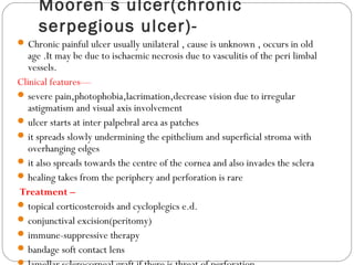
Dr. Mohamed Najmussadiq Khan
CORNEA
It is a transparent a vascular tissue that forms the anterior 1/6 of eye globe .
Dimension of cornea :-
10.6 mm vertically and 11.7 mm horizontally .
thickness is 0.5 mm at the center (0.49 – 0.56 ) and 0.7 mm at the periphery .
histology :-from anterior to posterior is :
a- epithelium : stratified Squamous non keratinized about 5-6 layers continuous at
limbus with the conjunctival epithelium and it rests on basement membrane and there
are single basal columnar cells attached to their basement membrane .
b- bowman s membrane(anterior elastic lamina)- : formed of collagen fibers
does not generation if it destroyed .
c- substantia propria(stroma)- : form 90% of thickness formed of lamellae
(collagen and elastic fibers) + cells (keratocytes and histocytes ).stroma composed of
collagen producing fibroblast (keratocyte)+ collagen fibrils + ground substance .
d- descement s membrane(posterior elastic lamina)- : elastic continuous with
trabecular meshwork at the limbus .It increases in thickness with age and it is basal
lamina of corneal endothelium .It is very resistant membrane and regenerates after
injury.In old age some warty elevations called Hassall-henle bodies at corneal
periphery.
e- endothelium : monolayer of hexagonal cells continuous around the angle
chamber with the endothelium of iris and no generation if it effected .
nutrition :-
diffusion form Limbal vessels .
diffusion form aqueous humor posteriorly and tears anteriorly limbus
(corneal sclera junction )
At limbus(corneosclral juncion)the following changes occur :
corneal epithelium become continuous with conjunctival epithelium
and becomes more thicker ( 10 layers).
Bowman s membrane ends .
Fibers of substantia propria become irregular and continuous with the
sclera .
Descement s membrane becomes continuous with trabecular
meshwork.
Endothelium is continuous with that of the angle and on iris .
Presence of blood vessels and lymphatics and mylenated nerve
branches of trigeminal .
* Importance of limbus :-
Contains exit channels of aqueous .
Landmark for eye surgery as glaucoma and cataract .
* Transparence of cornea is due to :-
absence of blood vessels .
non medullated nerve fibers .
corneal lamellae are uniform in size and running parallel to each others .
state of relative dehydration of the cornea maitained by bicarbonate
pump of endothelium and water evaporation from precorneal tear film .
* nerve supply comes from 1st
division of trigeminal nerve .
corneal epithelium contains rich network of non medullated nerve fibers
so severe pain results even from minor abrasions of corneal
epithelium .They are derived from long and short ciliary
nerves(ophthalmic division of trigeminal nerve).
* drugs penetration to cornea is biphasic ---:-
fat soluble drugs can pass through intact epithelium.
Water soluble drugs can pass through intact stroma .
So it s pass through the cornea drugs must be both lipids and water
soluble .
* Precorneal tear film :-A protective film formed of :
Outer lipid layer to prevent evaporation of tear produced by
Meibomian gland .
Intermediate watery (aqueous) layer produced by Lacrimal
and accessory Lacrimal glands .
Inner mucoprotein layer produced by goblet cell of
conjunctiva and Lacrimal glands
corneal epithelium is a good barrier to entrance of
microorganisms except gonococci and diphtheria bacilli
Pneumococci is the only true (bacteria) corneal pathogen and
all of others are opportunistic that require heavy inoculation
or compromised host e.g trauma to epithelium to cause
infection
SYMPTOMS OF CORNEAL DISEASES
Pain : worsened by lid movements persists until healing .
Blurred vision : because of high refractive power of cornea
(+40dioptes ) due to loss of central corneal transparency .
Haloes : are due to the diffraction of light as a results of
epithelial and sub-epithelial edema .
Photophobia : due to painful contraction of hyperemic iris
(inflamed)or due to abnormal strong light induced vision of
inflamed iris .Its severe in most corneal diseases except
herpetic keratitis .
No discharge : except in purulent bacteria ulcers .
Lacrimation : degree parallel to the severity of photophobia .
Clinical exam of cornea :-
shape : normally anterior surface is convex .
diameter : 11.7 mm . micorcornea = < 10mm and macrocornea
12 mm .
curvature (radius): normally is 7.7 mm and regular surface .
vascularization : normally cornea is not .
transparency : most important may be lost by opacities (describe
shape , size , site ).
Scars , edema , K.P , pannus , ulcers .
Corneal sensation : by touching the cornea with fine cotton wool
---- normally there will be reflex lid closure .
cornea)
Classification to :
ULCERATIVE NON – ULCERATIVE
 Central peripheral -epithelium
 - Bacterial - ring ulcer -superficial punctate
 - Fungal - marginal catarrh ulcer -superficial limbic keratoconjunctivitis
 - Viral - phlyctecular keratoconjunctivitis -viral
 - Neurotropic - marginal keratitis autoimmune disease -stromal
 - Exposure - mooren s ulcer
 - Nutritional
ulcerative sometimes classified as :
INFECTIVE NON-INFECTIVE
 Bacterial -Traumatic -Exposure - Nutritional
 Fungal -Allergic -Neurotropic - Degeneration
 Viral -Mooner s ulcer (TEN AND M).
 Acanthamoeba
Deep keratitis—
 Interstitial keratitis
 Disciform keratitis
 Sclerosing keratitis
CAUSES of keratitis :-
Exogenous infections-most common
Endogenous infections-rare like allergic in nature
From ocular tissues- conjunctiva,sclera,uveal tract e.g
gonococci conjunctivitis and uveitis .
ULCERATIVE KERATITIS--
lesions in ulcerative keratitis involve the epithelium , stroma
or both causing necrosis of tissue affected .
general sign and symptoms of ulcerative keratitis :-
Lacrimation.
blepharosoasia.
photophobia.
edema and redness of eyelids.
ciliary injection slight conjunctival injection .
loss of transparency & stromal of cornea due to edema ,
cellular infiltration and vascularization .
Bacterial keratitis
Predisposing factor :-The intact corneal epithelium can not be
penetrated by any organism except Neisseria
gonorrhoea,N.meningitidis,C.diphtheriae,Haemophyllus.
1-Trauma to the corneal epithelium by injury ,foreign
body,trichiasis,entropion,contact lens etc.
2- underlying corneal disease (herpetic keratitis , bullous keratopathy
,keratomalacia).
3-Inflammation of adjacent Adenxa
4- trauma to cornea & dry eye
5-contact lenses wear.
6- exposure keratopathy or neurotropic
7- administration of topical or systemic
8-decreased resistance like diabetes,AIDS,immunosuppressive drugs
Common causative agents : (affecting
corneal epithelial integrity)
Staph. epidermidis
Staph. Aureus
Strept. Pneumonia
H.infleunza
P.aeruginosa
N.gonorrhea
Predisposing factors:
Contact lens users
Keratoconjunctivitis sicca (dry eye)
Prolonged use of topical steroids
Trauma (breach in a corneal epithelium)
Use of contaminated ocular medications
Recent corneal disease (herpetic keratitis,
neurotrophic keratopathy)
Pathogenesis—
Stage of infiltration and progression with edema ,localised
necrosis ,ulcer ,iritis ,leucocytosis in aqueous ,hypopyon
Stage of regression with digestion of necrotic tissue by
polymorphs,demarcation and polymorphs are replaced by
mononuclear cells
Stage of cicatrisation with healing and fibrous tissue
formation
Symptoms—
Acute pain,redness,lacrimation
Photophobia,decreased visual acuity
Signs—
Marked blepharospasm,lid edema
Ciliary congestion
Corneal ulcer with or without hypopyon
Fluorescein staining
Features of iritis
IOP may be raised due to hypopyon but low after
perforation
Streptococcus pneumoniae & Staphylococcus aureus
both cause acute hypopyon ulcer and sever bacterial
inflammation of cornea associated with pus in the
anterior chamber and iridocyclitis and the ulcer is oval
yellow white dense opaque .
Pseudomonas aeruginosa this organism produce protease
enzyme which destroy corneal proteoglycans and also
produce collagenase .Ulcer with mucopurulent greenish
exudates with ground glass type ulcer
Enterobacteriaceae like E.coli, Proteus, Klebsiella
produce a shallow ulcer with pleomorphic greyish white
necrotic area.
Treatment should be as soon as
possible :-
 Immediately with a broad spectrum antibiotic until the identification of causative organism
and either drops or subconjunctival injection .For gram +ve
chloremphenicol,cephazoline,ciprofloxacin,penicillin and for gram-ve
gentamycin,tobramicin,polymyxin ,norfloxacin
 Systemic antibiotics are used if marginal ulcer,perforating ulcer or scleral involvement
 Atropine to avoid anterior uveitis and do cycloplegia to prevent formation of posterior
synechiae form 2ry anterior uveitis and decrease pain from ciliary spasm .
 Steroids are contraindication because they cause reactive of infection .
 Tobramycin used in case of resist progressive ulcer (topical , subconjunctival injection ).
 Hot fomentation to improve blood circulation and to relieve pain
 Analgesics with antacids to relieve pain
 Tab acetazolamide to control I.O.P.
 In case of non healing ulcer debridement(removal of necrotic material by scrapping) and
cauterization(by a swab of carbolic acid or iodine)
 Therapeutic keratoplasty
Diagnosis by swab , culture , scrap .
HYPOPYON ULCER(Ulcus serpens)-
It is a typical bacterial ulcer which creeps over the cornea in a
serpegious fashion and associated with hypopyon and violent
iridocyclitis.Most severe and destructive type.Cornea is infiltrated by
polymorphonuclear leukocytes and epithelium is damaged . Corneal
collagen is destroyed by collagenase produced by PMN leukocytes and
cornea itself . The main cause is pneumococci .chronic dacryocystitis
may be the source of infection
Symptoms and signs—
Same as in bacterial corneal ulcer except pain little initially so treatment
is delayed
Greyish white disc shaped ulcer near the centre of the cornea
Ulcer is greater at the edges than at the centre
On one side the ulcer spreads and on the other side it may undergo
cicaterization
Whole cornea hazy with conjunctival and ciliary congestion and lids
edematous
Complications—
Early perforation,panophthalmitis
Increased hypopyon with secondary glaucoma
Treatment—
Same as bacterial corneal ulcer except scrapping of floor and
excision of overhanging edges
Antiglaucoma treatment
Other corneal ulcers with hypopyon are
Pneumococcal infection
Pseudomonas pyocyanea
Gonococcal,staphylococcal,streptococcal,moraxella infection
Fungal ulcer with aspergillus and candida
Fungal keratitis
Usually caused by
Filametous fungi-aspergillus(commonest) and
fusarium.common in agricultural areas
opportunistic fungi(yeast)- Candida
predisposing factors—
same as bacterial keratitis
indiscriminate use of topical steroids
immuno-compromised patients
symptoms and signs—
the symptoms are same as bacterial ulcer but less prominent than
equal sized bacterial ulcer
fungal ulcer appears as dry gray white lesion with indistinct
margin and delicate finger like projection and
surrounded by greyish halo and multiple satellite lesions
overlying epithelium elevated and may be intact.
Severe ciliary injection .
Hypopyon (pus in anterior chamber )is frequent and iridocyclitis
Unlike bacterial ulcer the hypopyon may not be sterile as fungus
can penetrate into anterior chamber even without perforation.
Corneal vascularisation usually absent
Investigation—K O H preparation and culture in sabourauds
media
Fungus has infiltrated and
destroyed much of the
deep stroma to involve
anterior chamber
Fungus has penetrated to
the ant chamber
The high magnification
image shows a fungus with
hyphal structures Aspergillus.
Treatment :
Scrapping and debridement of the ulcer
topical atropine eye ointment 3 times daily
topcal natamycin 5% eye drop 1 hourly
topical and subconjunctival Miconazole and oral
ketoconazole as antifungal .
Yeast infection is treated by flucytosine .
Topical amphotericin-B 0.25% for aspergillus and candida
Therapeutic full thickness keratoplasty
Viral keratitis
Herpes simplex keratitis :-2 type of herpes virus exist .
type I in facial , oral , ocular lesion (above waist).
type II in genital lesion in newborn (below waist).
most infection is caused by type I .
herpes simplex keratitis may be: primary infection in which
virus travels up to the trigeminal ganglion and lies in a latent
state or recurrent infection in which virus travels down along
the sensory nerve to its target tissues and causes recurrent
lesions.
Etiology ;-
latent infection of the child by H.S.V is activated in adult
age by :-
-infectious diseases that decrease immunity .
-labial herpes ,mild trauma,mensuration
-exposure to hot or cold climate .
-loss of corneal sensation .
-topical and systemic corticosteroids
primary herpes simplex keratitis :-it is common between
6 month to 5 year of age and transmitted form person
with acute herpes of lip or mouth .Caused mainly by
type I and few case have clinical sign but all cases
become carrier so subjected to recurrent infection.
Clinically the 1ry infection is mild self limited in about 2
weeks
Sign :- -usually unilateral. -ulcerative blepharitis.
 -skin of eyelids with vesicles. - no hypopyon
 -Acute follicular conjunctivitis
 - preauricular Lymphadenopathy.
 -dendritic corneal ulcer
 -Fine epithelial punctate keratitis
Treatment : antiviral ointment like acyclovir , Idoxuridine 5
times a day for 3 weeks.
Because the virus invades and compromises the
epithelial cells surrounding the ulcer, the leading
edges (the so-called "terminal end-bulbs") will stain
with rose bengal or lissamine green.
Recurrent herpes simplex keratitis :
Etiology :- occurs after 1ry infection with type I due to
reactivation of virus which dormant in trigeminal ganglia by
--Poor general health ,topical corticosteroid,exposure to hot
or cold state, Trauma ,stress,menstruation ,psychiatric
disturbance ,immunosuppression .
clinically :- photophobia , tearing , irritation , decrease
vision if central cornea affected .The corneal involvement is
common in the form of epithelial and stromal lesion .
classification in to : active epithelial (dendritic and
geographical ulcer )and stromal (necrotic and disciform
ulcer)
Dendritic ulcer :
has linear pattern with branches and terminal bulbs at the ends
(resembling a tree braches ).The vesicles on epithelium unite to
form the linear pattern .The bed of the ulcer stains with
fluorescein and the diseased cells at the margin takes up rose
Bengal stain.corneal sensation is diminished or absent.
Geographical ulcer :
this occurs due to extension of dendritic ulcer mainly due to use
of corticosteroid treatment .It is geographical or amoeboid in
shape.
Trophic ulcer (meta herpetic keratitis):
its not due to active viral infection but mainly due to
persistent defect in basement membrane .Sub epithelial
opacities seen as ghost like image ; remains superficial may
be enlarged by use antiviral drugs like Idoxuridine and
regress after 1 year .treated by artificial
tears,patching,bandage contact lens
Disciform keratitis :
gray disc shape edematous area. It’s a cell mediated
hypersensitivity reaction to the virus .Edema of stroma is the
prominent sign it may heal or cause scarring and
vascularization of the cornea and hypopyon is rare .
Mild form—only focal disc shaped stromal edema with fine
Kps.
Severe form—stromal edema more diffuse,descemets
membrane folds,deep vascularisation,focal bullous
keratopathy,Wesselys immune ring,marked anterior
uveitis,absent or diminished corneal sensation.In the
treatment topical corticosteroids eye drops are used 4 times
a day under antiviral cover (acyclovir oint 2 times a day)
Stromal keratitis:
It is caused by active viral invasion and destruction.Presents
with cheesy and necrotic appearance of the stroma,associated
with anterior uveitis(herpetic
keratouveitis),vascularisation,scarring and even
perforation.In treatment corticosteroids are usedwith
antiviral therapy.
Treatment
Mechanical removal of affected epithelium called debridement
with wound cotton tipped application .
Antiviral therapy : acyclovir 3%, Idoxuridine0.5% , Vidarabine
ointments 5times a day, triflurothymidine (1%) drops .
Atropine eye ointment 3 times a day
Cryo-cautery or iodine cautery in non healing cases
Corticosteriods are contraindicated herpes simplex
keratitis because they :-
Increase destruction action of collagenase produced by damaged
corneal epithelium or PMN cells .
Increase virus activity and susceptibility to secondary infection by
immunosuppressive action .
Varicella zoster keratitis (herpes zoster
ophthalmicus)
The varicella zoster virus (herpes zoster) infection occur in 2
forms either .
Primary (varicella ).
Recurrent (zoster ).
The virus lies dormant in trigeminal ganglion and under stress it
migrates down to ophthalmic division to cause ocular
complications. More common in elderly and immuno
compromised patients.
Hutchinsons rule—when the tip of the nose is involved, the
eye will also be involved since both are supplied by nasociliary
nerve. It causes lid swelling (maybe bilateral), keratitis ,iritis,
secondary glaucoma.
Varicela zoster virus -affect the ophthalmic
division of the trigeminal N. (15% )
Increases with age (6th
-7th
decades).
Other ocular manifest. :ptosis, mucus
secreting conjunctivitis, neuralgia& scleritis
which may lead to scleral atrophy.
Clinical picture of ophthalmic zoster
Always unilateral ,severe neuralgic pain followed by redness
, edema ,vesicles ,pustules along the distribution of affected
ophthalmic branches of trigeminal nerve .there are three
stages
Hutchinson’s Sign
Lesions at tip, side, root of
nose
Innervated by ant ethmoidal
branch of nasociliary N.
Nasociliary N. also innervates
cornea and ciliary body
Symptoms:
Have prodromal period, typically presents with
nondescript facial pain, fever and general
malaise.
About four days after onset, a unilateral
vesicular skin rash over forehead, upper
eyelid, nose (1st
div of 5th
CN), characteristically
respecting the vertical midline. The vesicles will
discharge fluid and begin to scab over after about
one week. The pain is extreme during the
inflammatory stage, and patients are
tremendously symptomatic.
1.acute ocular lesions—which develops
within 3 weeks
Lids –redness,edema,vesicular eruptions
Mucopurulent conjunctivitis
Episcleritis and scleritis
Cornea—
 .punctate epithelial keratitis
 .small fine multiple micro dendrites or satellite lesions
 .nummular keratitis with multiple granular lesions surrounded by a halo
of stromal haze.
 .disciform keratitis
 .corneal sensations are diminished or absent
Acute iridocyclitis with hyphaema and patches of iris atrophy
I.O.P. may be raised
Acute retinal necrosis
Optic neuritis and cranial nerve palsies affecting 3rd
(commonest),4th
and 6th
nerves
2.chronic ocular lesions—may persist
fof upto 10 years
Ptosis due to lid scarring,trichiasis,entropion,lid notching
Mucus secreting conjunctivitis,scleritis
Nummular and disciform keratitis
7% patients present with post herpetic neuralgia(wrost at
night)
3.recurrent ocular lesions—may
reappear after 10 years
Episcleritis
Scleritis
Mucus plaque keratitis
Nummular keratitis
Iritis
Secondary glaucoma.
Complication
Post-herpetic Trigemina Neuralgia
Scleritis
Uveitis
Optic Neuritis
2ry Glaucoma
Opportunistic infection
Treatment
oral acyclovir 800 mg 5 times a day for 7 days
strong analgesic to relieve pain
antibiotic and corticosteroid cream for skin lesions.calamine lotion promotes
crust formation
topical acyclovir eye ointment 5 times a day and antibiotic for secondary
infection
topical steroids in presence of corneal involvement and anterior uveitis.
Cycloplegics e.d. for iritis and severe keratitis
Artificial tears and bandage soft contact lens
Penetrating keratoplasty for dense corneal scar
Systemic steroids if severe post herpetic neuralgia,optic neuritis,cranial nerve
palsy.
note in case of varicella corneal ( pseudo- dendrites ) from mucus deposition
may form , this can be distinguished from dendrite of HSV by their
eliminated appearance and peripheral location , poor storm by fluorescin
,ability to be wiped from corneal surface .
Some viral disease cause non
ulcerative keratitis as :-
Molluscum contagiosum:- characterized by smooth waxy
papules if on eyelids ---- epithelial keratitis and follicular
conjunctivitis , pannus .
Treatment :- removal of papule .
Verruca :- caused by human papilloma virus usually disappear
with no treatment .
Adenovirus :- keratitis is associated with all types of adenoviral
conjunctivitis usually 5-7 days after onset of conjunctivitis .
Clinically :- its in the form of epithelial and subepithelium
keratitis .
 Lacrimation , photophobia , foreign body sensation , discharge .
 Conjunctiva show hyperemia , infiltration follicular and papillary hypertrophy ,
pseudomembrane (sometimes).
Treatment :- self-limited no therapy needed .
Acanthamoeba keratitis(protozoa)
Acanthamoeba is found in pond,swimming pool,contact lens
solution & cases.
Clinical features—
Generally misdiagnosed as herpetic or fungal keratitis
It may co-exist in ptn having herpetic keratitis
Severe pain which is out of proportion to the degree of
inflammation
Progressive stromal keratitis with recurrent corneal
epithelium breakdown
Paracentral corneal ulcer which may perforate
Nodular scleritis
Diagnosed by - by scraping of the amoeba from the
cornea & culture on a special plate with E.coli and
calcofluor white staining
Treatment—
Treatment :
Is long, involves toxic medications, and may be
unsuccessful in curing the infection if involves the
posterior cornea. 
A combination of topical anti-amoebic agents,
including biguanides (eg, PHMB
(polyhexamethylene biguanide) and
chlorhexidine), diamides (eg, propamidine)
and aminoglycosides (eg, neomycin) are
typically used. 
The use of topical steroids is controversial.  It clearly
improves patient comfort, but may potentiate the
infection by conversion of the cyst to trophozoites.
Therapeutic penetrating keratoplasty
Acanthamoeba keratitis
Acanthamoebic Keratitis, Perineural Infiltrate:typical are an
irregular epithelium without defects, localized stromal edema
with infiltration. Note especially perineural infiltrate (at 2:30 and
3:00 o'clock, arrows) outlining the course of the nerves,
explaining pronounced pain
NUROTROPIC KERATITIS--
Corneal inflammation due to insensitivity of cornea which
permits trauma of corneal epithelium without reflex protection
and cornea shows punctuate epithelial erosions, central ulceration
and exfoliation . In early stage fluorescin produces punctate
staining of superficial epithelium then patchy areas of denudation
appear .Epithelium may be absent in some areas .
Congenital—very rare.Riley day syndrome,andydrotic ectodermal
dysplasia,congenital insensitivity to pain
Acquired—5th
nerve trauma,HSV and HZV keratitis,diabetes mellitus
usual cause is interruption of trigeminal nerve by trauma , inflammation ,
surgery , tumor
Treatment :- tarsorrhaphy ( suture of upper lid to lower lid to
protect cornea and routine corneal ulcer management &
patching.Buller shield , keep cornea moist .
Exposure keratitis
(Lagophthalmic keratitis)--
Its an inflammation caused by failure of the eyelids to cover
the globe lead to dry cornea .Its usually associated with facial
nerve disorders where orbicularis oculi muscle is paralyzed ,
also in Proptosis , thyroid eye disease , ectropion .
Clinically :- pain and ciliary infection ( keratitis is sterile
unless 2ry infection ).minimal epithelial erosions to severe
ulceration and even perforation.lower third cornea is
commonly affected.
Treatment :- prevent corneal dryness by lubrication drops
and ointments.Blepharoplasly for facial nerve paralysis.
Tarsorrhaphy for Proptosis .Soft contact lens .
Keratomalacia (Nutritional
keratitis )--
Softening of the cornea .Occur in children due to vit A
deficiency .Conjunctiva is dry [xerosis conjunctiva]
(Xeropthalmia) with Bitot Spots .Cornea is soft and insensitive
and no or minimal inflammatory reaction
Treatment :- general treatment by fluids , plasma proteins , vit A
drops .Local treatment and treatment or ulcer and vit A
ointment
Peripheral ulcerative
keratitis
Inflammation of peripheral cornea is less common than
central ulcerative keratitis .types are-
Marginal catarrhal ulcer-
Due to hypersensitivity to exotoxin of staphylococcus aureus
and other substance .There may be associated conjunctivitis
or chronic staphylococcal blepharitis .
Sub epithelial infiltrates at the periphery mostly at 4-8 or
10-12 o,clock and they are separated from the limbus by a
clear zone
It spreads circumfrentially,corneal sensation is normal
Treatment :-
topical corticosteriods 4 /day
mild cycloplegics like cyclopentolate
treatment of blepharitis to prevent recurrence
Ring ulcer-
Mucopurulent inflammation of the peripheral cornea .Starts
as a ring abscess at the limbus .
Usual cause bacteria or fungus .
Then is circles the whole cornea panophthalmitis .It maybe
occur in acute leukemia , influenza , bacillary dysentery ,
porphyria.
Note :- may be cause ( E coli , klebsiella ) .
Mooren s ulcer(chronic
serpegious ulcer)-
Chronic painful ulcer usually unilateral , cause is unknown , occurs in old
age .It may be due to ischaemic necrosis due to vasculitis of the peri limbal
vessels.
Clinical features—
severe pain,photophobia,lacrimation,decrease vision due to irregular
astigmatism and visual axis involvement
ulcer starts at inter palpebral area as patches
it spreads slowly undermining the epithelium and superficial stroma with
overhanging edges
it also spreads towards the centre of the cornea and also invades the sclera
healing takes from the periphery and perforation is rare
Treatment –
topical corticosteroids and cycloplegics e.d.
conjunctival excision(peritomy)
immune-suppressive therapy
bandage soft contact lens
Marginal keratitis in
autoimmune disease
The peripheral cornea is involved often in systemic
autoimmune disease may be because of deposition of
immune complexes on basement membranes of Limbal
capillary network .
There’s infiltration , ulceration , thinning of peripheral
cornea .
some examples -------- rheumatoid arthritis , SLE , Crohn s
disease , scleroderma .
Phlyctenular keratitis
commonly found at limbus and mainly affects
children.Pathology is
A typical nodule is formed on the bowman membrane
Epithelium is readily destroyed and a phlyctenular ulcer is
formed
Infected usually by staphylococci
Some time blood vessels also grow from limbus towards the
nodule forming phlyctenular pannus
Clinical features—
Pain,photophobia,lacrimation,blepharosmasm
Mucopurulent discharge due to secondary infection
A greyish nodule (corneal phlycten) is present
Generally resolve spontaneously but sometime severe
ulceration or perforation
Complications
Fascicular ulcer—the phlyctenular ulcer slowly migrates
from the limbus to the centre of cornea in a serpegious
way.It carries a leash of blood vessels but never perforates
and after healing blood vessels attenuated forming corneal
opacity.
Ring ulcer—formed by multiple phlyctens which unite
together at the corneal periphery.
Phlyctenular pannus
Perforation of cornea-- due to sec. Infections
Treatment—
Topical corticosteroids e.d. 4/day
Atropine eye oint. 2/day
Topical antibiotic e.d. for sec. Infection
Interstitial keratitis
inflammation affecting the corneal stroma without primary
involvement of the epithelium or endothelium.The causes
are
Congenitatal syphilis,tuberculosis,cogans syndrome,acquired
syphilis
Clinical features
It is a late menifeststion of congenital syphilis between 5-15 years
Usually bilateral but unilateral cases are of delayed type after age
of 30 year particularly in acquired syphilis
Initially corneal irritation and haziness followed by pain
,lacrimation,photophobia,severe blepharospasm
Profound loss of vision
Circum ciliary congestion with ground glass corneal appearance
Deep vacularisation covered by hazy cornea develops known as
Salmon patch of Hutchinson
Some degree of iridocyclitis
After weeks to months cornea clears with some useful vision
General signs
Hutchins triad-interstitial keratitis,permanent
deafness,Hutchinson teeth(notched upper 2 incisors)
Prominent frontal eminence
Flat nasal bridge
Rhagades at the mouth angle
Shotty cervical lymph nodes
Periosteal nodules on the tibia
Treatment—
Systemic penicillin
Topical corticosteroids e.d. 4/day
Atropine eye oint. 2/day
Penetrating keratoplasty
Complication of corneal ulcer
iritis , acute anterior uveitis ,
2ry glaucoma,
residual opacities
Keratocele (protrusion of descement s membrane through a
defect in outer layer of cornea .
Keratoctasia protrusion of cornea from its normal site .
If perforation occurs
Anterior synechiae : adhesion of iris to cornea .
Iris prolapse .
Leukoma adherent : iris adheres to corneal scar .
Corneal fistula .
2ry glaucoma .
anterior staphyloma : bulging of cornea .
anterior polar cataract .
subluxation of lens due to sudden drop of I.O.P.
anterior dislocation of lens .
intraocular hemorrhage .
panophthalmitis .
Corneal opacities
corneal opacities may be central or peripheral .
when located in the visual axis they may impair vision
(disturbing curvature of corneal refractive surface ).
There are three types of opacities :-
a)Leukoma :-(adherent and non adherent)
The involved portion of cornea is totally opaque .May be
localized as white scar surrounded by normal cornea .
Generalized whole cornea is white .Leukoma may be
congenital .
b)Macula :- opacity is more transparent than in leukoma .
c)Nebula :- mild loss of transparency .
Corneal opacity
Epithelial defect heal with no scaring .
Bowman s membrane and stroma heal with permanent
opacities .
Endothelium dose not regenerate .
Leukoma adherent : iris sticks to the opaque part .
Resent corneal opacity --- combined by inflammatory
reaction .
Central opacity ---- visual defect .
Peripheral ---- astigmatism .
Corneal vascularisation--
Cornea has no blood vessels but depends on atmospheric
oxygen for its aerobic metabolism .
Corneal neovascularization is a normal inflammatory
response may inflammation and infections resolve after
vascularization .
They are associated with ciliary injection and corneal
clouding .
Most serious complication is loss of corneal transparency
with biochemical changes of corneal tissue resulting in
immune reaction .
Neovascularization occur in wide verity of corneal disorder ,
the new B.V may be superficial or deep .A pannus is an
ingrowths of fibro vascular tissue from the limbus into the
superficial cornea usually between epithelial and bowman s
layers .
Superior pannus is associated with contact lens and trachoma
Inferior pannus is associated with exposure keratitis , rosacae
and long standing irritation of cornea by trichiasis
Corneal vascularisation
Generalized pannus may develop in :-
Cicatricial pemphigoid .
Steven s Johnson syndrome .
Mooren s ulcer .
Chemical burns .
Corneal edema
Both epithelium and endothelium are necessary to maintain the
cornea in its relatively dehydrated state.
Epithelium --- a barrier for external aqueous fluids.
Endothelium ---- removes fluids from corneal stroma .
If I.O.P IS >50 mmHg or using hard contact lens ----
( epithelial and subepithelial edema ).
Cornea looks dull and hazy , decrease visual acuity .
Its prevented from spreading to stroma by bowman s zone
Stromal edema indicates inability of corneal endothelium to
pump adequate fluid from cornea into aqueous humor.In
sever cases the epithelium is edematous also this condition is
called bullous keratopathy here vision is severely depressed .
Corneal pigmentation
Iron deposition-
Hudson stahli line ---- horizontal , corresponds to line of lid
closure . normal in old age .
Fleischer ring ---- at the base of cone in keratoconus .
Ferry line ---- anterior to a filtering bleb .
Stocker line ----- vertical at the head of pterygium .
Siderosis & blood staining of cornea
Heavy metals :-
Silver (argyria ) --- local medication .
Iron (siderosis ) ---- foreign body , blood .
Copper (chalcosis) ---- hepatolenticular degeneration
(Wilson disease , Kayser- Fleischer ring ).
Gold (chrysiasis ) ---- systemic therapy .
Krukenberg s spindle :-Brown pigment endothelial
surface of cornea usually in myopic women.
Blood staining :-Usually following trauma due to
hemosiderin in stroma cornea is golden brown .
Keratic precipitates (K.P):-These are inflammatory cells
adherent to the endothelium may become pigmented .
Kayser- Fleischer ring :-Is 1-3 mm in diameter and
located just inside the limbus posteriorly , occur in case of
Wilson s disease.
Degenerative corneal
conditions
Keratoconus (conical cornea ):-
Keratoconus is an abnormality in which the corneal curvature
becomes thin with forward bulging of central part of cornea
Etiology
uncommon inherited diseases (autosomal recessive or
dominant ).
Commoner in females , adults , 85%bilateral .
Found to be associated with many diseases as :-
Marfan s syndrome , Retinitis pigmentosa Down s syndrome
,Aniridia , Spring catarrh (MRDAS )
 atopic dermatitis , Apert s syndrome , Ehler- Danlos
syndrome
vernal catarrh , Turner's Synd,ectopia lentis
Clinically
Symptoms : gradual decrease in visual acuity because of severe astigmatism .
Sign :-
center of the cornea is thin and bulging forward as a cone. The apex of the
cone is below and nasal to center of the cornea.
 -Munson's sign :- patient looks down --- forward angulations of lower lid (V
shape ).
 -By ophthalmoscope :- red reflex is divided by dark shadow into central and peripheral area
A brown ring (Fleischer s ring ) is seen sometimes at the base of the cone due
to hemosiderin deposition.
Acute hydrops may occur --- sudden diminution vision associated with corneal
edema due to entrance of aqueous humor into stroma through breaks in
descement s membrane .
Vertical folds at the level of the deep stroma and descemets membrane(Vogts
lines)
Prominent corneal nerves
Intolerance to contact lens after some time
Keratoconus does not rupture
Diagnosis :-
examine the cornea from above by looking down from
behind the patient and over the brow ---- corneal cone .
patient of placido disk is distorted (for corneal reflection ).
Irregular light reflex in retinoscopy .
Fundus not clearly seen because of corneal distortion
(keratometry ).
Treatment :-
Hard contact lenses in early stage .
5% sodium chloride e.d. for acute hydrops
Penetrating keratoplasty has 95% success rate
Thermokeratoplasty to shrink the corneal collegen by heat
Epikeratophakia-not popular
Keratoglobus—a very rare condition in which thinning and
protrusion of the entire cornea.
Arcus senilis :-Bilateral deposition of lipids on superficial and
deep layers of cornea .More common in old people but it may
occur in adults called arcus juvenile which is usually associated
with hyperlipidemia.No symptoms & does not affect vision .
It appears as gray ring with clear space between it and the limbus
It appears first superior and inferior then surrounds whole
cornea .
No complication .
No treatment .
Calcific band keratopathy (band
shaped keratopathy)-
Deposition of calcium salt in Bowman s membrane and
anterior part of stroma in a horizontal band shape (in
interpalpral area).This deposition is in the from of a band
with clear holes --- (Swiss cheese appearance ).the band
begins at 3 and 9 o clock position and affects central area last
Symptoms ----
blurred vision , irritation , injection .
Its usually associated with juvenile rheumatoid arthritis , still
s disease,childhood chronic iridocyclitis,phthisis
bulbi,absolute glaucoma,interstitial keratitis,systemic
hypercalcaemia,idiopathic in elderly .
the epithelium is irregular and its breakdown is very painful.
Treatment ‘:-
Scrapping of corneal epithelium and 0.01M solution of EDTA
(Ethylene Diamine Tetra Acetate ) dropsfor 10 min .Then eye is
patched with antibiotic and cycloplegics
Lamellar keratoplasty
Chrneal dystrophies
This is a groups of rare hereditary disorders of cornea with
unknown cause .
Characterized by abnormal deposition of substance with
alteration of corneal structure
They are classified as
Anterior dystrophies :-Affect corneal epithelium and Bowman s layers .
 Meesman s dystrophy .
 Cogan s dystrophy .
 Finger print dystrophy
 Reis Bucklers dystrophy .
 Recurrent corneal erosion .
Stromal dystrophy:-
 Granular
 macular
 lattice .
Posterior dystrophies :-Affect corneal endothelial and descement s membrane
 Fuchs s dystrophy (post polymorphous dystrophy).
 Cornea guttata
 Congenital hereditary endothelial dystrophy
Ectatic (bulging) dystrophies:-
 Keratoconus (anterior and posterior)
 keratoglobus
Keratoplasty (corneal transplantation)
Its replacement of diseased corneal tissue by healthy cornea
from human donor .
There are 2 types of keratoplasty
Penetrating keratoplasty :- the whole thickening of cornea is
removed and replaced by transparent corneal tissue .The graft
tissues should be used within 48 hrs after death.
Lamellar keratoplasty :- a superficial layer is removed and
replaced without entry into the anterior chamber .Graft tissue
can be kept for weeks .
Donor cornea is obtained from non infected adult preferably 25-
35 Y who has died from an acute disease or injury .
Eye should not be used if donor had leukemia , septicemia ,
hepatitis , tumor of anterior ocular segment
Absence of blood vessels and lymphatics in the normal cornea
prevent the donor cornea from sensitizing the recipient
Indication for keratoplasty
Optical keratoplasty : to remove vision in case of corneal
opacities , keratoconus .
Structural keratoplasty : to restore corneal continuity i.e
fuchs s endothelial dystrophy .
Therapeutic keratoplasty : to assist healing as corneal
ulcer .
Cosmetic keratoplasty : to replace a blind opaque
cornea.

Cornea

  • 1.
  • 2.
    It is atransparent a vascular tissue that forms the anterior 1/6 of eye globe . Dimension of cornea :- 10.6 mm vertically and 11.7 mm horizontally . thickness is 0.5 mm at the center (0.49 – 0.56 ) and 0.7 mm at the periphery . histology :-from anterior to posterior is : a- epithelium : stratified Squamous non keratinized about 5-6 layers continuous at limbus with the conjunctival epithelium and it rests on basement membrane and there are single basal columnar cells attached to their basement membrane . b- bowman s membrane(anterior elastic lamina)- : formed of collagen fibers does not generation if it destroyed . c- substantia propria(stroma)- : form 90% of thickness formed of lamellae (collagen and elastic fibers) + cells (keratocytes and histocytes ).stroma composed of collagen producing fibroblast (keratocyte)+ collagen fibrils + ground substance . d- descement s membrane(posterior elastic lamina)- : elastic continuous with trabecular meshwork at the limbus .It increases in thickness with age and it is basal lamina of corneal endothelium .It is very resistant membrane and regenerates after injury.In old age some warty elevations called Hassall-henle bodies at corneal periphery. e- endothelium : monolayer of hexagonal cells continuous around the angle chamber with the endothelium of iris and no generation if it effected .
  • 7.
    nutrition :- diffusion formLimbal vessels . diffusion form aqueous humor posteriorly and tears anteriorly limbus (corneal sclera junction ) At limbus(corneosclral juncion)the following changes occur : corneal epithelium become continuous with conjunctival epithelium and becomes more thicker ( 10 layers). Bowman s membrane ends . Fibers of substantia propria become irregular and continuous with the sclera . Descement s membrane becomes continuous with trabecular meshwork. Endothelium is continuous with that of the angle and on iris . Presence of blood vessels and lymphatics and mylenated nerve branches of trigeminal . * Importance of limbus :- Contains exit channels of aqueous . Landmark for eye surgery as glaucoma and cataract .
  • 8.
    * Transparence ofcornea is due to :- absence of blood vessels . non medullated nerve fibers . corneal lamellae are uniform in size and running parallel to each others . state of relative dehydration of the cornea maitained by bicarbonate pump of endothelium and water evaporation from precorneal tear film . * nerve supply comes from 1st division of trigeminal nerve . corneal epithelium contains rich network of non medullated nerve fibers so severe pain results even from minor abrasions of corneal epithelium .They are derived from long and short ciliary nerves(ophthalmic division of trigeminal nerve). * drugs penetration to cornea is biphasic ---:- fat soluble drugs can pass through intact epithelium. Water soluble drugs can pass through intact stroma . So it s pass through the cornea drugs must be both lipids and water soluble .
  • 9.
    * Precorneal tearfilm :-A protective film formed of : Outer lipid layer to prevent evaporation of tear produced by Meibomian gland . Intermediate watery (aqueous) layer produced by Lacrimal and accessory Lacrimal glands . Inner mucoprotein layer produced by goblet cell of conjunctiva and Lacrimal glands corneal epithelium is a good barrier to entrance of microorganisms except gonococci and diphtheria bacilli Pneumococci is the only true (bacteria) corneal pathogen and all of others are opportunistic that require heavy inoculation or compromised host e.g trauma to epithelium to cause infection
  • 10.
    SYMPTOMS OF CORNEALDISEASES Pain : worsened by lid movements persists until healing . Blurred vision : because of high refractive power of cornea (+40dioptes ) due to loss of central corneal transparency . Haloes : are due to the diffraction of light as a results of epithelial and sub-epithelial edema . Photophobia : due to painful contraction of hyperemic iris (inflamed)or due to abnormal strong light induced vision of inflamed iris .Its severe in most corneal diseases except herpetic keratitis . No discharge : except in purulent bacteria ulcers . Lacrimation : degree parallel to the severity of photophobia .
  • 11.
    Clinical exam ofcornea :- shape : normally anterior surface is convex . diameter : 11.7 mm . micorcornea = < 10mm and macrocornea 12 mm . curvature (radius): normally is 7.7 mm and regular surface . vascularization : normally cornea is not . transparency : most important may be lost by opacities (describe shape , size , site ). Scars , edema , K.P , pannus , ulcers . Corneal sensation : by touching the cornea with fine cotton wool ---- normally there will be reflex lid closure .
  • 12.
    cornea) Classification to : ULCERATIVENON – ULCERATIVE  Central peripheral -epithelium  - Bacterial - ring ulcer -superficial punctate  - Fungal - marginal catarrh ulcer -superficial limbic keratoconjunctivitis  - Viral - phlyctecular keratoconjunctivitis -viral  - Neurotropic - marginal keratitis autoimmune disease -stromal  - Exposure - mooren s ulcer  - Nutritional ulcerative sometimes classified as : INFECTIVE NON-INFECTIVE  Bacterial -Traumatic -Exposure - Nutritional  Fungal -Allergic -Neurotropic - Degeneration  Viral -Mooner s ulcer (TEN AND M).  Acanthamoeba Deep keratitis—  Interstitial keratitis  Disciform keratitis  Sclerosing keratitis
  • 13.
    CAUSES of keratitis:- Exogenous infections-most common Endogenous infections-rare like allergic in nature From ocular tissues- conjunctiva,sclera,uveal tract e.g gonococci conjunctivitis and uveitis .
  • 14.
    ULCERATIVE KERATITIS-- lesions inulcerative keratitis involve the epithelium , stroma or both causing necrosis of tissue affected . general sign and symptoms of ulcerative keratitis :- Lacrimation. blepharosoasia. photophobia. edema and redness of eyelids. ciliary injection slight conjunctival injection . loss of transparency & stromal of cornea due to edema , cellular infiltration and vascularization .
  • 19.
    Bacterial keratitis Predisposing factor:-The intact corneal epithelium can not be penetrated by any organism except Neisseria gonorrhoea,N.meningitidis,C.diphtheriae,Haemophyllus. 1-Trauma to the corneal epithelium by injury ,foreign body,trichiasis,entropion,contact lens etc. 2- underlying corneal disease (herpetic keratitis , bullous keratopathy ,keratomalacia). 3-Inflammation of adjacent Adenxa 4- trauma to cornea & dry eye 5-contact lenses wear. 6- exposure keratopathy or neurotropic 7- administration of topical or systemic 8-decreased resistance like diabetes,AIDS,immunosuppressive drugs
  • 20.
    Common causative agents: (affecting corneal epithelial integrity) Staph. epidermidis Staph. Aureus Strept. Pneumonia H.infleunza P.aeruginosa N.gonorrhea
  • 21.
    Predisposing factors: Contact lensusers Keratoconjunctivitis sicca (dry eye) Prolonged use of topical steroids Trauma (breach in a corneal epithelium) Use of contaminated ocular medications Recent corneal disease (herpetic keratitis, neurotrophic keratopathy)
  • 22.
    Pathogenesis— Stage of infiltrationand progression with edema ,localised necrosis ,ulcer ,iritis ,leucocytosis in aqueous ,hypopyon Stage of regression with digestion of necrotic tissue by polymorphs,demarcation and polymorphs are replaced by mononuclear cells Stage of cicatrisation with healing and fibrous tissue formation
  • 23.
    Symptoms— Acute pain,redness,lacrimation Photophobia,decreased visualacuity Signs— Marked blepharospasm,lid edema Ciliary congestion Corneal ulcer with or without hypopyon Fluorescein staining Features of iritis IOP may be raised due to hypopyon but low after perforation
  • 26.
    Streptococcus pneumoniae &Staphylococcus aureus both cause acute hypopyon ulcer and sever bacterial inflammation of cornea associated with pus in the anterior chamber and iridocyclitis and the ulcer is oval yellow white dense opaque . Pseudomonas aeruginosa this organism produce protease enzyme which destroy corneal proteoglycans and also produce collagenase .Ulcer with mucopurulent greenish exudates with ground glass type ulcer Enterobacteriaceae like E.coli, Proteus, Klebsiella produce a shallow ulcer with pleomorphic greyish white necrotic area.
  • 29.
    Treatment should beas soon as possible :-  Immediately with a broad spectrum antibiotic until the identification of causative organism and either drops or subconjunctival injection .For gram +ve chloremphenicol,cephazoline,ciprofloxacin,penicillin and for gram-ve gentamycin,tobramicin,polymyxin ,norfloxacin  Systemic antibiotics are used if marginal ulcer,perforating ulcer or scleral involvement  Atropine to avoid anterior uveitis and do cycloplegia to prevent formation of posterior synechiae form 2ry anterior uveitis and decrease pain from ciliary spasm .  Steroids are contraindication because they cause reactive of infection .  Tobramycin used in case of resist progressive ulcer (topical , subconjunctival injection ).  Hot fomentation to improve blood circulation and to relieve pain  Analgesics with antacids to relieve pain  Tab acetazolamide to control I.O.P.  In case of non healing ulcer debridement(removal of necrotic material by scrapping) and cauterization(by a swab of carbolic acid or iodine)  Therapeutic keratoplasty Diagnosis by swab , culture , scrap .
  • 30.
    HYPOPYON ULCER(Ulcus serpens)- Itis a typical bacterial ulcer which creeps over the cornea in a serpegious fashion and associated with hypopyon and violent iridocyclitis.Most severe and destructive type.Cornea is infiltrated by polymorphonuclear leukocytes and epithelium is damaged . Corneal collagen is destroyed by collagenase produced by PMN leukocytes and cornea itself . The main cause is pneumococci .chronic dacryocystitis may be the source of infection Symptoms and signs— Same as in bacterial corneal ulcer except pain little initially so treatment is delayed Greyish white disc shaped ulcer near the centre of the cornea Ulcer is greater at the edges than at the centre On one side the ulcer spreads and on the other side it may undergo cicaterization Whole cornea hazy with conjunctival and ciliary congestion and lids edematous
  • 31.
    Complications— Early perforation,panophthalmitis Increased hypopyonwith secondary glaucoma Treatment— Same as bacterial corneal ulcer except scrapping of floor and excision of overhanging edges Antiglaucoma treatment Other corneal ulcers with hypopyon are Pneumococcal infection Pseudomonas pyocyanea Gonococcal,staphylococcal,streptococcal,moraxella infection Fungal ulcer with aspergillus and candida
  • 32.
    Fungal keratitis Usually causedby Filametous fungi-aspergillus(commonest) and fusarium.common in agricultural areas opportunistic fungi(yeast)- Candida predisposing factors— same as bacterial keratitis indiscriminate use of topical steroids immuno-compromised patients
  • 33.
    symptoms and signs— thesymptoms are same as bacterial ulcer but less prominent than equal sized bacterial ulcer fungal ulcer appears as dry gray white lesion with indistinct margin and delicate finger like projection and surrounded by greyish halo and multiple satellite lesions overlying epithelium elevated and may be intact. Severe ciliary injection . Hypopyon (pus in anterior chamber )is frequent and iridocyclitis Unlike bacterial ulcer the hypopyon may not be sterile as fungus can penetrate into anterior chamber even without perforation. Corneal vascularisation usually absent Investigation—K O H preparation and culture in sabourauds media
  • 34.
    Fungus has infiltratedand destroyed much of the deep stroma to involve anterior chamber Fungus has penetrated to the ant chamber The high magnification image shows a fungus with hyphal structures Aspergillus.
  • 37.
    Treatment : Scrapping anddebridement of the ulcer topical atropine eye ointment 3 times daily topcal natamycin 5% eye drop 1 hourly topical and subconjunctival Miconazole and oral ketoconazole as antifungal . Yeast infection is treated by flucytosine . Topical amphotericin-B 0.25% for aspergillus and candida Therapeutic full thickness keratoplasty
  • 39.
    Viral keratitis Herpes simplexkeratitis :-2 type of herpes virus exist . type I in facial , oral , ocular lesion (above waist). type II in genital lesion in newborn (below waist). most infection is caused by type I . herpes simplex keratitis may be: primary infection in which virus travels up to the trigeminal ganglion and lies in a latent state or recurrent infection in which virus travels down along the sensory nerve to its target tissues and causes recurrent lesions.
  • 41.
    Etiology ;- latent infectionof the child by H.S.V is activated in adult age by :- -infectious diseases that decrease immunity . -labial herpes ,mild trauma,mensuration -exposure to hot or cold climate . -loss of corneal sensation . -topical and systemic corticosteroids primary herpes simplex keratitis :-it is common between 6 month to 5 year of age and transmitted form person with acute herpes of lip or mouth .Caused mainly by type I and few case have clinical sign but all cases become carrier so subjected to recurrent infection.
  • 42.
    Clinically the 1ryinfection is mild self limited in about 2 weeks Sign :- -usually unilateral. -ulcerative blepharitis.  -skin of eyelids with vesicles. - no hypopyon  -Acute follicular conjunctivitis  - preauricular Lymphadenopathy.  -dendritic corneal ulcer  -Fine epithelial punctate keratitis Treatment : antiviral ointment like acyclovir , Idoxuridine 5 times a day for 3 weeks.
  • 44.
    Because the virusinvades and compromises the epithelial cells surrounding the ulcer, the leading edges (the so-called "terminal end-bulbs") will stain with rose bengal or lissamine green.
  • 45.
    Recurrent herpes simplexkeratitis : Etiology :- occurs after 1ry infection with type I due to reactivation of virus which dormant in trigeminal ganglia by --Poor general health ,topical corticosteroid,exposure to hot or cold state, Trauma ,stress,menstruation ,psychiatric disturbance ,immunosuppression . clinically :- photophobia , tearing , irritation , decrease vision if central cornea affected .The corneal involvement is common in the form of epithelial and stromal lesion . classification in to : active epithelial (dendritic and geographical ulcer )and stromal (necrotic and disciform ulcer)
  • 46.
    Dendritic ulcer : haslinear pattern with branches and terminal bulbs at the ends (resembling a tree braches ).The vesicles on epithelium unite to form the linear pattern .The bed of the ulcer stains with fluorescein and the diseased cells at the margin takes up rose Bengal stain.corneal sensation is diminished or absent.
  • 48.
    Geographical ulcer : thisoccurs due to extension of dendritic ulcer mainly due to use of corticosteroid treatment .It is geographical or amoeboid in shape.
  • 50.
    Trophic ulcer (metaherpetic keratitis): its not due to active viral infection but mainly due to persistent defect in basement membrane .Sub epithelial opacities seen as ghost like image ; remains superficial may be enlarged by use antiviral drugs like Idoxuridine and regress after 1 year .treated by artificial tears,patching,bandage contact lens
  • 51.
    Disciform keratitis : graydisc shape edematous area. It’s a cell mediated hypersensitivity reaction to the virus .Edema of stroma is the prominent sign it may heal or cause scarring and vascularization of the cornea and hypopyon is rare . Mild form—only focal disc shaped stromal edema with fine Kps. Severe form—stromal edema more diffuse,descemets membrane folds,deep vascularisation,focal bullous keratopathy,Wesselys immune ring,marked anterior uveitis,absent or diminished corneal sensation.In the treatment topical corticosteroids eye drops are used 4 times a day under antiviral cover (acyclovir oint 2 times a day)
  • 54.
    Stromal keratitis: It iscaused by active viral invasion and destruction.Presents with cheesy and necrotic appearance of the stroma,associated with anterior uveitis(herpetic keratouveitis),vascularisation,scarring and even perforation.In treatment corticosteroids are usedwith antiviral therapy.
  • 58.
    Treatment Mechanical removal ofaffected epithelium called debridement with wound cotton tipped application . Antiviral therapy : acyclovir 3%, Idoxuridine0.5% , Vidarabine ointments 5times a day, triflurothymidine (1%) drops . Atropine eye ointment 3 times a day Cryo-cautery or iodine cautery in non healing cases Corticosteriods are contraindicated herpes simplex keratitis because they :- Increase destruction action of collagenase produced by damaged corneal epithelium or PMN cells . Increase virus activity and susceptibility to secondary infection by immunosuppressive action .
  • 59.
    Varicella zoster keratitis(herpes zoster ophthalmicus) The varicella zoster virus (herpes zoster) infection occur in 2 forms either . Primary (varicella ). Recurrent (zoster ). The virus lies dormant in trigeminal ganglion and under stress it migrates down to ophthalmic division to cause ocular complications. More common in elderly and immuno compromised patients. Hutchinsons rule—when the tip of the nose is involved, the eye will also be involved since both are supplied by nasociliary nerve. It causes lid swelling (maybe bilateral), keratitis ,iritis, secondary glaucoma.
  • 60.
    Varicela zoster virus-affect the ophthalmic division of the trigeminal N. (15% ) Increases with age (6th -7th decades). Other ocular manifest. :ptosis, mucus secreting conjunctivitis, neuralgia& scleritis which may lead to scleral atrophy.
  • 61.
    Clinical picture ofophthalmic zoster Always unilateral ,severe neuralgic pain followed by redness , edema ,vesicles ,pustules along the distribution of affected ophthalmic branches of trigeminal nerve .there are three stages
  • 63.
    Hutchinson’s Sign Lesions attip, side, root of nose Innervated by ant ethmoidal branch of nasociliary N. Nasociliary N. also innervates cornea and ciliary body
  • 64.
    Symptoms: Have prodromal period,typically presents with nondescript facial pain, fever and general malaise. About four days after onset, a unilateral vesicular skin rash over forehead, upper eyelid, nose (1st div of 5th CN), characteristically respecting the vertical midline. The vesicles will discharge fluid and begin to scab over after about one week. The pain is extreme during the inflammatory stage, and patients are tremendously symptomatic.
  • 65.
    1.acute ocular lesions—whichdevelops within 3 weeks Lids –redness,edema,vesicular eruptions Mucopurulent conjunctivitis Episcleritis and scleritis Cornea—  .punctate epithelial keratitis  .small fine multiple micro dendrites or satellite lesions  .nummular keratitis with multiple granular lesions surrounded by a halo of stromal haze.  .disciform keratitis  .corneal sensations are diminished or absent Acute iridocyclitis with hyphaema and patches of iris atrophy I.O.P. may be raised Acute retinal necrosis Optic neuritis and cranial nerve palsies affecting 3rd (commonest),4th and 6th nerves
  • 67.
    2.chronic ocular lesions—maypersist fof upto 10 years Ptosis due to lid scarring,trichiasis,entropion,lid notching Mucus secreting conjunctivitis,scleritis Nummular and disciform keratitis 7% patients present with post herpetic neuralgia(wrost at night)
  • 68.
    3.recurrent ocular lesions—may reappearafter 10 years Episcleritis Scleritis Mucus plaque keratitis Nummular keratitis Iritis Secondary glaucoma.
  • 71.
  • 74.
    Treatment oral acyclovir 800mg 5 times a day for 7 days strong analgesic to relieve pain antibiotic and corticosteroid cream for skin lesions.calamine lotion promotes crust formation topical acyclovir eye ointment 5 times a day and antibiotic for secondary infection topical steroids in presence of corneal involvement and anterior uveitis. Cycloplegics e.d. for iritis and severe keratitis Artificial tears and bandage soft contact lens Penetrating keratoplasty for dense corneal scar Systemic steroids if severe post herpetic neuralgia,optic neuritis,cranial nerve palsy. note in case of varicella corneal ( pseudo- dendrites ) from mucus deposition may form , this can be distinguished from dendrite of HSV by their eliminated appearance and peripheral location , poor storm by fluorescin ,ability to be wiped from corneal surface .
  • 75.
    Some viral diseasecause non ulcerative keratitis as :- Molluscum contagiosum:- characterized by smooth waxy papules if on eyelids ---- epithelial keratitis and follicular conjunctivitis , pannus . Treatment :- removal of papule . Verruca :- caused by human papilloma virus usually disappear with no treatment . Adenovirus :- keratitis is associated with all types of adenoviral conjunctivitis usually 5-7 days after onset of conjunctivitis . Clinically :- its in the form of epithelial and subepithelium keratitis .  Lacrimation , photophobia , foreign body sensation , discharge .  Conjunctiva show hyperemia , infiltration follicular and papillary hypertrophy , pseudomembrane (sometimes). Treatment :- self-limited no therapy needed .
  • 76.
    Acanthamoeba keratitis(protozoa) Acanthamoeba isfound in pond,swimming pool,contact lens solution & cases. Clinical features— Generally misdiagnosed as herpetic or fungal keratitis It may co-exist in ptn having herpetic keratitis Severe pain which is out of proportion to the degree of inflammation Progressive stromal keratitis with recurrent corneal epithelium breakdown Paracentral corneal ulcer which may perforate Nodular scleritis
  • 77.
    Diagnosed by -by scraping of the amoeba from the cornea & culture on a special plate with E.coli and calcofluor white staining Treatment— Treatment : Is long, involves toxic medications, and may be unsuccessful in curing the infection if involves the posterior cornea.  A combination of topical anti-amoebic agents, including biguanides (eg, PHMB (polyhexamethylene biguanide) and chlorhexidine), diamides (eg, propamidine) and aminoglycosides (eg, neomycin) are typically used. 
  • 78.
    The use oftopical steroids is controversial.  It clearly improves patient comfort, but may potentiate the infection by conversion of the cyst to trophozoites. Therapeutic penetrating keratoplasty
  • 79.
  • 80.
    Acanthamoebic Keratitis, PerineuralInfiltrate:typical are an irregular epithelium without defects, localized stromal edema with infiltration. Note especially perineural infiltrate (at 2:30 and 3:00 o'clock, arrows) outlining the course of the nerves, explaining pronounced pain
  • 82.
    NUROTROPIC KERATITIS-- Corneal inflammationdue to insensitivity of cornea which permits trauma of corneal epithelium without reflex protection and cornea shows punctuate epithelial erosions, central ulceration and exfoliation . In early stage fluorescin produces punctate staining of superficial epithelium then patchy areas of denudation appear .Epithelium may be absent in some areas . Congenital—very rare.Riley day syndrome,andydrotic ectodermal dysplasia,congenital insensitivity to pain Acquired—5th nerve trauma,HSV and HZV keratitis,diabetes mellitus usual cause is interruption of trigeminal nerve by trauma , inflammation , surgery , tumor Treatment :- tarsorrhaphy ( suture of upper lid to lower lid to protect cornea and routine corneal ulcer management & patching.Buller shield , keep cornea moist .
  • 85.
    Exposure keratitis (Lagophthalmic keratitis)-- Itsan inflammation caused by failure of the eyelids to cover the globe lead to dry cornea .Its usually associated with facial nerve disorders where orbicularis oculi muscle is paralyzed , also in Proptosis , thyroid eye disease , ectropion . Clinically :- pain and ciliary infection ( keratitis is sterile unless 2ry infection ).minimal epithelial erosions to severe ulceration and even perforation.lower third cornea is commonly affected. Treatment :- prevent corneal dryness by lubrication drops and ointments.Blepharoplasly for facial nerve paralysis. Tarsorrhaphy for Proptosis .Soft contact lens .
  • 88.
    Keratomalacia (Nutritional keratitis )-- Softeningof the cornea .Occur in children due to vit A deficiency .Conjunctiva is dry [xerosis conjunctiva] (Xeropthalmia) with Bitot Spots .Cornea is soft and insensitive and no or minimal inflammatory reaction Treatment :- general treatment by fluids , plasma proteins , vit A drops .Local treatment and treatment or ulcer and vit A ointment
  • 89.
    Peripheral ulcerative keratitis Inflammation ofperipheral cornea is less common than central ulcerative keratitis .types are-
  • 90.
    Marginal catarrhal ulcer- Dueto hypersensitivity to exotoxin of staphylococcus aureus and other substance .There may be associated conjunctivitis or chronic staphylococcal blepharitis . Sub epithelial infiltrates at the periphery mostly at 4-8 or 10-12 o,clock and they are separated from the limbus by a clear zone It spreads circumfrentially,corneal sensation is normal Treatment :- topical corticosteriods 4 /day mild cycloplegics like cyclopentolate treatment of blepharitis to prevent recurrence
  • 94.
    Ring ulcer- Mucopurulent inflammationof the peripheral cornea .Starts as a ring abscess at the limbus . Usual cause bacteria or fungus . Then is circles the whole cornea panophthalmitis .It maybe occur in acute leukemia , influenza , bacillary dysentery , porphyria. Note :- may be cause ( E coli , klebsiella ) .
  • 95.
    Mooren s ulcer(chronic serpegiousulcer)- Chronic painful ulcer usually unilateral , cause is unknown , occurs in old age .It may be due to ischaemic necrosis due to vasculitis of the peri limbal vessels. Clinical features— severe pain,photophobia,lacrimation,decrease vision due to irregular astigmatism and visual axis involvement ulcer starts at inter palpebral area as patches it spreads slowly undermining the epithelium and superficial stroma with overhanging edges it also spreads towards the centre of the cornea and also invades the sclera healing takes from the periphery and perforation is rare Treatment – topical corticosteroids and cycloplegics e.d. conjunctival excision(peritomy) immune-suppressive therapy bandage soft contact lens
  • 100.
    Marginal keratitis in autoimmunedisease The peripheral cornea is involved often in systemic autoimmune disease may be because of deposition of immune complexes on basement membranes of Limbal capillary network . There’s infiltration , ulceration , thinning of peripheral cornea . some examples -------- rheumatoid arthritis , SLE , Crohn s disease , scleroderma .
  • 105.
    Phlyctenular keratitis commonly foundat limbus and mainly affects children.Pathology is A typical nodule is formed on the bowman membrane Epithelium is readily destroyed and a phlyctenular ulcer is formed Infected usually by staphylococci Some time blood vessels also grow from limbus towards the nodule forming phlyctenular pannus
  • 106.
    Clinical features— Pain,photophobia,lacrimation,blepharosmasm Mucopurulent dischargedue to secondary infection A greyish nodule (corneal phlycten) is present Generally resolve spontaneously but sometime severe ulceration or perforation
  • 108.
    Complications Fascicular ulcer—the phlyctenularulcer slowly migrates from the limbus to the centre of cornea in a serpegious way.It carries a leash of blood vessels but never perforates and after healing blood vessels attenuated forming corneal opacity. Ring ulcer—formed by multiple phlyctens which unite together at the corneal periphery. Phlyctenular pannus Perforation of cornea-- due to sec. Infections
  • 109.
    Treatment— Topical corticosteroids e.d.4/day Atropine eye oint. 2/day Topical antibiotic e.d. for sec. Infection
  • 110.
    Interstitial keratitis inflammation affectingthe corneal stroma without primary involvement of the epithelium or endothelium.The causes are Congenitatal syphilis,tuberculosis,cogans syndrome,acquired syphilis
  • 111.
    Clinical features It isa late menifeststion of congenital syphilis between 5-15 years Usually bilateral but unilateral cases are of delayed type after age of 30 year particularly in acquired syphilis Initially corneal irritation and haziness followed by pain ,lacrimation,photophobia,severe blepharospasm Profound loss of vision Circum ciliary congestion with ground glass corneal appearance Deep vacularisation covered by hazy cornea develops known as Salmon patch of Hutchinson Some degree of iridocyclitis After weeks to months cornea clears with some useful vision
  • 115.
    General signs Hutchins triad-interstitialkeratitis,permanent deafness,Hutchinson teeth(notched upper 2 incisors) Prominent frontal eminence Flat nasal bridge Rhagades at the mouth angle Shotty cervical lymph nodes Periosteal nodules on the tibia Treatment— Systemic penicillin Topical corticosteroids e.d. 4/day Atropine eye oint. 2/day Penetrating keratoplasty
  • 116.
    Complication of cornealulcer iritis , acute anterior uveitis , 2ry glaucoma, residual opacities Keratocele (protrusion of descement s membrane through a defect in outer layer of cornea . Keratoctasia protrusion of cornea from its normal site .
  • 117.
    If perforation occurs Anteriorsynechiae : adhesion of iris to cornea . Iris prolapse . Leukoma adherent : iris adheres to corneal scar . Corneal fistula . 2ry glaucoma . anterior staphyloma : bulging of cornea . anterior polar cataract . subluxation of lens due to sudden drop of I.O.P. anterior dislocation of lens . intraocular hemorrhage . panophthalmitis .
  • 119.
    Corneal opacities corneal opacitiesmay be central or peripheral . when located in the visual axis they may impair vision (disturbing curvature of corneal refractive surface ). There are three types of opacities :-
  • 120.
    a)Leukoma :-(adherent andnon adherent) The involved portion of cornea is totally opaque .May be localized as white scar surrounded by normal cornea . Generalized whole cornea is white .Leukoma may be congenital . b)Macula :- opacity is more transparent than in leukoma . c)Nebula :- mild loss of transparency .
  • 121.
  • 122.
    Epithelial defect healwith no scaring . Bowman s membrane and stroma heal with permanent opacities . Endothelium dose not regenerate . Leukoma adherent : iris sticks to the opaque part . Resent corneal opacity --- combined by inflammatory reaction . Central opacity ---- visual defect . Peripheral ---- astigmatism .
  • 123.
    Corneal vascularisation-- Cornea hasno blood vessels but depends on atmospheric oxygen for its aerobic metabolism . Corneal neovascularization is a normal inflammatory response may inflammation and infections resolve after vascularization . They are associated with ciliary injection and corneal clouding . Most serious complication is loss of corneal transparency with biochemical changes of corneal tissue resulting in immune reaction .
  • 124.
    Neovascularization occur inwide verity of corneal disorder , the new B.V may be superficial or deep .A pannus is an ingrowths of fibro vascular tissue from the limbus into the superficial cornea usually between epithelial and bowman s layers . Superior pannus is associated with contact lens and trachoma Inferior pannus is associated with exposure keratitis , rosacae and long standing irritation of cornea by trichiasis
  • 125.
  • 126.
    Generalized pannus maydevelop in :- Cicatricial pemphigoid . Steven s Johnson syndrome . Mooren s ulcer . Chemical burns .
  • 128.
    Corneal edema Both epitheliumand endothelium are necessary to maintain the cornea in its relatively dehydrated state. Epithelium --- a barrier for external aqueous fluids. Endothelium ---- removes fluids from corneal stroma .
  • 129.
    If I.O.P IS>50 mmHg or using hard contact lens ---- ( epithelial and subepithelial edema ). Cornea looks dull and hazy , decrease visual acuity . Its prevented from spreading to stroma by bowman s zone Stromal edema indicates inability of corneal endothelium to pump adequate fluid from cornea into aqueous humor.In sever cases the epithelium is edematous also this condition is called bullous keratopathy here vision is severely depressed .
  • 130.
    Corneal pigmentation Iron deposition- Hudsonstahli line ---- horizontal , corresponds to line of lid closure . normal in old age . Fleischer ring ---- at the base of cone in keratoconus . Ferry line ---- anterior to a filtering bleb . Stocker line ----- vertical at the head of pterygium . Siderosis & blood staining of cornea
  • 131.
    Heavy metals :- Silver(argyria ) --- local medication . Iron (siderosis ) ---- foreign body , blood . Copper (chalcosis) ---- hepatolenticular degeneration (Wilson disease , Kayser- Fleischer ring ). Gold (chrysiasis ) ---- systemic therapy .
  • 132.
    Krukenberg s spindle:-Brown pigment endothelial surface of cornea usually in myopic women. Blood staining :-Usually following trauma due to hemosiderin in stroma cornea is golden brown . Keratic precipitates (K.P):-These are inflammatory cells adherent to the endothelium may become pigmented . Kayser- Fleischer ring :-Is 1-3 mm in diameter and located just inside the limbus posteriorly , occur in case of Wilson s disease.
  • 134.
  • 135.
    Keratoconus (conical cornea):- Keratoconus is an abnormality in which the corneal curvature becomes thin with forward bulging of central part of cornea
  • 136.
    Etiology uncommon inherited diseases(autosomal recessive or dominant ). Commoner in females , adults , 85%bilateral . Found to be associated with many diseases as :- Marfan s syndrome , Retinitis pigmentosa Down s syndrome ,Aniridia , Spring catarrh (MRDAS )  atopic dermatitis , Apert s syndrome , Ehler- Danlos syndrome vernal catarrh , Turner's Synd,ectopia lentis
  • 137.
    Clinically Symptoms : gradualdecrease in visual acuity because of severe astigmatism . Sign :- center of the cornea is thin and bulging forward as a cone. The apex of the cone is below and nasal to center of the cornea.  -Munson's sign :- patient looks down --- forward angulations of lower lid (V shape ).  -By ophthalmoscope :- red reflex is divided by dark shadow into central and peripheral area A brown ring (Fleischer s ring ) is seen sometimes at the base of the cone due to hemosiderin deposition. Acute hydrops may occur --- sudden diminution vision associated with corneal edema due to entrance of aqueous humor into stroma through breaks in descement s membrane . Vertical folds at the level of the deep stroma and descemets membrane(Vogts lines) Prominent corneal nerves Intolerance to contact lens after some time Keratoconus does not rupture
  • 142.
    Diagnosis :- examine thecornea from above by looking down from behind the patient and over the brow ---- corneal cone . patient of placido disk is distorted (for corneal reflection ). Irregular light reflex in retinoscopy . Fundus not clearly seen because of corneal distortion (keratometry ). Treatment :- Hard contact lenses in early stage . 5% sodium chloride e.d. for acute hydrops Penetrating keratoplasty has 95% success rate Thermokeratoplasty to shrink the corneal collegen by heat Epikeratophakia-not popular
  • 146.
    Keratoglobus—a very rarecondition in which thinning and protrusion of the entire cornea. Arcus senilis :-Bilateral deposition of lipids on superficial and deep layers of cornea .More common in old people but it may occur in adults called arcus juvenile which is usually associated with hyperlipidemia.No symptoms & does not affect vision . It appears as gray ring with clear space between it and the limbus It appears first superior and inferior then surrounds whole cornea . No complication . No treatment .
  • 148.
    Calcific band keratopathy(band shaped keratopathy)- Deposition of calcium salt in Bowman s membrane and anterior part of stroma in a horizontal band shape (in interpalpral area).This deposition is in the from of a band with clear holes --- (Swiss cheese appearance ).the band begins at 3 and 9 o clock position and affects central area last Symptoms ---- blurred vision , irritation , injection . Its usually associated with juvenile rheumatoid arthritis , still s disease,childhood chronic iridocyclitis,phthisis bulbi,absolute glaucoma,interstitial keratitis,systemic hypercalcaemia,idiopathic in elderly . the epithelium is irregular and its breakdown is very painful.
  • 149.
    Treatment ‘:- Scrapping ofcorneal epithelium and 0.01M solution of EDTA (Ethylene Diamine Tetra Acetate ) dropsfor 10 min .Then eye is patched with antibiotic and cycloplegics Lamellar keratoplasty
  • 153.
    Chrneal dystrophies This isa groups of rare hereditary disorders of cornea with unknown cause . Characterized by abnormal deposition of substance with alteration of corneal structure They are classified as
  • 154.
    Anterior dystrophies :-Affectcorneal epithelium and Bowman s layers .  Meesman s dystrophy .  Cogan s dystrophy .  Finger print dystrophy  Reis Bucklers dystrophy .  Recurrent corneal erosion . Stromal dystrophy:-  Granular  macular  lattice . Posterior dystrophies :-Affect corneal endothelial and descement s membrane  Fuchs s dystrophy (post polymorphous dystrophy).  Cornea guttata  Congenital hereditary endothelial dystrophy Ectatic (bulging) dystrophies:-  Keratoconus (anterior and posterior)  keratoglobus
  • 170.
    Keratoplasty (corneal transplantation) Itsreplacement of diseased corneal tissue by healthy cornea from human donor .
  • 171.
    There are 2types of keratoplasty Penetrating keratoplasty :- the whole thickening of cornea is removed and replaced by transparent corneal tissue .The graft tissues should be used within 48 hrs after death. Lamellar keratoplasty :- a superficial layer is removed and replaced without entry into the anterior chamber .Graft tissue can be kept for weeks . Donor cornea is obtained from non infected adult preferably 25- 35 Y who has died from an acute disease or injury . Eye should not be used if donor had leukemia , septicemia , hepatitis , tumor of anterior ocular segment Absence of blood vessels and lymphatics in the normal cornea prevent the donor cornea from sensitizing the recipient
  • 172.
    Indication for keratoplasty Opticalkeratoplasty : to remove vision in case of corneal opacities , keratoconus . Structural keratoplasty : to restore corneal continuity i.e fuchs s endothelial dystrophy . Therapeutic keratoplasty : to assist healing as corneal ulcer . Cosmetic keratoplasty : to replace a blind opaque cornea.