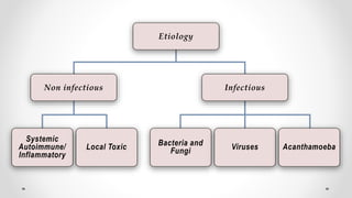
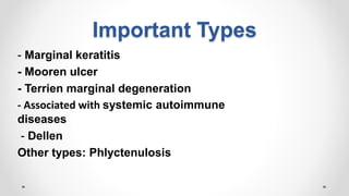
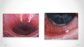
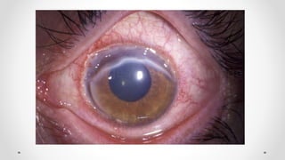
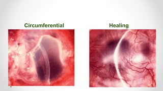
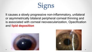
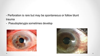
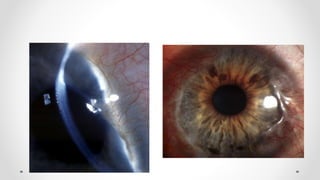
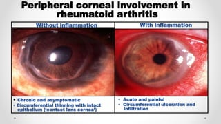
Non Infectious Corneal
Ulcers
Corneal Ulcers
Def: Corneal ulcers are defects in the corneal epithelium
with or without stromal infiltration.
Types:
A) Infectious ulcerative keratitis
B) Non infectious ulcerative keratitis
Bacteria and
Fungi Viruses Acanthamoeba
Systemic
Autoimmune/
Inflammatory
Local Toxic
InfectiousNon infectious
Etiology
Non Infectious Ulcerative Keratitis
Causes:
Local causes:
Punctate marginal keratitis: Staphylococci, Streptococci, hypersensitivity to
medications
Peripheral keratitis associated with blepharitis:
Systemic causes:
Generally manifestation of systemic, immune-mediated disease
Most common: Rheumatoid arthritis, Wegener’s granulomatosis and
polyarteritis nodosa
Non INFECTIOUSINFECTIOUS
No painPain
No dischargeDischarge
AC reaction: absentAC reaction: present
PeripheralCentral
Trauma: -------Trauma : ++++
Important Types
- Marginal keratitis
- Mooren ulcer
- Terrien marginal degeneration
- Associated with systemic autoimmune
diseases
- Dellen
Other types: Phlyctenulosis
Marginal keratitis
Marginal Keratitis
• Caused by hypersensitivity reaction against
Staphylococcal exotoxin and cell wall proteins with
deposition of Ag-Ab complex in peripheral cornea
• Lesions are culture negative but S. aureus can be
isolated from lid margin
Signs and symptoms
• Chronic Blepheritis
• Subepithelial marginal infiltrates seperated from
limbus by a clear zone
• Conjunctival Injection
• Coalescence and circumferential spread
• Little or no AC reaction
• Resolution usually occurs in 1-4 wks, occasionally
there is residual superficial scarring
Treatment
• Weak topical steroid
• May be combined with a topical antibiotic
• Tetracycline orally
• For children , breastfeeding and pregnancy
erythromycin
• Treatment of blepheritis
Mooren's Ulcer
• Rare autoimmune disease
• Characterized by
– Progressive,
– Peripheral,with Limbitis
– Circumferential, stromal corneal ulceration
Vascularization involving the bed of the ulcer up to its leading edge but not
beyond
– Later central spread
Risk factors for Mooren's ulcer include corneal surgery, previous trauma,
and infection.
Circumferential Healing
Complications
• Severe astigmatism due to extensive vascularization &
fibrosis
• Perforation following minor trauma
• Secondary bacterial infection
Management
• Topical
– Steroids
– Cyclosporin (weeks to show significant effect)
– Artificial tears
– Collagenase inhibitors (acetylcystine)
• bandage contact lenses
• Conjunctival resection
• Immunosuppression
• Systemic collagenase inhibitors such as doxycycline
Lamellar keratoplasty, keratoepithelioplasty and conjunctival flap and patch grafts
Terrien marginal degeneration
• Idiopathic thinning of the peripheral cornea
• Young adult to elderly patients
• Uncommon
• 75% males
• Usually bilateral
Symptoms
• Asymptomatic
• Gradual visual deterioration can occur due to
astigmatism
• A few patients experience episodic pain and
inflammation
Signs
It causes a slowly progressive non-inflammatory, unilateral
or asymmetrically bilateral peripheral corneal thinning and
is associated with corneal neovascularization, Opacification
and lipid deposition
- Perforation is rare but may be spontaneous or follow blunt
trauma
- Pseudopterygia sometimes develop
Management
• Safety spectacles if thinning is significant
• Contact lenses for astigmatism.
Scleral or soft lenses
with rigid gas permeable
• Surgery :- Cresentric excision of the gutter
with lamellar or full-thickness
corneoscleral patch grafts transplantation
Peripheral Ulcerative Keratitis
Associated With Systemic
Autoimmune Disease
Destructive inflammation of the peripheral cornea
associated with corneal epithelial sloughing and
Keratolysis
The mechanism includes immune complex
deposition in peripheral cornea, episcleral and
conjunctival capillary occlusion with secondary
cytokine release and inflammatory cell recruitment,
the upregulation of collagenases and reduced activity
of their inhibitors
Systemic associations
• Rheumatoid arthritis (most common)
– PUK is bilateral in 30% and tends to occur in advanced RA
• Wegener granulomatosis (2nd most common)
– In contrast to RA ocular complications are the initial presentation in
50%
• Other conditions include polyarteritis nodosa, relapsing
polychondritis ,SLE , Churg – Strauss ,Microscopic Polyangiitis,
Inflammatory Bowel Disease
Clinically
Crescentic ulceration Contact lens cornea
Perforation Limbitis, episcleritis or scleritis
Peripheral corneal involvement in
rheumatoid arthritis
• Chronic and asymptomatic
• Circumferential thinning with intact
epithelium (‘contact lens cornea’)
• Acute and painful
• Circumferential ulceration and
infiltration
Without inflammation With inflammation
Management
Principally with systemic immunosuppression in
collaboration with a rheumatologist
Topical
Artificial tears (preservative-free)
Antibiotics as prophylaxis
Steroids may worsen thinning so are generally avoided
Systemic
Steroids (via pulsed IV administration) are used to control
acute disease, with immunosuppressive therapy and
biological blockers for longer-term management
Tetracycline for its anticollagenase effect
Dellen
Localized corneal disturbance associated with
drying of a focal area
Usually associated with an adjacent elevated
lesion as pinguecula or large subconjunctival
haemorrhage that impairs physiological
lubrication
Treatment by lubricants and elimination of cause
Phlyctenulosis
Uncommon, unilateral - typically affects children
Severe photophobia, lacrimation and blepharospasm
Delayed hypersensitivity reaction to staphylococcal
antigen.
In developing countries, the majority are associated with
tuberculosis or helminthic infestation
• Small pinkish-white nodule
near limbus
• Usually transient and resolves
spontaneously
• Starts astride limbus
• Resolves spontaneously or extends
onto cornea
Conjunctival phlycten
Treatment - Topical steroids
Corneal phlycten
Thank you

Non infectious corneal ulcers

  • 1.
  • 2.
    Corneal Ulcers Def: Cornealulcers are defects in the corneal epithelium with or without stromal infiltration. Types: A) Infectious ulcerative keratitis B) Non infectious ulcerative keratitis
  • 3.
    Bacteria and Fungi VirusesAcanthamoeba Systemic Autoimmune/ Inflammatory Local Toxic InfectiousNon infectious Etiology
  • 4.
    Non Infectious UlcerativeKeratitis Causes: Local causes: Punctate marginal keratitis: Staphylococci, Streptococci, hypersensitivity to medications Peripheral keratitis associated with blepharitis: Systemic causes: Generally manifestation of systemic, immune-mediated disease Most common: Rheumatoid arthritis, Wegener’s granulomatosis and polyarteritis nodosa
  • 5.
    Non INFECTIOUSINFECTIOUS No painPain NodischargeDischarge AC reaction: absentAC reaction: present PeripheralCentral Trauma: -------Trauma : ++++
  • 6.
    Important Types - Marginalkeratitis - Mooren ulcer - Terrien marginal degeneration - Associated with systemic autoimmune diseases - Dellen Other types: Phlyctenulosis
  • 7.
  • 8.
    Marginal Keratitis • Causedby hypersensitivity reaction against Staphylococcal exotoxin and cell wall proteins with deposition of Ag-Ab complex in peripheral cornea • Lesions are culture negative but S. aureus can be isolated from lid margin
  • 9.
    Signs and symptoms •Chronic Blepheritis • Subepithelial marginal infiltrates seperated from limbus by a clear zone • Conjunctival Injection • Coalescence and circumferential spread • Little or no AC reaction • Resolution usually occurs in 1-4 wks, occasionally there is residual superficial scarring
  • 11.
    Treatment • Weak topicalsteroid • May be combined with a topical antibiotic • Tetracycline orally • For children , breastfeeding and pregnancy erythromycin • Treatment of blepheritis
  • 12.
    Mooren's Ulcer • Rareautoimmune disease • Characterized by – Progressive, – Peripheral,with Limbitis – Circumferential, stromal corneal ulceration Vascularization involving the bed of the ulcer up to its leading edge but not beyond – Later central spread Risk factors for Mooren's ulcer include corneal surgery, previous trauma, and infection.
  • 14.
  • 15.
    Complications • Severe astigmatismdue to extensive vascularization & fibrosis • Perforation following minor trauma • Secondary bacterial infection
  • 16.
    Management • Topical – Steroids –Cyclosporin (weeks to show significant effect) – Artificial tears – Collagenase inhibitors (acetylcystine) • bandage contact lenses • Conjunctival resection • Immunosuppression • Systemic collagenase inhibitors such as doxycycline Lamellar keratoplasty, keratoepithelioplasty and conjunctival flap and patch grafts
  • 17.
    Terrien marginal degeneration •Idiopathic thinning of the peripheral cornea • Young adult to elderly patients • Uncommon • 75% males • Usually bilateral
  • 18.
    Symptoms • Asymptomatic • Gradualvisual deterioration can occur due to astigmatism • A few patients experience episodic pain and inflammation
  • 19.
    Signs It causes aslowly progressive non-inflammatory, unilateral or asymmetrically bilateral peripheral corneal thinning and is associated with corneal neovascularization, Opacification and lipid deposition
  • 20.
    - Perforation israre but may be spontaneous or follow blunt trauma - Pseudopterygia sometimes develop
  • 22.
    Management • Safety spectaclesif thinning is significant • Contact lenses for astigmatism. Scleral or soft lenses with rigid gas permeable • Surgery :- Cresentric excision of the gutter with lamellar or full-thickness corneoscleral patch grafts transplantation
  • 23.
    Peripheral Ulcerative Keratitis AssociatedWith Systemic Autoimmune Disease
  • 24.
    Destructive inflammation ofthe peripheral cornea associated with corneal epithelial sloughing and Keratolysis The mechanism includes immune complex deposition in peripheral cornea, episcleral and conjunctival capillary occlusion with secondary cytokine release and inflammatory cell recruitment, the upregulation of collagenases and reduced activity of their inhibitors
  • 25.
    Systemic associations • Rheumatoidarthritis (most common) – PUK is bilateral in 30% and tends to occur in advanced RA • Wegener granulomatosis (2nd most common) – In contrast to RA ocular complications are the initial presentation in 50% • Other conditions include polyarteritis nodosa, relapsing polychondritis ,SLE , Churg – Strauss ,Microscopic Polyangiitis, Inflammatory Bowel Disease
  • 26.
  • 27.
  • 28.
    Peripheral corneal involvementin rheumatoid arthritis • Chronic and asymptomatic • Circumferential thinning with intact epithelium (‘contact lens cornea’) • Acute and painful • Circumferential ulceration and infiltration Without inflammation With inflammation
  • 29.
    Management Principally with systemicimmunosuppression in collaboration with a rheumatologist Topical Artificial tears (preservative-free) Antibiotics as prophylaxis Steroids may worsen thinning so are generally avoided Systemic Steroids (via pulsed IV administration) are used to control acute disease, with immunosuppressive therapy and biological blockers for longer-term management Tetracycline for its anticollagenase effect
  • 30.
    Dellen Localized corneal disturbanceassociated with drying of a focal area Usually associated with an adjacent elevated lesion as pinguecula or large subconjunctival haemorrhage that impairs physiological lubrication
  • 31.
    Treatment by lubricantsand elimination of cause
  • 32.
    Phlyctenulosis Uncommon, unilateral -typically affects children Severe photophobia, lacrimation and blepharospasm Delayed hypersensitivity reaction to staphylococcal antigen. In developing countries, the majority are associated with tuberculosis or helminthic infestation
  • 33.
    • Small pinkish-whitenodule near limbus • Usually transient and resolves spontaneously • Starts astride limbus • Resolves spontaneously or extends onto cornea Conjunctival phlycten Treatment - Topical steroids Corneal phlycten
  • 34.