Downloaded 123 times


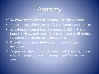


Peripheral ulcerative keratitis (PUK) is a destructive inflammatory disease of the peripheral cornea characterized by sloughing of the corneal epithelium and stromal melting. It begins with a crescent-shaped inflammatory lesion near the limbus and can progress circumferentially, leading to stromal thinning and potential perforation. PUK is often associated with autoimmune diseases and may be the initial presentation of an undiagnosed systemic vasculitis. Treatment involves topical immunosuppression with corticosteroids for mild cases or systemic corticosteroids and immunosuppressive drugs for more severe or progressive disease to halt inflammation and promote healing. Surgical interventions like conjunctival resection or grafting are


