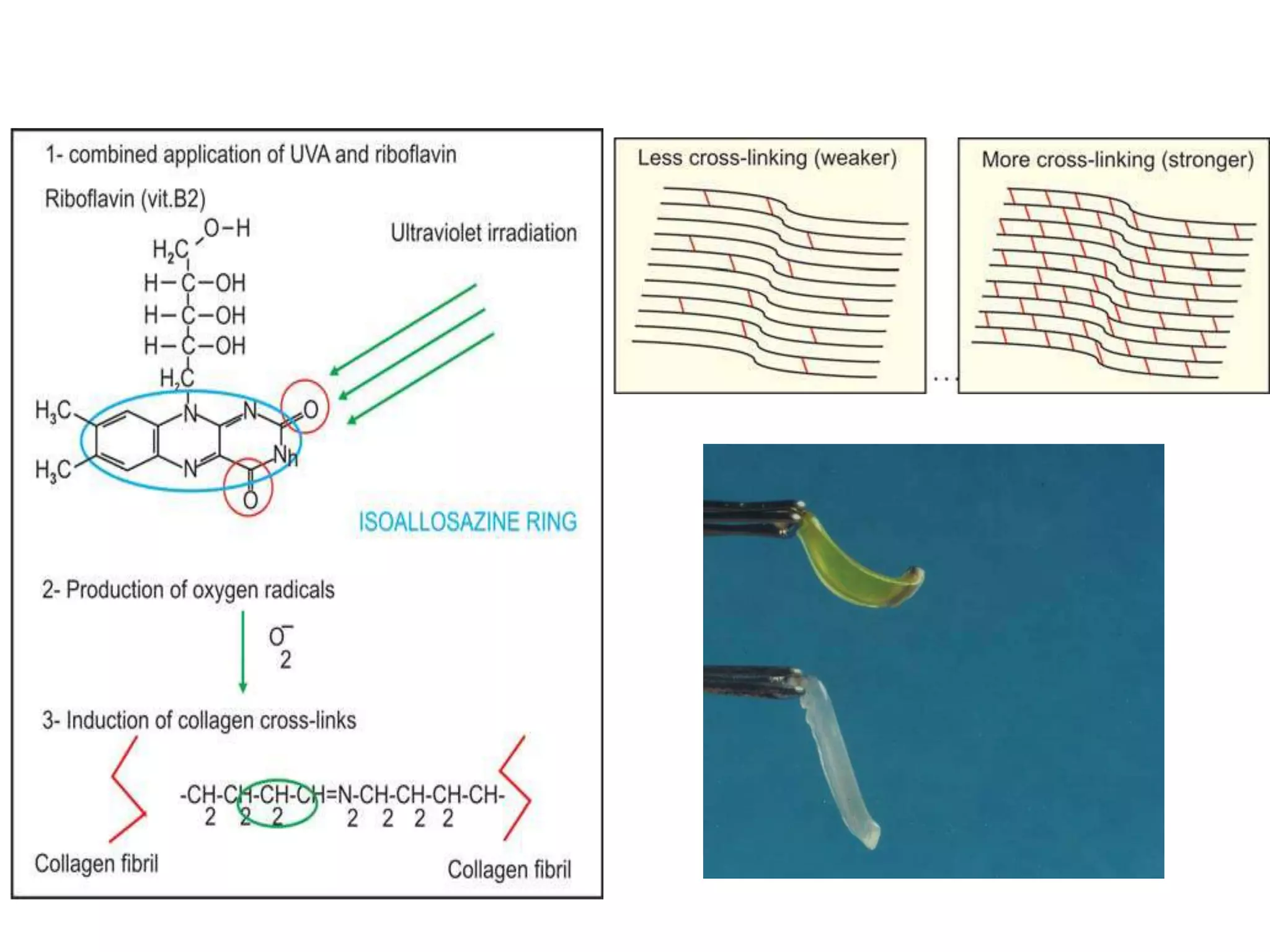
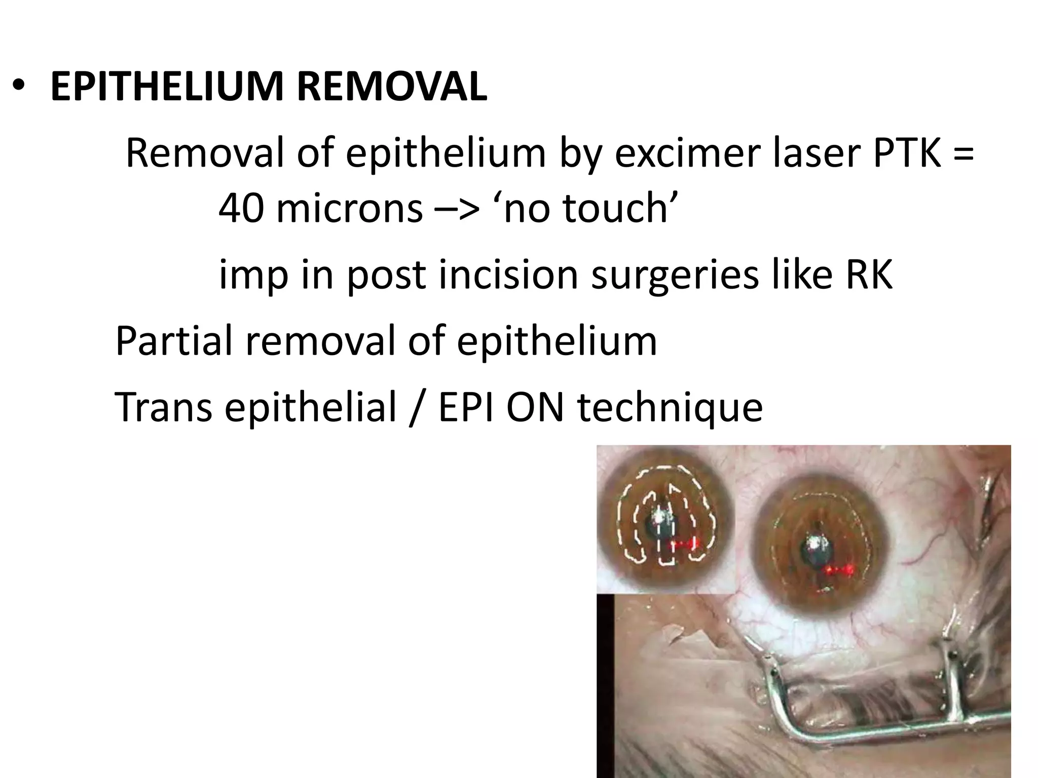
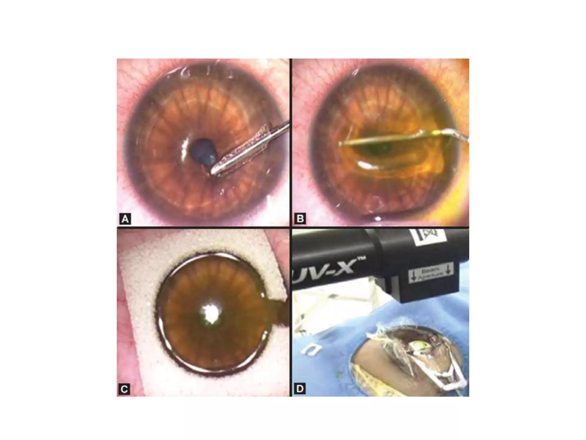
CORNEAL COLLAGEN
CROSS LINKING
Dr PAAVAN KALRA
Department of Ophthalmology
S P Medical College
Bikaner
KERATOCONUS
• Progressive, non inflammatory, ectatic disease of cornea
• 1 in 2000 to 1 in 500
• Most common indication for PKP in west
• Cornea becomes conical in shape with off centre apex
• Myopic and irregular astigmatism
• Begins at puberty, progresses eventually
stops by 40
• Worsening of ectasia during pregnancy
• Increase in proteolytic enzymes
• Decrease in enzyme inhibitors
• Decreased total proteins,
sulphated proteoglycans
• Decreased inter fibrillar bonds in collagen
• Uneven distribution of stromal lamellae.
• Central/ paracentral thinning of stroma.
• Slippage of lamellae and bowman’s layer
• Biomechanical strength in about half the normal
• Glasses
• Contact lenses – soft
-spherical RGP
-specialty RGP - rose k
-scleral fitted
• INTACS
• Lamellar keratoplasty – DALK
• PKP
LASER(ABLATIVE) REFRACTIVE SURGERY
• keratoconus – contraindication
– especially early, subclinical forms which tend to get
missed
• Post refractive keratectasia – LASIK / PRK
– One of the most dreaded complication for refractive
surgeon
– 0.12 to 0.66 %
– Central steeping due to weakened biomechanics of
cornea
– Myopic shift , astigmatic changes
– May require Penetrating Keratoplasty
• Risk factors
– Pre op pachymetry <500 microns
– High myopia
– Thick flap – flap later on doesn’t contribute to
biomechanical stability
– Low residual stromal thickness <250/300 microns
– Pre op topographic abnormalities – FFKC and PMD
Posterior float elevations > 40 mm
Asymmetric inferior corneal steeping
Skewed steep radial axes above and below horizontal meridian
Bow tie pattern
CROSS LINKING
• Increase in inter molecular bonds
Natural in corneal tissue
ageing
diabetes – advanced glycation end products
No keratoconus in type 1 DM
Artificial
Chemical  gluteraldehyde – prosthetic valves
Physical  UV in dentistry
Photo chemical  C3R
• THEO SEILER
• E SPOERL
• G WOLLENSAK
• CAPOROSSI
• PINELLI
• LECCISOTTI
• CHAN
• BRIAN BOXER WACHLER
• KANELLOPOULOS
• GULANI
Animal studies and experiments – 1993 – 1997
1st report on clinical trial - 2003
• RIBOFLAVIN –vitamin B2 – intermediary metabolism
Photo sensitizer and UV blocker
Non toxic
Absorption peak -> 365 – 370 mm
• UV - A
photo keratitis and skin cancer are caused by UV –B ( 290-
320mic)
UV - A transmittance = 7 % across cornea saturated with
riboflavin
• Release of ROS
• Ionization of amino groups
• Formation of new bonds
• Type 2 photo chemical reaction
Effect on keratocytes and corneal
nerves
• Loss of subepithelial and anterior stromal nerve fibres. Loss of
corneal sensations
• Loss in number and rarefaction of keratocytes upto depth of
300 microns.
• spongy honey comb like edema
• Activated keratocytes appear after 3 months with decrease in
edema
• Densely populated activated keratocytes, disappearance of
edema and reappearance of anterior stromal nerve fibres are
seen by 6 months
• Dense tissue around keratocytes upto 300 micron depth only
– observed as subclinical haze and demarcation line on
biomicroscopy
Effect on corneal stroma
• Effect only in anterior 250-300 microns
confirmed by histology, confocal microscopy
and AS OCT
• Increased intra and inter fibrillar bonds
• Increased collagen fiber diameter by 12%
• Increased resistance to collagenase digestion,
mercaptoethanol and heat
• Biomechanical effect -Microcomputer based
biomaterial testing machine - Increase in
rigidity 328.9% and young modulus by factor
of 4.5
• Thermo mechanical effect – heat dependent
shrinkage – increase in temperature from 700
to 750
OCULAR RESPONSE ANALYSER
Concern for endothelium, lens and
retina
INDICATIONS
• Documented Progressive keratoconus on
topography in young patient age < 30 years
Mainly - halt and delay progression and
hence need of PK
May lead to reversal of ectasia
• POST LASIK ECTASIA
CONTRAINDICATIONS
• Corneal Scarring - central
• Pachymetry < 400 microns at the thinnest point
• Vogt striae
• Dark micro striae in reticular pattern on confocal
microscopy
• Steep K > 58 D
• Poor BCVA with hard contact lenses
• High BCVA – 6/9
CONTRAINDICATIONS
• Active ocular disease - allergy
• Herpes keratitis
• Severe dry eye
• Children < 18 years
• Pregnancy and lactation
• Diabetes Mellitus
Pre op evaluation
UCVA, BCVA & BSCVA
REFRACTION – manifest spherical equivalent
SLIT LAMP EXAMINATION
KERATOMETRY
TOPOGRAPHY – elevation based – orbscan II / pentacam
PACHYMETRY
(contact lens free period upto 2 weeks)
AS - OCT and CONFOCAL MICROSCOPY give additional
information
• COUNSELLING and CONSENT – procedure not
for visual rehabilitation
• Under sterile conditions in OT
• Anti biotic eye drops
• Topical anesthesia
• Patching of the other eye
• Preparation and draping
• Insertion of eye speculum
ORIGINAL SEILER CROSSLINKING
(DRESDEN PROTOCOL)
• Complete EPITHELIUM SCRAPING manually by blunt
spatula– 7 mm zone
• RIBOFLAVIN 0.1% in 20% dextran T500 in NS (pH=7,
hyper osmolar) –1 drop every 3 mins for 30 mins
• Checking saturation of cornea with riboflavin :
SLE with blue light – green fluorescence in AC
• Checking calibration – irradiance
• Intra op pachymetry (USG) > 400 microns
• Patient instructed to look into centre of the
light
• UV-A irradiation from 5 cm - @ 3mw/sq cm X
30 mins ( 5.4 J/ sq cm)
• Proper focus and centration – loss of energy /
hot spots
• Riboflavin drops – every 5 mins
• BSS to prevent desiccation during procedure
• Solution protected from light
Protection of Sclera, goblet cells, limbus stem
cells during procedure
Cellulose sponge / merocel
RESULTS- DRESDON METHOD
• (2003) 22 eyes
steep K – flattening in 70 % patients by avg of 2D
stable in 5 patients
progressed in 1 by 0.28D
progression in 23% of control by 1.5D
improvement in refraction and BCVA by 1.26 lines
• (2008) 111 eyes longer follow up
steep K decreased by avg 2.29D, 3.27D, 4.34D in 1st,
2nd and 3rd year
vision improved or remained stable in 96 % by 3rd year
POST OP
• Healing of epithelial defect – 3-5 days
Bandage contact lens / patching
Cycloplegics, antibiotic , NSAID & artificial tears eye drops
Oral analgesic SOS
observed daily
• Mild steroid eye drops for 3 – 6 weeks (FML 0.1%)
• Review at 1 week, 4 weeks, 12 weeks, 24 weeks and 1
year
• Topography, keratometry after 12 weeks
• Patient can start using old RGP Lenses after 2 weeks
• Temporary blurring of vision, FB sensation, pain
• Prednsolone eye drops for persisting haze
COMPLICATIONS
• Transient Corneal edema – lasts few days
• CORNEAL HAZE
• HOT SPOTS
• CORNEAL SCARRING
• ENDOTHELIAL BURNS
• INFECTIONS
• CORNEAL MELT
• Persistence of epithelial defect
• failure
MODIFICATIONS
• More efficient delivery of energy
• Reduction of intra operative time
• Reduction in patient discomfort
• Reduction in chances of complications
infection/slow healing/sub epithelial haze
• Early rehabilitation
• Bilateral treatment in one sitting
• EPITHELIUM REMOVAL
Removal of epithelium by excimer laser PTK =
40 microns –> ‘no touch’
imp in post incision surgeries like RK
Partial removal of epithelium
Trans epithelial / EPI ON technique
• RIBOFLAVIN DELIVERY
use of epi lasik well
hypo osmolar solution for thinner corneas
– 0.5% solution diluted in BSS 1:4, applied
every 5 secs until intra op pachymetry
reaches 400 microns
Cornea should not be swollen > 80 microns
• IRRADIANCE
-CBM X linker VEGA
-higher irradiance and shorter duration –
7 mW/cm2 for 15 mins
CAPOROSSI CROSSLINKING TECHNIQUE
(SIENNA EYE CROSS PROJECT)
• Different illuminator - CBM(caporossi Baiocchi
mazotta) X linker VEGA
• 5 uv a LED array – sharper focus
• Low power red laser sources - proper aiming
1 mm defocus / 0.2 mm defocus = 10% loss of
energy
• Micro camera and automated control
TRANSEPITHELIAL/ EPI ON(PINELLI)
• Proparacaine – two drops every 5 mins for 15
mins
• Application of riboflavin via merocel sponge
for 5 mins – pre soak
• Every 3 mins during procedure
Penetration of riboflavin can be enhanced with
EDTA and topical gentamicin 3 hr pre
treatment – tensio actives
• Reported no endothelial toxicity
• Controversy regarding riboflavin penetration
and relatively higher delivery of energy to
endothelium
• Doubts about efficacy
• Punctate epithelial defects may occur
KANELLOPOULOS INTRALASE
CROSSLINKING
In early disease, IntraLase femtosecond laser is
used to create a corneal pocket at 100 microns
depth with 50 opening.
0.1% riboflavin is instilled one time in that
pocket for 2 mins
Cross-links, 60 microns anterior to the pocket
and 200 microns behind without having to
remove the corneal epithelium
• Follow up of 4- 8 months
• No endothelial damage
• K value reduced by 1.7 D and MSE by 1.5
SANCHEZ-LEON MODIFIED
TECHNIQUE FOR LASIK ECTASIA
Lasik flap edge is identified.
Opened gently 100 using a Sinsky hook.
IntraLase Lasik spatula is used to create an intra
stromal pocket, leaving the rest of the margin
of the flap intact.
The 0.1% Riboflavin is instilled direct in to the
stromal space.
OTHER USES
• PMD – 11 mm area needs to be debrided, limbus protected.
Single segment ICRS may have to be inserted
• Stabilizing corneal flattening and hyperopia in post RK
• PSEUDOPHAKIC BULLOUS KERATOPATHY – glycerol to de
swell the cornea upto 400 microns
• STERILE CORNEAL MELTS – resistence to digestive enzymes -
low surface irradiation = 2.5 mW/cm2 because of thinning
• INFECTIOUS CORNEAL ULCERS – e g acanthamoeba keratitis
CORNEOPLASTIQUES
• Corneal reshaping techniques without using invasive
procedures like PKP and LK
Mechanical -ICRS
-incisions
Elastic -Conductive keratoplasty
-C3R
LASER- Excimer – PRK / ASA
• Modifying the shape first and then solidifying by C3R
• Even in post PK / LK patients for reshaping the cornea
• Basically managing high irregular astigmatism
• Combined with ICRS and orthokeratology
• Trans epithelial cross linking after ICRS
• Combined with TOPO GUIDED PRK (Kanellopoulos)
min pachy at thinnest point = 450 microns
better than wavefront guided prk
persistent epithelial defects especially if
mitomycin c is used
25 % under correction required
C3R followed by PRK later more risk of scarring
loss of cross linked tissue
CONCLUSIONS
• Rapidly evolving procedure
– Techniques
– Indications
– combinations
• Main aim is to halt the/ delay the progress of ectasia
postponing PKP by strengthen the lamellar fibres
• LONG TERM STUDIES needed
-durability of stiffening as Collagen turn over is
known to occur in 2-3 years
-serious late complications
THANK YOU

Corneal collagen cross linking

  • 1.
    CORNEAL COLLAGEN CROSS LINKING DrPAAVAN KALRA Department of Ophthalmology S P Medical College Bikaner
  • 2.
    KERATOCONUS • Progressive, noninflammatory, ectatic disease of cornea • 1 in 2000 to 1 in 500 • Most common indication for PKP in west • Cornea becomes conical in shape with off centre apex • Myopic and irregular astigmatism • Begins at puberty, progresses eventually stops by 40 • Worsening of ectasia during pregnancy
  • 3.
    • Increase inproteolytic enzymes • Decrease in enzyme inhibitors • Decreased total proteins, sulphated proteoglycans • Decreased inter fibrillar bonds in collagen • Uneven distribution of stromal lamellae. • Central/ paracentral thinning of stroma. • Slippage of lamellae and bowman’s layer • Biomechanical strength in about half the normal
  • 4.
    • Glasses • Contactlenses – soft -spherical RGP -specialty RGP - rose k -scleral fitted • INTACS • Lamellar keratoplasty – DALK • PKP
  • 5.
    LASER(ABLATIVE) REFRACTIVE SURGERY •keratoconus – contraindication – especially early, subclinical forms which tend to get missed • Post refractive keratectasia – LASIK / PRK – One of the most dreaded complication for refractive surgeon – 0.12 to 0.66 % – Central steeping due to weakened biomechanics of cornea – Myopic shift , astigmatic changes – May require Penetrating Keratoplasty
  • 6.
    • Risk factors –Pre op pachymetry <500 microns – High myopia – Thick flap – flap later on doesn’t contribute to biomechanical stability – Low residual stromal thickness <250/300 microns – Pre op topographic abnormalities – FFKC and PMD Posterior float elevations > 40 mm Asymmetric inferior corneal steeping Skewed steep radial axes above and below horizontal meridian Bow tie pattern
  • 7.
    CROSS LINKING • Increasein inter molecular bonds Natural in corneal tissue ageing diabetes – advanced glycation end products No keratoconus in type 1 DM Artificial Chemical  gluteraldehyde – prosthetic valves Physical  UV in dentistry Photo chemical  C3R
  • 8.
    • THEO SEILER •E SPOERL • G WOLLENSAK • CAPOROSSI • PINELLI • LECCISOTTI • CHAN • BRIAN BOXER WACHLER • KANELLOPOULOS • GULANI Animal studies and experiments – 1993 – 1997 1st report on clinical trial - 2003
  • 9.
    • RIBOFLAVIN –vitaminB2 – intermediary metabolism Photo sensitizer and UV blocker Non toxic Absorption peak -> 365 – 370 mm • UV - A photo keratitis and skin cancer are caused by UV –B ( 290- 320mic) UV - A transmittance = 7 % across cornea saturated with riboflavin
  • 10.
    • Release ofROS • Ionization of amino groups • Formation of new bonds • Type 2 photo chemical reaction
  • 12.
    Effect on keratocytesand corneal nerves • Loss of subepithelial and anterior stromal nerve fibres. Loss of corneal sensations • Loss in number and rarefaction of keratocytes upto depth of 300 microns. • spongy honey comb like edema • Activated keratocytes appear after 3 months with decrease in edema • Densely populated activated keratocytes, disappearance of edema and reappearance of anterior stromal nerve fibres are seen by 6 months • Dense tissue around keratocytes upto 300 micron depth only – observed as subclinical haze and demarcation line on biomicroscopy
  • 14.
    Effect on cornealstroma • Effect only in anterior 250-300 microns confirmed by histology, confocal microscopy and AS OCT • Increased intra and inter fibrillar bonds • Increased collagen fiber diameter by 12% • Increased resistance to collagenase digestion, mercaptoethanol and heat
  • 15.
    • Biomechanical effect-Microcomputer based biomaterial testing machine - Increase in rigidity 328.9% and young modulus by factor of 4.5 • Thermo mechanical effect – heat dependent shrinkage – increase in temperature from 700 to 750
  • 16.
  • 17.
    Concern for endothelium,lens and retina
  • 19.
    INDICATIONS • Documented Progressivekeratoconus on topography in young patient age < 30 years Mainly - halt and delay progression and hence need of PK May lead to reversal of ectasia • POST LASIK ECTASIA
  • 20.
    CONTRAINDICATIONS • Corneal Scarring- central • Pachymetry < 400 microns at the thinnest point • Vogt striae • Dark micro striae in reticular pattern on confocal microscopy • Steep K > 58 D • Poor BCVA with hard contact lenses • High BCVA – 6/9
  • 21.
    CONTRAINDICATIONS • Active oculardisease - allergy • Herpes keratitis • Severe dry eye • Children < 18 years • Pregnancy and lactation • Diabetes Mellitus
  • 22.
    Pre op evaluation UCVA,BCVA & BSCVA REFRACTION – manifest spherical equivalent SLIT LAMP EXAMINATION KERATOMETRY TOPOGRAPHY – elevation based – orbscan II / pentacam PACHYMETRY (contact lens free period upto 2 weeks) AS - OCT and CONFOCAL MICROSCOPY give additional information
  • 23.
    • COUNSELLING andCONSENT – procedure not for visual rehabilitation • Under sterile conditions in OT • Anti biotic eye drops • Topical anesthesia • Patching of the other eye • Preparation and draping • Insertion of eye speculum
  • 24.
    ORIGINAL SEILER CROSSLINKING (DRESDENPROTOCOL) • Complete EPITHELIUM SCRAPING manually by blunt spatula– 7 mm zone • RIBOFLAVIN 0.1% in 20% dextran T500 in NS (pH=7, hyper osmolar) –1 drop every 3 mins for 30 mins
  • 25.
    • Checking saturationof cornea with riboflavin : SLE with blue light – green fluorescence in AC • Checking calibration – irradiance • Intra op pachymetry (USG) > 400 microns • Patient instructed to look into centre of the light
  • 26.
    • UV-A irradiationfrom 5 cm - @ 3mw/sq cm X 30 mins ( 5.4 J/ sq cm) • Proper focus and centration – loss of energy / hot spots • Riboflavin drops – every 5 mins • BSS to prevent desiccation during procedure • Solution protected from light
  • 28.
    Protection of Sclera,goblet cells, limbus stem cells during procedure Cellulose sponge / merocel
  • 29.
    RESULTS- DRESDON METHOD •(2003) 22 eyes steep K – flattening in 70 % patients by avg of 2D stable in 5 patients progressed in 1 by 0.28D progression in 23% of control by 1.5D improvement in refraction and BCVA by 1.26 lines • (2008) 111 eyes longer follow up steep K decreased by avg 2.29D, 3.27D, 4.34D in 1st, 2nd and 3rd year vision improved or remained stable in 96 % by 3rd year
  • 30.
    POST OP • Healingof epithelial defect – 3-5 days Bandage contact lens / patching Cycloplegics, antibiotic , NSAID & artificial tears eye drops Oral analgesic SOS observed daily • Mild steroid eye drops for 3 – 6 weeks (FML 0.1%) • Review at 1 week, 4 weeks, 12 weeks, 24 weeks and 1 year • Topography, keratometry after 12 weeks • Patient can start using old RGP Lenses after 2 weeks • Temporary blurring of vision, FB sensation, pain • Prednsolone eye drops for persisting haze
  • 31.
    COMPLICATIONS • Transient Cornealedema – lasts few days • CORNEAL HAZE • HOT SPOTS • CORNEAL SCARRING • ENDOTHELIAL BURNS • INFECTIONS • CORNEAL MELT • Persistence of epithelial defect • failure
  • 32.
    MODIFICATIONS • More efficientdelivery of energy • Reduction of intra operative time • Reduction in patient discomfort • Reduction in chances of complications infection/slow healing/sub epithelial haze • Early rehabilitation • Bilateral treatment in one sitting
  • 33.
    • EPITHELIUM REMOVAL Removalof epithelium by excimer laser PTK = 40 microns –> ‘no touch’ imp in post incision surgeries like RK Partial removal of epithelium Trans epithelial / EPI ON technique
  • 34.
    • RIBOFLAVIN DELIVERY useof epi lasik well hypo osmolar solution for thinner corneas – 0.5% solution diluted in BSS 1:4, applied every 5 secs until intra op pachymetry reaches 400 microns Cornea should not be swollen > 80 microns
  • 35.
    • IRRADIANCE -CBM Xlinker VEGA -higher irradiance and shorter duration – 7 mW/cm2 for 15 mins
  • 36.
    CAPOROSSI CROSSLINKING TECHNIQUE (SIENNAEYE CROSS PROJECT) • Different illuminator - CBM(caporossi Baiocchi mazotta) X linker VEGA • 5 uv a LED array – sharper focus • Low power red laser sources - proper aiming 1 mm defocus / 0.2 mm defocus = 10% loss of energy • Micro camera and automated control
  • 38.
    TRANSEPITHELIAL/ EPI ON(PINELLI) •Proparacaine – two drops every 5 mins for 15 mins • Application of riboflavin via merocel sponge for 5 mins – pre soak • Every 3 mins during procedure Penetration of riboflavin can be enhanced with EDTA and topical gentamicin 3 hr pre treatment – tensio actives
  • 40.
    • Reported noendothelial toxicity • Controversy regarding riboflavin penetration and relatively higher delivery of energy to endothelium • Doubts about efficacy • Punctate epithelial defects may occur
  • 41.
    KANELLOPOULOS INTRALASE CROSSLINKING In earlydisease, IntraLase femtosecond laser is used to create a corneal pocket at 100 microns depth with 50 opening. 0.1% riboflavin is instilled one time in that pocket for 2 mins Cross-links, 60 microns anterior to the pocket and 200 microns behind without having to remove the corneal epithelium
  • 43.
    • Follow upof 4- 8 months • No endothelial damage • K value reduced by 1.7 D and MSE by 1.5
  • 44.
    SANCHEZ-LEON MODIFIED TECHNIQUE FORLASIK ECTASIA Lasik flap edge is identified. Opened gently 100 using a Sinsky hook. IntraLase Lasik spatula is used to create an intra stromal pocket, leaving the rest of the margin of the flap intact. The 0.1% Riboflavin is instilled direct in to the stromal space.
  • 46.
    OTHER USES • PMD– 11 mm area needs to be debrided, limbus protected. Single segment ICRS may have to be inserted • Stabilizing corneal flattening and hyperopia in post RK • PSEUDOPHAKIC BULLOUS KERATOPATHY – glycerol to de swell the cornea upto 400 microns • STERILE CORNEAL MELTS – resistence to digestive enzymes - low surface irradiation = 2.5 mW/cm2 because of thinning • INFECTIOUS CORNEAL ULCERS – e g acanthamoeba keratitis
  • 47.
    CORNEOPLASTIQUES • Corneal reshapingtechniques without using invasive procedures like PKP and LK Mechanical -ICRS -incisions Elastic -Conductive keratoplasty -C3R LASER- Excimer – PRK / ASA • Modifying the shape first and then solidifying by C3R • Even in post PK / LK patients for reshaping the cornea • Basically managing high irregular astigmatism
  • 48.
    • Combined withICRS and orthokeratology • Trans epithelial cross linking after ICRS
  • 51.
    • Combined withTOPO GUIDED PRK (Kanellopoulos) min pachy at thinnest point = 450 microns better than wavefront guided prk persistent epithelial defects especially if mitomycin c is used 25 % under correction required C3R followed by PRK later more risk of scarring loss of cross linked tissue
  • 52.
    CONCLUSIONS • Rapidly evolvingprocedure – Techniques – Indications – combinations • Main aim is to halt the/ delay the progress of ectasia postponing PKP by strengthen the lamellar fibres • LONG TERM STUDIES needed -durability of stiffening as Collagen turn over is known to occur in 2-3 years -serious late complications
  • 53.