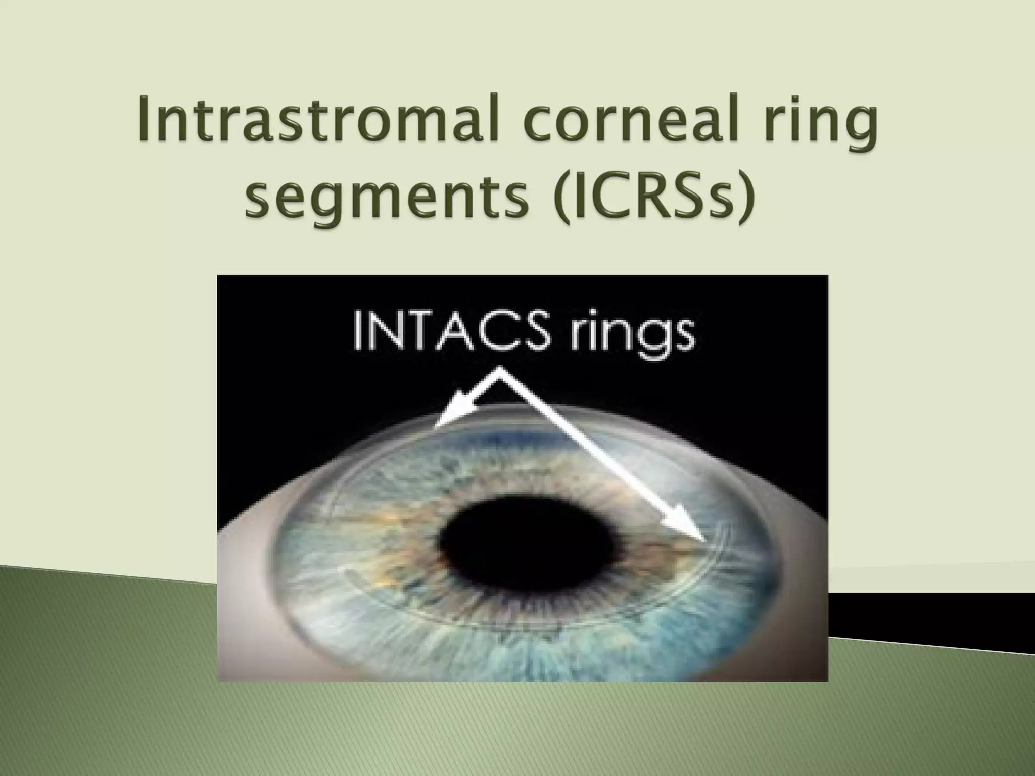
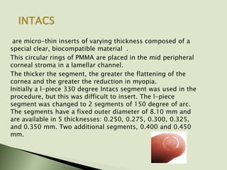
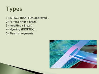
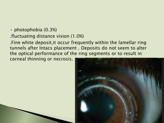
are micro-thin inserts of varying thickness composed of a
special clear, biocompatible material .
This circular rings of PMMA are placed in the mid peripheral
corneal stroma in a lamellar channel.
The thicker the segment, the greater the flattening of the
cornea and the greater the reduction in myopia.
Initially a l-piece 330 degree lntacs segment was used in the
procedure, but this was difficult to insert. The l-piece
segment was changed to 2 segments of 150 degree of arc.
The segments have a fixed outer diameter of 8.10 mm and
are available in 5 thicknesses: 0.250, 0.275, 0.300, 0.325,
and 0.350 mm. Two additional segments, 0.400 and 0.450
mm.
The ring segment procedure involves creating a lamellar
channel at approximately 68%-70% stromal depth by
mechanical or Femtosecond laser procedure and then
inserting the ring segments.The procedure usually takes
10-15 minutes per eye to complete
Imagine the cornea as compressed arcs of fiber covering the
eye. If a spacer element is inserted between those fibers,
effectively pushing them apart, then the arc must flatten to
accommodate the inserted element. An addition in the cornea
periphery results in its flattening, and the ring diameter
determines how much the cornea will be flattened. In other
words, the more tissue is added (increasing ring thickness)
and the smaller the diameter, the greater will be the myopia
correction obtained. Intracorneal ring segments inserts
shorten the corneal arc length, produce flattening across the
entire cornea, and maintain the positive asphericity of the
cornea
1) INTACS (USA) FDA approved .
2) Ferrara rings ( Brazil)
3) KeraRing ( Brazil)
4) Myoring (DIOPTEX).
5) Bisantis segments
The ring segments can be explanted, making the refractive
result of the procedure potentially reversible.
The central clear zone of the cornea is not directly treated
The normal cornea is generally prolate, or steeper centrally than
peripherally: after placement of ring segments has been
shown to maintain an aspheric, prolate shape because the
ring segments flatten the peripheral cornea more the central
than the central cornea.
Specialized equipment and training are required to create the
lamellar channels and to insert the rings.
The procedure generally takes longer time to perform
Patients can experience ocular discomfort after surgery, and
some patients, especially those with large pupils, may
complain of visual disturbances such as glare.
1. Low levels of Myopia ( FDA approved )
2. Progressive Keratoconus (FDA approved )
3. Ectasia after LASIK
4. Residual Myopia after LASIK
5. Pellucide marginal degeneration
• 21 years or older
• with documented stability of refraction, as demonstrated by
change of 0.50 D for at least 12 months prior to the
preoperative examination
• with 1.00 D of astigmatism or less
Prescribing
Range
Correction
Recommended
Ring
Seg.Thickness
-1.00-1.630-1.3000.250mm
-1.625-1.750-1.6500.275mm
-1.75-2.250-2.0000.300mm
-2.25-2.50
-2.375-3.00
-2.350
-2.700
0.325mm
0.350mm
Intacs are typically contraindicated in
• patients with collagen vascular, autoimmune, or
immunodeficiency diseases
• pregnant or nursing women
• the presence of ocular conditions (such as recurrent corneal
erosion syndrome, corneal dystrophy) that may predispose
the patient to future complications
• patients with a low-light pupil diameter of 7.0 mm or larger
because of the predisposition to low-light visual symptoms
(the inner segment diameter is 6.8 mm)
• patients with systemic diseases likely to affect wound healing,
such as insulin-dependent diabetes or severe atopic disease
• patients with a history of ophthalmic herpes simplex or
herpes zoster
• eyes with a central corneal thickness of less than 480 or a
peripheral thickness of less than 570 mm
• eyes with a corneal keratometry steeper than 46.00 0 or
flatter than 40.00 D Instrumentation
used in reducing or eliminating myopia and astigmatism in certain pa­
tients with keratoconus, specifically those who are no longer able to
achieve adequate vision with their contact lenses or spectacles.
AIM is to restore functional vision and defer the need for a corneal
transplant.
• experience with a progressive deterioration in vision such that the
patient can no longer achieve adequate functional vision on a daily basis
with contact lenses or spectacles
• age 21 years or older
• clear central cornea
• a corneal thickness of 450 mm or greater at the proposed incision site
. corneal transplantation is the only remaining option for improving
functional vision
The visual improvement was significant. With an average follow-up of 9
months, the mean UCVA improved from approximately 20/200 to 20/80
. The mean BCVA also improved, from approximately 20/50 to 20/30
Most patients still required optical correction to achieve their
best-corrected vision. Eyes with corneal scarring had a similar
improvement UCVA and BCVA. Inferior steepening was
reduced on topography.
New data indicate that when the Keratoconus is peripheral, not
central, it may be preferable to place a single segment
instead of 2 segments. The reason is that the keratoconic
cornea has 2 optical areas of distortion within the pupil: a
steep lower and flat upper area. For peripheral
keratconus,instead of flattening the entire cornea .
better to flatten the steep area and steepen the flat area.
Single-segment placement achieve that result . When a single
segment is placed, it flattens the adjacent cornea but causes
steepening of the cornea 180° away-the "bean bag effect"
yields a more physiologic improvement than a global
flattening effect from double segments.
Intacs can also be combined with C3-R (corneal collagen cross
linking with riboflavin) to yield additional flattening and
improved corneal strength.
In limited early trials using Intacs to treat post-LASIK ectasia,
myopia was reduced and UCVA was improved ( Off-label )
Uses for Ring Segments After LASIK
This procedure may be useful in patients whose stromal bed
would not support repeat excimer laser ablation.conversely,
after ring segments have been removed from patients whose
vision did not improve to a satisfactory level (eg, due to
undercorrection or induced astigmatism), LASIK has been
performed with good success
• anterior chamber perforation
• microbial keratitis
• implant extrusion
• shallow ring segment placement
• corneal thinning over Intacs
Ocular complications have been reported in 11 % of patients at
12 months postoperatively. This include:
• reduced corneal sensitivity (5.5%)
• induced astigmatism between 1 and 2 D (3.7%)
• deep neovascularization at the incision ite (1.2%)
• persistent epithelial defect (0.2%)
• iritis/uveitis (0.2%)
Visual symptom have been reported in approximately 14% of
patients and include
• difficulty with night vision (4.8%)
• blurred vision (2.9%)
• diplopia (1.6%)
• glare (1.3%)
• halos (1.2)
• photophobia (0.3%)
.fluctuating distance vision (1.0%)
.Fine white deposit,it occur frequently within the lamellar ring
tunnels after Intacs placement . Deposits do not seem to alter
the optical performance of the ring segments or to result in
corneal thinning or necrosis.
Ferrara Ring is made of yellow PMMA, an inert and
biocompatible, used in intracorneal implants. The apical
diameters are 5 mm and 6 mm. It has a flat base with a
variable arch length of 90 and 210 and a variable thickness
of 0.15 mm to 0.30 mm.
Ferrara rings have a smaller optical zone and more of a
flattening effect than lntacs. Ferrara rings are not FDA
approved in the United States
.
Characteristic INTACS Ferrara Rings
Arc length (degrees) 150 160
Cross-section Hexagonal Triangular
Thickness (mm) 0.25 – 0.45 (0.05 increments) 0.20-0.35 (0.05 increments)
Inner Radius (mm) 6.77 4.40
Outer Radius (mm) 8.10 5.60
offers 40 different thickness, arc length and diameter options,
allowing for greater customization of corneal remodeling and
refractive correction
INDICATIONS
• Keratoconus with corrected low visual acuity and
intolerance to the use of contact lenses
• Pellucid marginal degeneration
• Post-refractive surgery corneal ectasia (PRK, LASIK)
• Post radial keratotomy (RK) irregular astigmatism
• High astigmatism post-penetrating cornea transplant
• Post-trauma corneal irregularities
CONTRAINDICATIONS
• Acute keratoconus with keratometry > 70 D.
• Major central leucoma
• Edema
• Post-penetrating transplant, if the button is off-center
• Severe atopy
• Recurrent corneal erosion syndrome
• High expectation of emmetropia without correction
Indications:
Myopia:
All grades of Myopia between 1 dioptre and 20 dioptre, which are
not eligible to Excimer Laser treatment such as:
thin cor neas
irregular cor neal surface
forme fruste keratoconus
high myopia
the patient denies LASIK but wants a minimally-invasive and reversible
myopia treatment
Keratoconus:
All grades of non-central and central Keratoconus, PMD as well as
post-LASIK Keratectasia as long as the minimal corneal thickness is
larger than 350 microns.1 In very advanced cases, the MyoRing
implantation should be combined with corneal crosslinking.
THANK YOU

Intrastromal Corneal Ring Segment (ICRSs)

  • 2.
    are micro-thin insertsof varying thickness composed of a special clear, biocompatible material . This circular rings of PMMA are placed in the mid peripheral corneal stroma in a lamellar channel. The thicker the segment, the greater the flattening of the cornea and the greater the reduction in myopia. Initially a l-piece 330 degree lntacs segment was used in the procedure, but this was difficult to insert. The l-piece segment was changed to 2 segments of 150 degree of arc. The segments have a fixed outer diameter of 8.10 mm and are available in 5 thicknesses: 0.250, 0.275, 0.300, 0.325, and 0.350 mm. Two additional segments, 0.400 and 0.450 mm.
  • 3.
    The ring segmentprocedure involves creating a lamellar channel at approximately 68%-70% stromal depth by mechanical or Femtosecond laser procedure and then inserting the ring segments.The procedure usually takes 10-15 minutes per eye to complete
  • 5.
    Imagine the corneaas compressed arcs of fiber covering the eye. If a spacer element is inserted between those fibers, effectively pushing them apart, then the arc must flatten to accommodate the inserted element. An addition in the cornea periphery results in its flattening, and the ring diameter determines how much the cornea will be flattened. In other words, the more tissue is added (increasing ring thickness) and the smaller the diameter, the greater will be the myopia correction obtained. Intracorneal ring segments inserts shorten the corneal arc length, produce flattening across the entire cornea, and maintain the positive asphericity of the cornea
  • 6.
    1) INTACS (USA)FDA approved . 2) Ferrara rings ( Brazil) 3) KeraRing ( Brazil) 4) Myoring (DIOPTEX). 5) Bisantis segments
  • 7.
    The ring segmentscan be explanted, making the refractive result of the procedure potentially reversible. The central clear zone of the cornea is not directly treated The normal cornea is generally prolate, or steeper centrally than peripherally: after placement of ring segments has been shown to maintain an aspheric, prolate shape because the ring segments flatten the peripheral cornea more the central than the central cornea.
  • 8.
    Specialized equipment andtraining are required to create the lamellar channels and to insert the rings. The procedure generally takes longer time to perform Patients can experience ocular discomfort after surgery, and some patients, especially those with large pupils, may complain of visual disturbances such as glare.
  • 9.
    1. Low levelsof Myopia ( FDA approved ) 2. Progressive Keratoconus (FDA approved ) 3. Ectasia after LASIK 4. Residual Myopia after LASIK 5. Pellucide marginal degeneration
  • 10.
    • 21 yearsor older • with documented stability of refraction, as demonstrated by change of 0.50 D for at least 12 months prior to the preoperative examination • with 1.00 D of astigmatism or less Prescribing Range Correction Recommended Ring Seg.Thickness -1.00-1.630-1.3000.250mm -1.625-1.750-1.6500.275mm -1.75-2.250-2.0000.300mm -2.25-2.50 -2.375-3.00 -2.350 -2.700 0.325mm 0.350mm
  • 11.
    Intacs are typicallycontraindicated in • patients with collagen vascular, autoimmune, or immunodeficiency diseases • pregnant or nursing women • the presence of ocular conditions (such as recurrent corneal erosion syndrome, corneal dystrophy) that may predispose the patient to future complications
  • 12.
    • patients witha low-light pupil diameter of 7.0 mm or larger because of the predisposition to low-light visual symptoms (the inner segment diameter is 6.8 mm) • patients with systemic diseases likely to affect wound healing, such as insulin-dependent diabetes or severe atopic disease • patients with a history of ophthalmic herpes simplex or herpes zoster • eyes with a central corneal thickness of less than 480 or a peripheral thickness of less than 570 mm • eyes with a corneal keratometry steeper than 46.00 0 or flatter than 40.00 D Instrumentation
  • 13.
    used in reducingor eliminating myopia and astigmatism in certain pa­ tients with keratoconus, specifically those who are no longer able to achieve adequate vision with their contact lenses or spectacles. AIM is to restore functional vision and defer the need for a corneal transplant. • experience with a progressive deterioration in vision such that the patient can no longer achieve adequate functional vision on a daily basis with contact lenses or spectacles • age 21 years or older • clear central cornea • a corneal thickness of 450 mm or greater at the proposed incision site . corneal transplantation is the only remaining option for improving functional vision The visual improvement was significant. With an average follow-up of 9 months, the mean UCVA improved from approximately 20/200 to 20/80 . The mean BCVA also improved, from approximately 20/50 to 20/30
  • 14.
    Most patients stillrequired optical correction to achieve their best-corrected vision. Eyes with corneal scarring had a similar improvement UCVA and BCVA. Inferior steepening was reduced on topography.
  • 15.
    New data indicatethat when the Keratoconus is peripheral, not central, it may be preferable to place a single segment instead of 2 segments. The reason is that the keratoconic cornea has 2 optical areas of distortion within the pupil: a steep lower and flat upper area. For peripheral keratconus,instead of flattening the entire cornea . better to flatten the steep area and steepen the flat area. Single-segment placement achieve that result . When a single segment is placed, it flattens the adjacent cornea but causes steepening of the cornea 180° away-the "bean bag effect" yields a more physiologic improvement than a global flattening effect from double segments. Intacs can also be combined with C3-R (corneal collagen cross linking with riboflavin) to yield additional flattening and improved corneal strength.
  • 16.
    In limited earlytrials using Intacs to treat post-LASIK ectasia, myopia was reduced and UCVA was improved ( Off-label ) Uses for Ring Segments After LASIK This procedure may be useful in patients whose stromal bed would not support repeat excimer laser ablation.conversely, after ring segments have been removed from patients whose vision did not improve to a satisfactory level (eg, due to undercorrection or induced astigmatism), LASIK has been performed with good success
  • 17.
    • anterior chamberperforation • microbial keratitis • implant extrusion • shallow ring segment placement • corneal thinning over Intacs Ocular complications have been reported in 11 % of patients at 12 months postoperatively. This include: • reduced corneal sensitivity (5.5%) • induced astigmatism between 1 and 2 D (3.7%) • deep neovascularization at the incision ite (1.2%)
  • 18.
    • persistent epithelialdefect (0.2%) • iritis/uveitis (0.2%) Visual symptom have been reported in approximately 14% of patients and include • difficulty with night vision (4.8%) • blurred vision (2.9%) • diplopia (1.6%) • glare (1.3%) • halos (1.2)
  • 19.
    • photophobia (0.3%) .fluctuatingdistance vision (1.0%) .Fine white deposit,it occur frequently within the lamellar ring tunnels after Intacs placement . Deposits do not seem to alter the optical performance of the ring segments or to result in corneal thinning or necrosis.
  • 22.
    Ferrara Ring ismade of yellow PMMA, an inert and biocompatible, used in intracorneal implants. The apical diameters are 5 mm and 6 mm. It has a flat base with a variable arch length of 90 and 210 and a variable thickness of 0.15 mm to 0.30 mm. Ferrara rings have a smaller optical zone and more of a flattening effect than lntacs. Ferrara rings are not FDA approved in the United States
  • 23.
    . Characteristic INTACS FerraraRings Arc length (degrees) 150 160 Cross-section Hexagonal Triangular Thickness (mm) 0.25 – 0.45 (0.05 increments) 0.20-0.35 (0.05 increments) Inner Radius (mm) 6.77 4.40 Outer Radius (mm) 8.10 5.60
  • 24.
    offers 40 differentthickness, arc length and diameter options, allowing for greater customization of corneal remodeling and refractive correction INDICATIONS • Keratoconus with corrected low visual acuity and intolerance to the use of contact lenses • Pellucid marginal degeneration • Post-refractive surgery corneal ectasia (PRK, LASIK) • Post radial keratotomy (RK) irregular astigmatism • High astigmatism post-penetrating cornea transplant • Post-trauma corneal irregularities
  • 25.
    CONTRAINDICATIONS • Acute keratoconuswith keratometry > 70 D. • Major central leucoma • Edema • Post-penetrating transplant, if the button is off-center • Severe atopy • Recurrent corneal erosion syndrome • High expectation of emmetropia without correction
  • 26.
    Indications: Myopia: All grades ofMyopia between 1 dioptre and 20 dioptre, which are not eligible to Excimer Laser treatment such as: thin cor neas irregular cor neal surface forme fruste keratoconus high myopia the patient denies LASIK but wants a minimally-invasive and reversible myopia treatment Keratoconus: All grades of non-central and central Keratoconus, PMD as well as post-LASIK Keratectasia as long as the minimal corneal thickness is larger than 350 microns.1 In very advanced cases, the MyoRing implantation should be combined with corneal crosslinking.
  • 27.