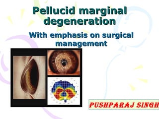
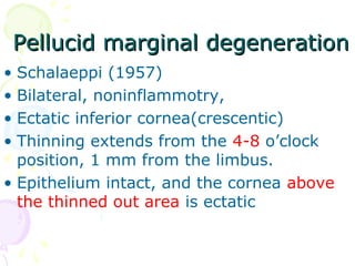
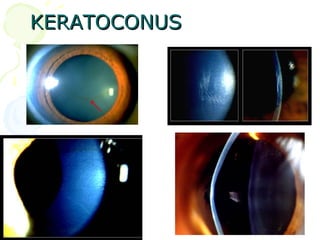
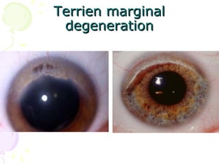
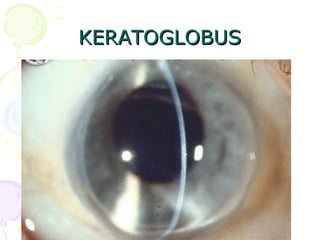
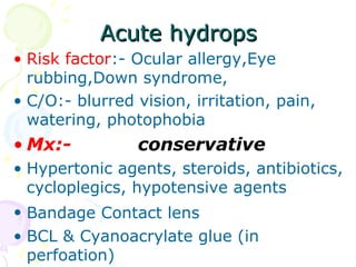
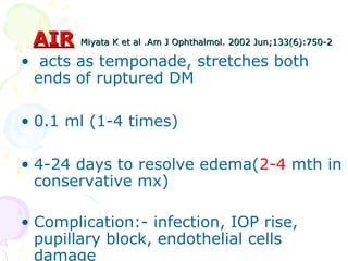
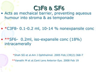
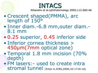
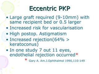
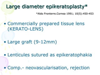
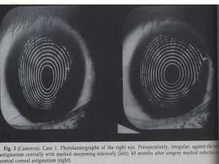
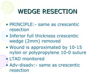
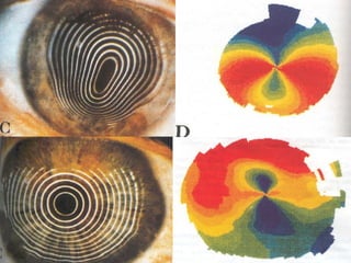
Pellucid marginalPellucid marginal
degenerationdegeneration
With emphasis on surgicalWith emphasis on surgical
managementmanagement
PushParaj singh
Pellucid marginal degenerationPellucid marginal degeneration
• Schalaeppi (1957)
• Bilateral, noninflammotry,
• Ectatic inferior cornea(crescentic)
• Thinning extends from the 4-8 o’clock
position, 1 mm from the limbus.
• Epithelium intact, and the cornea above
the thinned out area is ectatic
• The area between
the limbus and
thinning is
1. Clear,
2. NO Scarring
3. NO Lipid deposition,
4. NO vascularization.
• Age group- 30-40
yr
• Flattening of the cornea along a vertical
axis
• Steepening of the inferior cornea
peripheral to the site of the lesion (ATR)
DiffrentialDiffrential
DiagnosisDiagnosis
KERATOCONUSKERATOCONUS
Terrien marginalTerrien marginal
degenerationdegeneration
KERATOGLOBUSKERATOGLOBUS
MOORENS ULCERMOORENS ULCER
MANAGEMENTMANAGEMENT
• Spectacles:-small horizontal eye
sizes, & high refractive index
lenses with antireflective coating
• Contact lenses:- RGP, Toric lenses,
scleral lenses
• Verisye/ Artisan phakic iol*
*De verrien NE. cornea 2008;27:241-45
Acute hydropsAcute hydrops
• Risk factor:- Ocular allergy,Eye
rubbing,Down syndrome,
• C/O:- blurred vision, irritation, pain,
watering, photophobia
• Mx:- conservative
• Hypertonic agents, steroids, antibiotics,
cycloplegics, hypotensive agents
• Bandage Contact lens
• BCL & Cyanoacrylate glue (in
perfoation)
AIRAIR Miyata K et al .Miyata K et al .Am J Ophthalmol. 2002 Jun;133(6):750-2Am J Ophthalmol. 2002 Jun;133(6):750-2
• acts as temponade, stretches both
ends of ruptured DM
• 0.1 ml (1-4 times)
• 4-24 days to resolve edema(2-4 mth in
conservative mx)
• Complication:- infection, IOP rise,
pupillary block, endothelial cells
damage
CC33FF8 &8 & SFSF66
• Acts as mechaical barrier, preventing aqueous
humour into stroma & as temponade
• *C3F8- 0.1-0.2 ml, 10-14 % nonexpansile conc
• **SF6- 0.2ml, iso-expansile conc (18%)
intracamerally
*Shah SG et al.Am J Ophthalmol. 2005 Feb;139(2):368-7
**Vanathi M et al.Cont Lens Anterior Eye. 2008 Feb 19
SURGICAL MAMAGEMENTSURGICAL MAMAGEMENT
•INTACS
•Eccentric PKP
•Full thickness wedge resection
•Lamellar crescentic resection
•Large diameter epikeratoplasty
•lamellar crescentic keratoplasty
•Comb. Of lamellar crescentic +
PKP
INTACSINTACS
Allsandro et al.ophthalomology 2005;112:660-66Allsandro et al.ophthalomology 2005;112:660-66
• Crescent shaped(PMMA), arc
length of 150°
• Inner diam.-6.8 mm,outer diam.-
8.1 mm
• 0.25 superior, 0.45 inferior side
• Inferior cornea thickness >
450ÎĽm(7mm optical zone)
• Temporal 1.8 mm incision (70%
depth)
• FM lasers:- used to create intra
stromal tunnel (Ertan A.JCRS,2006;32:1710-16)
principleprinciple
• New optical zone, separates the
ectatic area from central zone
• Spherical equivalent, reduced by
flattening action of 2 opposite ring
• Inferior ring causes a barrier effect
against high astigmatism induced by
PMD towards central cornea
• Stabilization & elimination of the
progression of ectatic disease
Eccentric PKPEccentric PKP
• Large graft required (9-10mm) with
same recipient bed or 0.5 larger
• Increased risk for vascularisation
• High postop. Astigmatism
• Increased rejection(64% >
keratoconus)
• In one study 7 out 11 eyes,
endothelial rejection occurred*
* Gary A. Am.J.Ophthalmol 1990,110:149
Large diameter epikeratoplasty*Large diameter epikeratoplasty*
*Aldo Fronterre.Cornea 1991; 10(5):450-453*Aldo Fronterre.Cornea 1991; 10(5):450-453
• Commercially prepared tissue lens
(KERATO-LENS)
• Large graft (9-12mm)
• Lenticules sutured as epikeratophakia
• Comp.- neovascularisation, rejection
Lamellar crescentic keratoplastyLamellar crescentic keratoplasty
schanzlin J.am jschanzlin J.am j
ophthalmol.1983;96(2)253-254ophthalmol.1983;96(2)253-254
• Tectonic graft
• Limited to thinned area
• Sutured with nylon 10-0
Lamellar crescentic+ PKPLamellar crescentic+ PKP
Robinwitz et al.ophthalomology 2000 oct.107(10)1836-40Robinwitz et al.ophthalomology 2000 oct.107(10)1836-40
• Simultaneous peripheral crescentic LK &
central PKP
• Tendency for increased WRA Noted in
long term
Lamellar crescentic resectionLamellar crescentic resection
Principle
• Excise abnormal crescentic thinned stroma
• Approximate normal thickness stroma to
normal thickness stroma
• Aim for overcorrection of astigmatism
(WRA), Upto 50 % (Troutman)
• Adv:- localised to abnormal area
incision smaller
no donor tissue- no rejection
Disadv:- visual acuity poor for 6Disadv:- visual acuity poor for 6
monthmonth
long term astigmatism driftlong term astigmatism drift
WEDGE RESECTIONWEDGE RESECTION
• PRINCIPLE:- same as crescentic
resection
• Inferior full thickness crescentic
wedge (2mm) removed
• Wound is approximated by 10-15
nylon or polypropylene 10-0 suture
• LTAD monitored
• Adv-disadv:- same as crescentic
resection
Pellucid marginal degeneration

Pellucid marginal degeneration

  • 1.
    Pellucid marginalPellucid marginal degenerationdegeneration Withemphasis on surgicalWith emphasis on surgical managementmanagement PushParaj singh
  • 2.
    Pellucid marginal degenerationPellucidmarginal degeneration • Schalaeppi (1957) • Bilateral, noninflammotry, • Ectatic inferior cornea(crescentic) • Thinning extends from the 4-8 o’clock position, 1 mm from the limbus. • Epithelium intact, and the cornea above the thinned out area is ectatic
  • 4.
    • The areabetween the limbus and thinning is 1. Clear, 2. NO Scarring 3. NO Lipid deposition, 4. NO vascularization. • Age group- 30-40 yr
  • 5.
    • Flattening ofthe cornea along a vertical axis • Steepening of the inferior cornea peripheral to the site of the lesion (ATR)
  • 6.
  • 7.
  • 8.
  • 9.
  • 10.
  • 11.
    MANAGEMENTMANAGEMENT • Spectacles:-small horizontaleye sizes, & high refractive index lenses with antireflective coating • Contact lenses:- RGP, Toric lenses, scleral lenses • Verisye/ Artisan phakic iol* *De verrien NE. cornea 2008;27:241-45
  • 12.
    Acute hydropsAcute hydrops •Risk factor:- Ocular allergy,Eye rubbing,Down syndrome, • C/O:- blurred vision, irritation, pain, watering, photophobia • Mx:- conservative • Hypertonic agents, steroids, antibiotics, cycloplegics, hypotensive agents • Bandage Contact lens • BCL & Cyanoacrylate glue (in perfoation)
  • 13.
    AIRAIR Miyata Ket al .Miyata K et al .Am J Ophthalmol. 2002 Jun;133(6):750-2Am J Ophthalmol. 2002 Jun;133(6):750-2 • acts as temponade, stretches both ends of ruptured DM • 0.1 ml (1-4 times) • 4-24 days to resolve edema(2-4 mth in conservative mx) • Complication:- infection, IOP rise, pupillary block, endothelial cells damage
  • 14.
    CC33FF8 &8 &SFSF66 • Acts as mechaical barrier, preventing aqueous humour into stroma & as temponade • *C3F8- 0.1-0.2 ml, 10-14 % nonexpansile conc • **SF6- 0.2ml, iso-expansile conc (18%) intracamerally *Shah SG et al.Am J Ophthalmol. 2005 Feb;139(2):368-7 **Vanathi M et al.Cont Lens Anterior Eye. 2008 Feb 19
  • 15.
    SURGICAL MAMAGEMENTSURGICAL MAMAGEMENT •INTACS •EccentricPKP •Full thickness wedge resection •Lamellar crescentic resection •Large diameter epikeratoplasty •lamellar crescentic keratoplasty •Comb. Of lamellar crescentic + PKP
  • 16.
    INTACSINTACS Allsandro et al.ophthalomology2005;112:660-66Allsandro et al.ophthalomology 2005;112:660-66 • Crescent shaped(PMMA), arc length of 150° • Inner diam.-6.8 mm,outer diam.- 8.1 mm • 0.25 superior, 0.45 inferior side • Inferior cornea thickness > 450μm(7mm optical zone) • Temporal 1.8 mm incision (70% depth) • FM lasers:- used to create intra stromal tunnel (Ertan A.JCRS,2006;32:1710-16)
  • 17.
    principleprinciple • New opticalzone, separates the ectatic area from central zone • Spherical equivalent, reduced by flattening action of 2 opposite ring • Inferior ring causes a barrier effect against high astigmatism induced by PMD towards central cornea • Stabilization & elimination of the progression of ectatic disease
  • 18.
    Eccentric PKPEccentric PKP •Large graft required (9-10mm) with same recipient bed or 0.5 larger • Increased risk for vascularisation • High postop. Astigmatism • Increased rejection(64% > keratoconus) • In one study 7 out 11 eyes, endothelial rejection occurred* * Gary A. Am.J.Ophthalmol 1990,110:149
  • 19.
    Large diameter epikeratoplasty*Largediameter epikeratoplasty* *Aldo Fronterre.Cornea 1991; 10(5):450-453*Aldo Fronterre.Cornea 1991; 10(5):450-453 • Commercially prepared tissue lens (KERATO-LENS) • Large graft (9-12mm) • Lenticules sutured as epikeratophakia • Comp.- neovascularisation, rejection
  • 20.
    Lamellar crescentic keratoplastyLamellarcrescentic keratoplasty schanzlin J.am jschanzlin J.am j ophthalmol.1983;96(2)253-254ophthalmol.1983;96(2)253-254 • Tectonic graft • Limited to thinned area • Sutured with nylon 10-0
  • 21.
    Lamellar crescentic+ PKPLamellarcrescentic+ PKP Robinwitz et al.ophthalomology 2000 oct.107(10)1836-40Robinwitz et al.ophthalomology 2000 oct.107(10)1836-40 • Simultaneous peripheral crescentic LK & central PKP • Tendency for increased WRA Noted in long term
  • 22.
    Lamellar crescentic resectionLamellarcrescentic resection Principle • Excise abnormal crescentic thinned stroma • Approximate normal thickness stroma to normal thickness stroma • Aim for overcorrection of astigmatism (WRA), Upto 50 % (Troutman) • Adv:- localised to abnormal area incision smaller no donor tissue- no rejection
  • 23.
    Disadv:- visual acuitypoor for 6Disadv:- visual acuity poor for 6 monthmonth long term astigmatism driftlong term astigmatism drift
  • 25.
    WEDGE RESECTIONWEDGE RESECTION •PRINCIPLE:- same as crescentic resection • Inferior full thickness crescentic wedge (2mm) removed • Wound is approximated by 10-15 nylon or polypropylene 10-0 suture • LTAD monitored • Adv-disadv:- same as crescentic resection