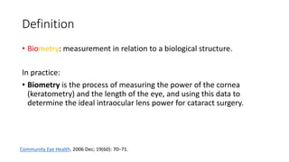
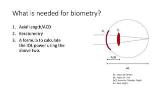
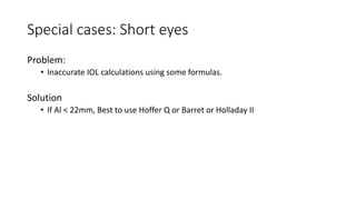
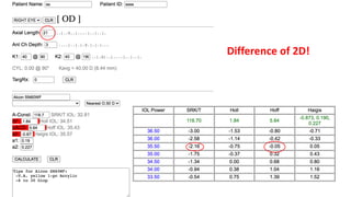
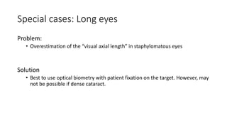
Biometry Made Easy
Dr Haitham Al-Mahrouqi, BMedSc (Hons) MB ChB
Definition
• Biometry: measurement in relation to a biological structure.
In practice:
• Biometry is the process of measuring the power of the cornea
(keratometry) and the length of the eye, and using this data to
determine the ideal intraocular lens power for cataract surgery.
Community Eye Health. 2006 Dec; 19(60): 70–71.
What is needed for biometry?
1. Axial length/ACD
2. Keratometry
3. A formula to calculate
the IOL power using the
above two.
Dc
DL
ACD
AL
Dc: Power of Cornea
DL: Power of Lens
ACD: Anterior Chamber Depth
AL: Axial length
Axial Length
Axial length: Introduction
Measured using two main methods:
1. Ultrasound method:
a) Contact
b) Non-contact (Immersion)
2. Optical method
Axial length: Principal of ultrasound
• Ultrasound echoes are reflected at
the interface of tissue with
different densities.
• Using the transit time between
transduction and receipt of the US
waves, the length of structures in
between the interfaces can be
calculated if the speed is known in
that structure.
Axial length: US contact A-scan
• Disadvantages:
• Compression of the cornea: thus
underestimates the axial length. This
will give a myopic surprise.
• Patient cannot fixate to align their
visual axis.
Axial length: Non-contact (immersion) A-scan
• Disadvantage:
• Still patient cannot fixate on a target
to align their visual axis.
• Requires a ”water bath” or
immersion shell
• Can miss the centre of the pupil but
is generally comparable to the optical
biometry.
However avoid compression.
Axial length: The optical method
• Uses light waves (780nm) instead of
Ultrasound waves.
• Partial coherence interferometry to
estimate the axial length.
Axial length: The optical method
• Advantages:
• More accurate: Patient can fixate
on a target and thus the “visual
axial length” is measured.
• No compression of the cornea.
• Measures the K values at the same
time.
• Disadvantages:
• Cannot penetrate dense opacities
(e.g. dense cataract).
US method
Regarding axial length: Unlike A-scan ultrasound, which reflects off the surface
of the retina, the IOLMaster beam passes through the translucent surface of
the retina and back further to the more opaque pigment layer. The computer
corrects for this difference, but because everyone has different tissue thick-
nesses, results from the IOLMaster and immersion ultrasound may be slightly
different.
In addition, the use of contact ultrasound will indent the cornea, thus decreas-
ing the distance between the cornea and the retina and artificially shortening
the axial length measured. Because of this, axial lengths measured on the IOL-
Master are almost always longer than those obtained through contact ultra-
sound.
Regarding corneal curvature: First, manual keratometry measures the corneal
curvature more peripherally—perhaps at 3.0 mm or 3.2 mm, depending on
your manual system—while the IOLMaster measures the more relevant central
curvature at 2.5 mm. Thus on a typical eye, IOLMaster K’s will be slightly
Conclusion for axial length
• Use optical biometry whenever possible
• In case of a dense cataract: It is best to use immersion US as opposed
to contact US.
What is needed for biometry?
1. Axial length
2. Keratometry
3. A formula to calculate the IOL power using the above two.
Keratometry
• Measure the power of the cornea
• The k-readings over the central 3mm zone is usually measured.
Keratometry: Methods
• Measurement using:
• Manual keratometers
• Automated keratometers
• IOLmaster/Lenstar
• Others: topographers, tomographers
Negligible differences in regular corneas
Keratometry
• Older machines measure only anterior corneal curvature (K-values)
• Assume a constant relationship between the anterior and corneal
curvature (Rpos/Rant: 82% and thus use n=1.3375)
• Newer technology also measures the posterior curvature and gives
more accurate net K. Important in toric iols, post refractive surgery
and keratoconus.
Conclusion for Keratometry readings
• If regular virgin cornea, use optical biometer K-readings.
• If keratoconus, refractive surgery, toric iol calculations best to use
total corneal power where the posterior corneal curvature is also
measured (using tomography, AS-OCT).
Trouble-shooting for wrong biometry
What is needed for biometry?
1. Axial length
2. Keratometry
3. A formula to calculate the IOL power using the above two.
IOL calculation formulas
In history, the formulas started as
theoretical formulas based on geometric
optics. However, there were problems:
1. Estimated lens position cannot be
determined from the pre-operative
anterior chamber depth.
2. The refractive index used to estimate the
corneal power from the anterior corneal
curvature is hypothetical.
3. The shape of the cornea and eye can be
altered by the surgery
IOL calculation: Formulas
• The first regression formula was introduced by Sanders, Retzlaff and Kraff
(SRK I) in 1980s
P = A – 0.9K – 2.5AL
• Regression: Modification of formulas based on a large number of post op
refraction.
• Since then many generations of theoratical and regression formulas have
been introduced. SRK-T (T for theoratical) combined theoratical optics and
regression data. Ray tracing formulas (Oslen) are also now introduced.
Available formulas
• SRK
• Haigis
• Holladay
• HofferQ
• Oslen
• Barret
SRK I formula
P = A – 0.9K – 2.5AL
P = Power of the lens for a particular target refraction
A = Constant relating to the lens design and their estimated position in
the eye.
K: Average corneal power
AL: Axial length.
Accuracy
Barret II and Hill-RBF work well for all AL
https://www.doctor-hill.com/iol-main/formulas.htm
Conclusion: formulas
• If AL <22mm use Hoffer Q
• If AL >22mm use SRK-T
OR
• Use Barret II universal or Hill-RBF for all AL
Example on how
to read IOLmaster
printout
Quality check:
Axial length
1) Composite SNR > 2
2) AL SD within 0.1
3) Inter-eye AL difference within 0.7
Keratometry
1) Make sure that not many X dots are
displayed.
Special cases
Special cases: Short eyes
Problem:
• Inaccurate IOL calculations using some formulas.
Solution
• If Al < 22mm, Best to use Hoffer Q or Barret or Holladay II
Difference of 2D!
Special cases: Long eyes
Problem:
• Overestimation of the “visual axial length” in staphylomatous eyes
Solution
• Best to use optical biometry with patient fixation on the target. However, may
not be possible if dense cataract.
Special cases: Dense cataract
Problem:
• The laser in optical biometry
will penetrate a dense cataract
Solution:
• Use US method to get a
reading of the AL and then
feed it into the optical
biometer.
Special cases: Regular astigmatism
Problem
• Patient might not attain
6/6 vision post op if
implanting a standard
monofocal lens.
Solution:
If corneal astigmatism is
0.75D, implant a toric IOL
Special cases: Irregular astigmatism
(Keratoconus)
Problem:
• Inaccurate K-readings by most devices. Can lead to upredictable post op surprise especially
hyperopic.
• Others: Irregular tear film, Long AL, deep AC, unpredictable ELP
Solution:
• If Kmax >55: Use standard Ks (because most devices are less repeatable and reliable) and
target for -1.5D post op refraction. Use SRK-T formula (most accurate and tend to have a
myopic error which counteract the hyperopic surprises).
• if Kmax <55: Use actual Ks (still the biometry prediction error is between -3 to +3D). Must
calculate the anterior and posterior corneal power without assumption. E.g. EKR-4.5 in
Pentacam with Hollday II or Barret True K formula (found online or in IOLmaster 700).
Br J Ophthalmol 2014; 98(3): 361-4.
Special cases: Refractive surgery
Problems:
• Instruments error:
• Most older devices (e.g. IOLmaster 500, lenstar, Placido devices) assume a constant
relationship between the anterior and posterior corneal curvature (Rpos/Rant: 82% and thus
use n=1.3375)
• Irregular corneal surfaces especially in decentered ablations, therefore wrong Ks.
• Formula error:
• ELP is estimated from the K values
Solution
• History or contact lens methods (old methods)
• Use actual posterior K values (obtained from Pentacam or IOLmaster 700).
Incorporate into the Holladay II or Barret true K formulas (calculators found
online at ASCRS, Barret-II suit)
Special cases: Silicone oil
Problem
• US waves travel slower in SO and thus
gives erranous long AL.
Solution
• AL x 2/3 (1000/1532 = 0.65)
• Optical biometer is not affected as SO
option is chosen.
Special cases: Macular pathology
Problem:
• Wrong AL if CME, RD, submacular hmg
(underestimated).
Solution:
• Always examine the retina!
• Newer devices (IOLmaster 700) give a
retinal image.
Thank you

Biometry made easy

  • 1.
    Biometry Made Easy DrHaitham Al-Mahrouqi, BMedSc (Hons) MB ChB
  • 2.
    Definition • Biometry: measurementin relation to a biological structure. In practice: • Biometry is the process of measuring the power of the cornea (keratometry) and the length of the eye, and using this data to determine the ideal intraocular lens power for cataract surgery. Community Eye Health. 2006 Dec; 19(60): 70–71.
  • 3.
    What is neededfor biometry? 1. Axial length/ACD 2. Keratometry 3. A formula to calculate the IOL power using the above two. Dc DL ACD AL Dc: Power of Cornea DL: Power of Lens ACD: Anterior Chamber Depth AL: Axial length
  • 4.
  • 5.
    Axial length: Introduction Measuredusing two main methods: 1. Ultrasound method: a) Contact b) Non-contact (Immersion) 2. Optical method
  • 6.
  • 7.
    Axial length: Principalof ultrasound • Ultrasound echoes are reflected at the interface of tissue with different densities. • Using the transit time between transduction and receipt of the US waves, the length of structures in between the interfaces can be calculated if the speed is known in that structure.
  • 8.
    Axial length: UScontact A-scan • Disadvantages: • Compression of the cornea: thus underestimates the axial length. This will give a myopic surprise. • Patient cannot fixate to align their visual axis.
  • 9.
    Axial length: Non-contact(immersion) A-scan • Disadvantage: • Still patient cannot fixate on a target to align their visual axis. • Requires a ”water bath” or immersion shell • Can miss the centre of the pupil but is generally comparable to the optical biometry. However avoid compression.
  • 10.
    Axial length: Theoptical method • Uses light waves (780nm) instead of Ultrasound waves. • Partial coherence interferometry to estimate the axial length.
  • 11.
    Axial length: Theoptical method • Advantages: • More accurate: Patient can fixate on a target and thus the “visual axial length” is measured. • No compression of the cornea. • Measures the K values at the same time. • Disadvantages: • Cannot penetrate dense opacities (e.g. dense cataract).
  • 12.
    US method Regarding axiallength: Unlike A-scan ultrasound, which reflects off the surface of the retina, the IOLMaster beam passes through the translucent surface of the retina and back further to the more opaque pigment layer. The computer corrects for this difference, but because everyone has different tissue thick- nesses, results from the IOLMaster and immersion ultrasound may be slightly different. In addition, the use of contact ultrasound will indent the cornea, thus decreas- ing the distance between the cornea and the retina and artificially shortening the axial length measured. Because of this, axial lengths measured on the IOL- Master are almost always longer than those obtained through contact ultra- sound. Regarding corneal curvature: First, manual keratometry measures the corneal curvature more peripherally—perhaps at 3.0 mm or 3.2 mm, depending on your manual system—while the IOLMaster measures the more relevant central curvature at 2.5 mm. Thus on a typical eye, IOLMaster K’s will be slightly
  • 13.
    Conclusion for axiallength • Use optical biometry whenever possible • In case of a dense cataract: It is best to use immersion US as opposed to contact US.
  • 14.
    What is neededfor biometry? 1. Axial length 2. Keratometry 3. A formula to calculate the IOL power using the above two.
  • 15.
    Keratometry • Measure thepower of the cornea • The k-readings over the central 3mm zone is usually measured.
  • 16.
    Keratometry: Methods • Measurementusing: • Manual keratometers • Automated keratometers • IOLmaster/Lenstar • Others: topographers, tomographers Negligible differences in regular corneas
  • 17.
    Keratometry • Older machinesmeasure only anterior corneal curvature (K-values) • Assume a constant relationship between the anterior and corneal curvature (Rpos/Rant: 82% and thus use n=1.3375) • Newer technology also measures the posterior curvature and gives more accurate net K. Important in toric iols, post refractive surgery and keratoconus.
  • 18.
    Conclusion for Keratometryreadings • If regular virgin cornea, use optical biometer K-readings. • If keratoconus, refractive surgery, toric iol calculations best to use total corneal power where the posterior corneal curvature is also measured (using tomography, AS-OCT).
  • 19.
  • 20.
    What is neededfor biometry? 1. Axial length 2. Keratometry 3. A formula to calculate the IOL power using the above two.
  • 21.
    IOL calculation formulas Inhistory, the formulas started as theoretical formulas based on geometric optics. However, there were problems: 1. Estimated lens position cannot be determined from the pre-operative anterior chamber depth. 2. The refractive index used to estimate the corneal power from the anterior corneal curvature is hypothetical. 3. The shape of the cornea and eye can be altered by the surgery
  • 23.
    IOL calculation: Formulas •The first regression formula was introduced by Sanders, Retzlaff and Kraff (SRK I) in 1980s P = A – 0.9K – 2.5AL • Regression: Modification of formulas based on a large number of post op refraction. • Since then many generations of theoratical and regression formulas have been introduced. SRK-T (T for theoratical) combined theoratical optics and regression data. Ray tracing formulas (Oslen) are also now introduced.
  • 24.
    Available formulas • SRK •Haigis • Holladay • HofferQ • Oslen • Barret
  • 25.
    SRK I formula P= A – 0.9K – 2.5AL P = Power of the lens for a particular target refraction A = Constant relating to the lens design and their estimated position in the eye. K: Average corneal power AL: Axial length.
  • 26.
  • 27.
    Barret II andHill-RBF work well for all AL https://www.doctor-hill.com/iol-main/formulas.htm
  • 28.
    Conclusion: formulas • IfAL <22mm use Hoffer Q • If AL >22mm use SRK-T OR • Use Barret II universal or Hill-RBF for all AL
  • 29.
    Example on how toread IOLmaster printout Quality check: Axial length 1) Composite SNR > 2 2) AL SD within 0.1 3) Inter-eye AL difference within 0.7 Keratometry 1) Make sure that not many X dots are displayed.
  • 33.
  • 34.
    Special cases: Shorteyes Problem: • Inaccurate IOL calculations using some formulas. Solution • If Al < 22mm, Best to use Hoffer Q or Barret or Holladay II
  • 35.
  • 36.
    Special cases: Longeyes Problem: • Overestimation of the “visual axial length” in staphylomatous eyes Solution • Best to use optical biometry with patient fixation on the target. However, may not be possible if dense cataract.
  • 37.
    Special cases: Densecataract Problem: • The laser in optical biometry will penetrate a dense cataract Solution: • Use US method to get a reading of the AL and then feed it into the optical biometer.
  • 38.
    Special cases: Regularastigmatism Problem • Patient might not attain 6/6 vision post op if implanting a standard monofocal lens. Solution: If corneal astigmatism is 0.75D, implant a toric IOL
  • 39.
    Special cases: Irregularastigmatism (Keratoconus) Problem: • Inaccurate K-readings by most devices. Can lead to upredictable post op surprise especially hyperopic. • Others: Irregular tear film, Long AL, deep AC, unpredictable ELP Solution: • If Kmax >55: Use standard Ks (because most devices are less repeatable and reliable) and target for -1.5D post op refraction. Use SRK-T formula (most accurate and tend to have a myopic error which counteract the hyperopic surprises). • if Kmax <55: Use actual Ks (still the biometry prediction error is between -3 to +3D). Must calculate the anterior and posterior corneal power without assumption. E.g. EKR-4.5 in Pentacam with Hollday II or Barret True K formula (found online or in IOLmaster 700). Br J Ophthalmol 2014; 98(3): 361-4.
  • 40.
    Special cases: Refractivesurgery Problems: • Instruments error: • Most older devices (e.g. IOLmaster 500, lenstar, Placido devices) assume a constant relationship between the anterior and posterior corneal curvature (Rpos/Rant: 82% and thus use n=1.3375) • Irregular corneal surfaces especially in decentered ablations, therefore wrong Ks. • Formula error: • ELP is estimated from the K values Solution • History or contact lens methods (old methods) • Use actual posterior K values (obtained from Pentacam or IOLmaster 700). Incorporate into the Holladay II or Barret true K formulas (calculators found online at ASCRS, Barret-II suit)
  • 41.
    Special cases: Siliconeoil Problem • US waves travel slower in SO and thus gives erranous long AL. Solution • AL x 2/3 (1000/1532 = 0.65) • Optical biometer is not affected as SO option is chosen.
  • 42.
    Special cases: Macularpathology Problem: • Wrong AL if CME, RD, submacular hmg (underestimated). Solution: • Always examine the retina! • Newer devices (IOLmaster 700) give a retinal image.
  • 44.