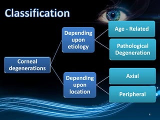
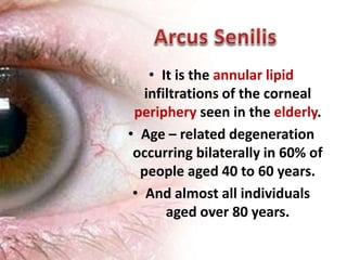
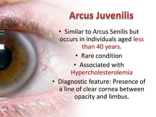
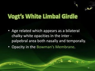
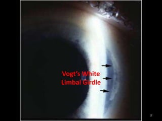
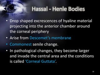
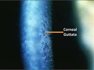
Apoorva Kottary 28
• Degenerative changes in the cornea.
• Definition: Corneal degeneration refers to the
conditions in which normal cells undergo
some degenerative changes under the
influence of age or some pathological
condition.
2
As being:
• Non - Hereditary and Non - Familial
• Usually Unilateral
• Mostly Peripheral
• More frequently seen
• Vascularity and Inflammation is seen.
3
4
Corneal
degenerations
Depending
upon
etiology
Age - Related
Pathological
Degeneration
Depending
upon
location
Axial
Peripheral
• Depending upon Location
I. Axial Corneal Degenerations
a) Fatty Degenerations
b) Hyaline Degenerations
c) Amyloidosis
d) Calcific Degenerations (Band
Keratopathy)
e) Salzmann’s Nodular Degeneration
5
II. Peripheral Degenerations
a) Arcus Senilis
b) Vogt’s White Limbal Girdle
c) Hassall – Henle Bodies
d) Terriens’s Marginal Degeneration
e) Mooren’s Ulcer
f) Pellucid Marginal Degeneration
g) Furrow Degeneration (Senile
Marginal Degeneration)
6
• Depending upon Etiology
I.Age Related Degenerations
a) Arcus Senilis
b) Vogts White Limbal Girdle
c) Hassal - Henle Bodies
d) Mosaic Degenerations
7
II. Pathological Degenerations:
a) Fatty Degeneration
b) Amyloidosis
c) Calcific Degenerations
d) Salzmann’s Nodular Degeneration
e) Terrien’s Marginal Degeneration
f) Mooren’s Ulcer
g) Pellucid Marginal Degeneration
h) Furrow Degenerations
i) Spheroidal Degeneration
8
9
Arcus Senilis
Vogt’s White Limbal Girdle
Hassal - Henle Bodies
• It is the annular lipid
infiltrations of the corneal
periphery seen in the elderly.
• Age – related degeneration
occurring bilaterally in 60% of
people aged 40 to 60 years.
• And almost all individuals
aged over 80 years.
10
• Commences as a crescentric grey or white arc
in the superior and inferior quadrant and
progresses to form a ring around the cornea,
• 1mm wide ring
• Lucid interval of Vogt’s – the clear zone which
separates the ring of opacity from the limbus.
• Peripheral border is sharp and inner border is
diffuse.
• Rarely double ring of Arcus is seen.
11
Arcus Senilis
Lucid Interval
of Vogt’s
12
• It is not of importance, as it does not decrease
vision or the vitality of the cornea.
• Unrelated to metabolic conditions such as
hypercholesterolemia.
13
• Similar to Arcus Senilis but
occurs in individuals aged less
than 40 years.
• Rare condition
• Associated with
Hypercholesterolemia
• Diagnostic feature: Presence of
a line of clear cornea between
opacity and limbus.
14
• Age related which appears as a bilateral
chalky white opacities in the inter -
palpebral area both nasally and temporally.
• Opacity in the Bowman's Membrane.
15
16
Vogt’s White
Limbal Girdle
17
Vogt’s White
Limbal Girdle
Hassal - Henle Bodies
• Drop shaped excrescences of hyaline material
projecting into the anterior chamber around
the corneal periphery
• Arise from Descemet’s membrane
• Commonest senile change.
• In pathological changes, they become larger
and invade the central area and the conditions
is called ‘Corneal Guttata’.
18
19
Corneal
Guttata
References
1. Parson’s Diseases of the Eye
2. Comprehensive Ophthalmology – A.K. Khurana
3. Pictures Courtesy – Online Journals
20
Thank you
21

Corneal Degeneration's, Ophthalmology

  • 1.
  • 2.
    • Degenerative changesin the cornea. • Definition: Corneal degeneration refers to the conditions in which normal cells undergo some degenerative changes under the influence of age or some pathological condition. 2
  • 3.
    As being: • Non- Hereditary and Non - Familial • Usually Unilateral • Mostly Peripheral • More frequently seen • Vascularity and Inflammation is seen. 3
  • 4.
  • 5.
    • Depending uponLocation I. Axial Corneal Degenerations a) Fatty Degenerations b) Hyaline Degenerations c) Amyloidosis d) Calcific Degenerations (Band Keratopathy) e) Salzmann’s Nodular Degeneration 5
  • 6.
    II. Peripheral Degenerations a)Arcus Senilis b) Vogt’s White Limbal Girdle c) Hassall – Henle Bodies d) Terriens’s Marginal Degeneration e) Mooren’s Ulcer f) Pellucid Marginal Degeneration g) Furrow Degeneration (Senile Marginal Degeneration) 6
  • 7.
    • Depending uponEtiology I.Age Related Degenerations a) Arcus Senilis b) Vogts White Limbal Girdle c) Hassal - Henle Bodies d) Mosaic Degenerations 7
  • 8.
    II. Pathological Degenerations: a)Fatty Degeneration b) Amyloidosis c) Calcific Degenerations d) Salzmann’s Nodular Degeneration e) Terrien’s Marginal Degeneration f) Mooren’s Ulcer g) Pellucid Marginal Degeneration h) Furrow Degenerations i) Spheroidal Degeneration 8
  • 9.
    9 Arcus Senilis Vogt’s WhiteLimbal Girdle Hassal - Henle Bodies
  • 10.
    • It isthe annular lipid infiltrations of the corneal periphery seen in the elderly. • Age – related degeneration occurring bilaterally in 60% of people aged 40 to 60 years. • And almost all individuals aged over 80 years. 10
  • 11.
    • Commences asa crescentric grey or white arc in the superior and inferior quadrant and progresses to form a ring around the cornea, • 1mm wide ring • Lucid interval of Vogt’s – the clear zone which separates the ring of opacity from the limbus. • Peripheral border is sharp and inner border is diffuse. • Rarely double ring of Arcus is seen. 11
  • 12.
  • 13.
    • It isnot of importance, as it does not decrease vision or the vitality of the cornea. • Unrelated to metabolic conditions such as hypercholesterolemia. 13
  • 14.
    • Similar toArcus Senilis but occurs in individuals aged less than 40 years. • Rare condition • Associated with Hypercholesterolemia • Diagnostic feature: Presence of a line of clear cornea between opacity and limbus. 14
  • 15.
    • Age relatedwhich appears as a bilateral chalky white opacities in the inter - palpebral area both nasally and temporally. • Opacity in the Bowman's Membrane. 15
  • 16.
  • 17.
  • 18.
    Hassal - HenleBodies • Drop shaped excrescences of hyaline material projecting into the anterior chamber around the corneal periphery • Arise from Descemet’s membrane • Commonest senile change. • In pathological changes, they become larger and invade the central area and the conditions is called ‘Corneal Guttata’. 18
  • 19.
  • 20.
    References 1. Parson’s Diseasesof the Eye 2. Comprehensive Ophthalmology – A.K. Khurana 3. Pictures Courtesy – Online Journals 20
  • 21.