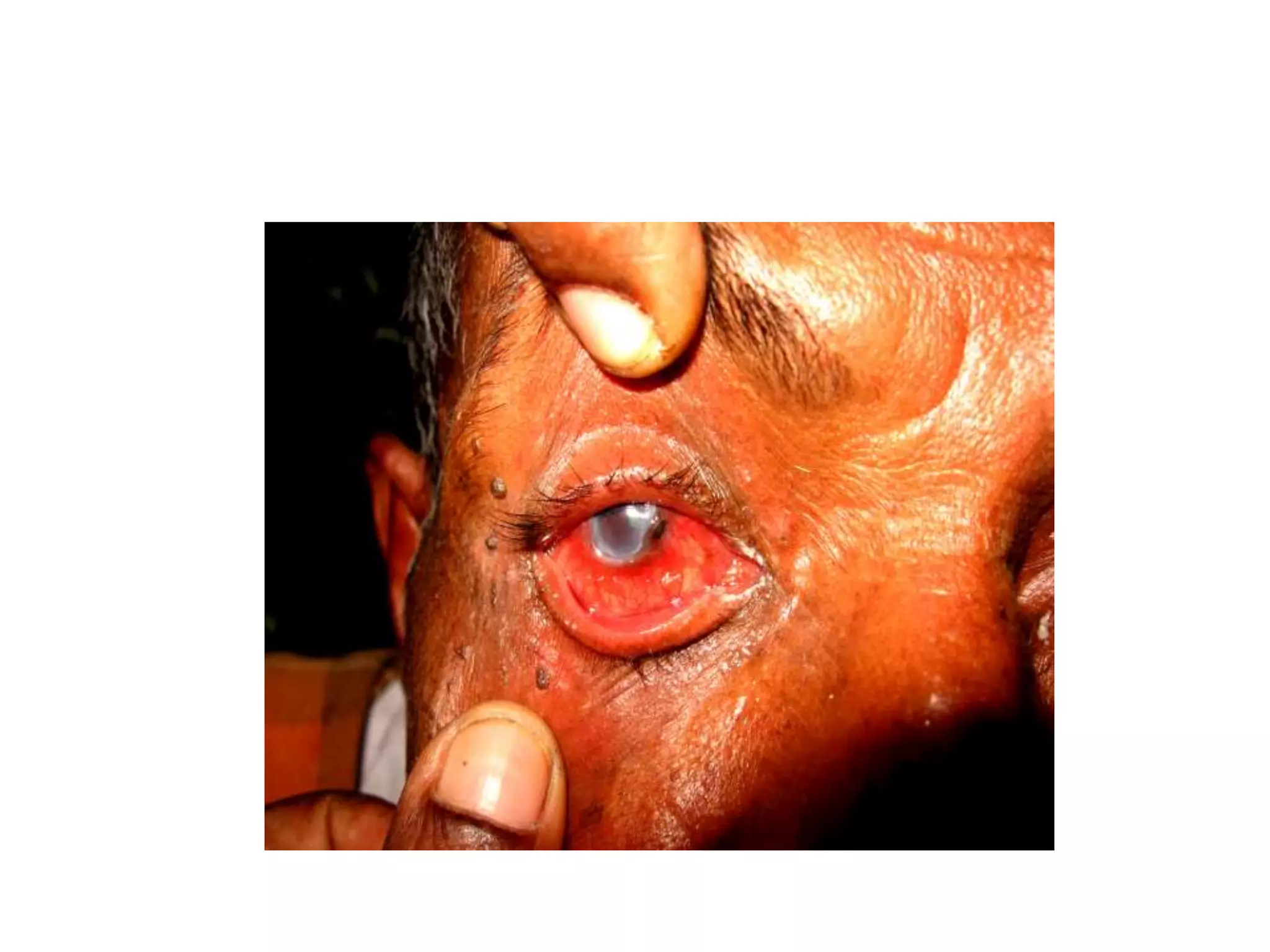
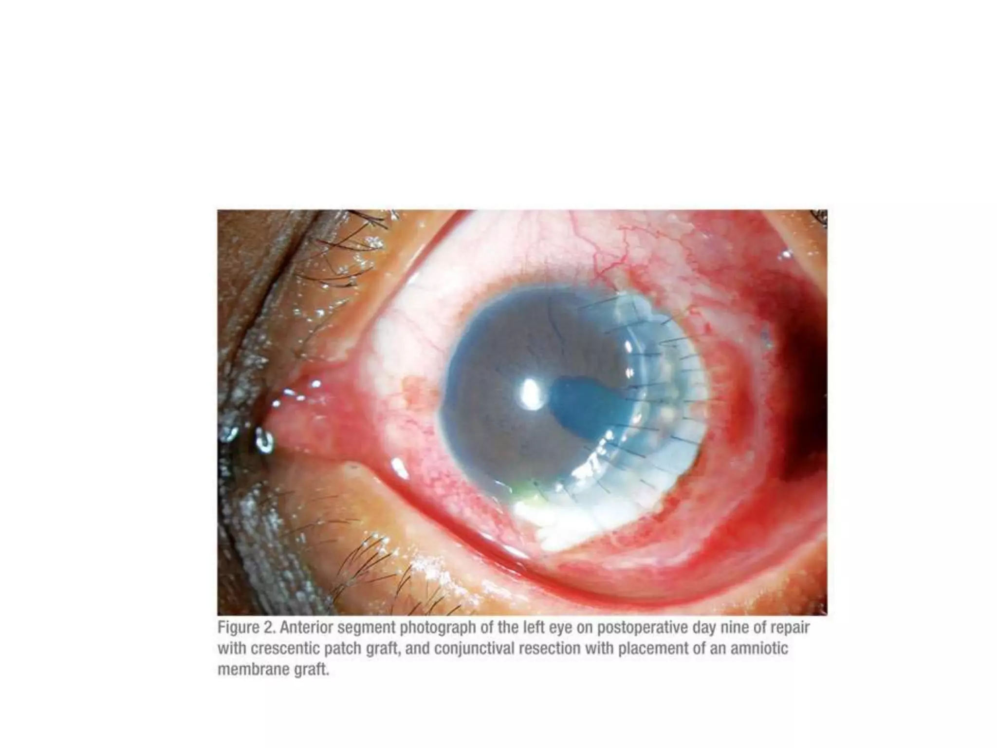
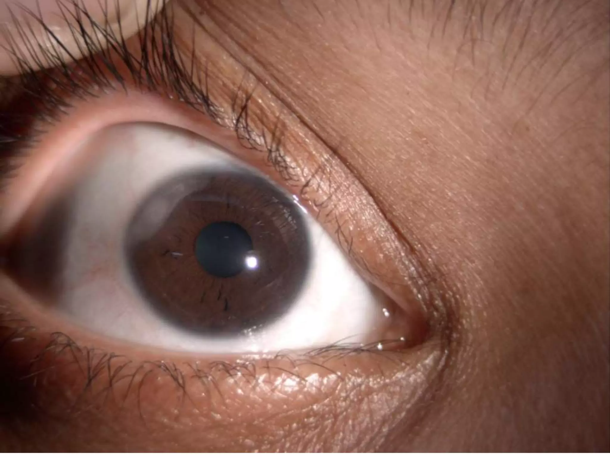
PERIPHERAL ULCERATIVE
KERATITIS
DR. K. VASANTHA M.S.,F.R.C.S. Ed
Director RIO Chennai (Rtd)
Definition
• Crescent shaped destructive inflammation of
the juxta limbal corneal stroma
• associated with epithelial defect,
• stromal inflammatory cells
• and stromal degradation
• Usually conjunctiva, episclera and sclera are
also involved.
• Biopsy will show micro angiopathy.
• Antibodies to corneal epithelium is also found.
• In ANCA +ve vasculitides cell mediated
cytotoxicity is also found
DIFFERENCIAL DIAGNOSIS
• Peripheral degenerations like
• Arcus, White limbal girdle of Vogt
• Idiopathic furrow degeneration – thinning in
the area of arcus
• Marginal keratitis
D.D
• Post irradiation esp. Beta radiation
• Terrien’s marginal degeneration
• Bilateral, young males, superior, stromal
thinning, ectasia, lucid interval between the
lesion and the limbus.
• Yellow lipid border at the advancing edge.
CLINICAL ASSOCIATIONS
AUTOIMMUNE
DISORDERS
RA, PAN,
WG,SLE,
Sjogrens dis
Behcets dis,
relapsing
polychondritis
SYSTEMIC
INFECTIONS
Hepatitis C
Syphilis
salmonella
OCULAR
CONDITIONS
Herpes simplex
Fungi
varicella zoster
acanthamoeba
Why limbus is involved?
• Limbal arcades
• Inflamed vessels draw inflammatory cells
which cause destruction
• Tight collagen bundles in the periphery
facilitates collection of IgM, C1 and other high
molecular weight molecules
• Immune complex deposits at the limbus and
corneal periphery
Why?
• Presence of Langerhans cells in the conj., and
corneal periphery
• Limbal macrophages (antigen presenting cells)
will be up regulated when there is inf., and
immune mediated events occur
• Channels from sub conj lymphatics enter the
periphery of the cornea- afferent arm
• Limbus is a reservoir of effector cells of
immune sys., cytokines
RHEUMATOID ARTHRITIS
• Autoimmune
• Typical joint deformities
• Ocular surface disorders more common
• Corneal melting can occur
• Following surgery PUK more common
Systemic Lupus Erythymatosis
• Dysfunction of immune regulation by T cells
and B cells
• Ocular surface disorder is more common
Wegener’s
• Necrotizing granulomatous vasculitis
• Bilateral PUK
• Lead on to ring ulcer
• Scleral inflammation is severe, may lead on to
necrosis
• Steroids not effective
Relapsing poly chondritis
• Cartilages involved
• Auditory impairment, tinnitus and vertigo
• Other collagen vascular disorders may co exist
• Ring ulcer with scleritis, bilateral – rare
• Lid edema, iritis, retinopathy, optic neuritis
• Cornea has glycosamino glycans- same as
sclera, aortic wall, cartilage and inner ear
Poly arteritis nodosa
• Non granumatous necrotizing vasculitis both
small and medium
• URI, drug abuse, hepatitis b and c
• Pseudo tumor, papilledema, papillitis, muscle
involvement
• Mooren’s like PUK
• Interferon alfa 2b. Steroids alone not effective,
cyclophosmamide to be added
MICRO POLYANGIITIS
• ANCA against myeloperoxidase
• Mooren’s like PUK, often bilateral with scleral
involvement
• Uveitis, ischemic optic neuropathy, muscle
palsies
MOOREN’S ULCER
• First described by Bowman in 1949
• Mooren published a paper later
• Type iii hypersensitivity ?
• Diagnosis by exclusion
• PUK may be the presenting feature of collagen
vascular diseases
• Primary – classic, idiopathic
• Secondary – trauma, surgical, chemical injury,
Herpes, syphilis, tuberculosis, Hepatitis C,
helminthiasis both ascariasis and ancylostoma
• Fulminating,
• centrally progressive,
• painful
• more common in males
• Under mining edge
• Sclera not usually involved
• Absence of any systemic disorder
Benign
• Older patients
• Unilateral
• Responds to steroids
Severe
• Younger
• Bilateral
• Progressive
• Sclera and episclera may be involved
Histology
• Necrosis of collagen tissue
• Chronic inflammatory cells
• Vascularisation of limbal margin
• Auto anti bodies to epithelium
• Polymorphs which are responsible for
collagenolysis
TREATMENT
• Steroids every hour
• Conjunctival excision- not just peritomy
• This helps by removing the causative factors
• Tissue adhesives – excludes neutrophils from
reaching the involved area
• Tetra cyclines to prevent collagenolysis
Immuno suppression
• Methotrexate 7.5 to 15 mg/week
• Azathioprine 2 mg/kg/day
• Cyclophosphamide 2 mg/kg/day
• The above three must be given for 6 months
and then tapered
• For Mooren’s topical cyclosporine can be used
• Before cataract surgery immune suppression
surgical
• Amniotic membrane graft
• Tectonic kp to prevent the immune complexes
from reaching the cornea
• Palliative, temporizing, inadequate to prevent
recurrences as immunologic process continues
THANK YOU

PERIPHERAL ULCERATIVE KERATITIS

  • 1.
    PERIPHERAL ULCERATIVE KERATITIS DR. K.VASANTHA M.S.,F.R.C.S. Ed Director RIO Chennai (Rtd)
  • 2.
    Definition • Crescent shapeddestructive inflammation of the juxta limbal corneal stroma • associated with epithelial defect, • stromal inflammatory cells • and stromal degradation
  • 3.
    • Usually conjunctiva,episclera and sclera are also involved. • Biopsy will show micro angiopathy. • Antibodies to corneal epithelium is also found. • In ANCA +ve vasculitides cell mediated cytotoxicity is also found
  • 4.
    DIFFERENCIAL DIAGNOSIS • Peripheraldegenerations like • Arcus, White limbal girdle of Vogt • Idiopathic furrow degeneration – thinning in the area of arcus • Marginal keratitis
  • 5.
    D.D • Post irradiationesp. Beta radiation • Terrien’s marginal degeneration • Bilateral, young males, superior, stromal thinning, ectasia, lucid interval between the lesion and the limbus. • Yellow lipid border at the advancing edge.
  • 6.
    CLINICAL ASSOCIATIONS AUTOIMMUNE DISORDERS RA, PAN, WG,SLE, Sjogrensdis Behcets dis, relapsing polychondritis SYSTEMIC INFECTIONS Hepatitis C Syphilis salmonella OCULAR CONDITIONS Herpes simplex Fungi varicella zoster acanthamoeba
  • 8.
    Why limbus isinvolved? • Limbal arcades • Inflamed vessels draw inflammatory cells which cause destruction • Tight collagen bundles in the periphery facilitates collection of IgM, C1 and other high molecular weight molecules • Immune complex deposits at the limbus and corneal periphery
  • 9.
    Why? • Presence ofLangerhans cells in the conj., and corneal periphery • Limbal macrophages (antigen presenting cells) will be up regulated when there is inf., and immune mediated events occur • Channels from sub conj lymphatics enter the periphery of the cornea- afferent arm • Limbus is a reservoir of effector cells of immune sys., cytokines
  • 10.
    RHEUMATOID ARTHRITIS • Autoimmune •Typical joint deformities • Ocular surface disorders more common • Corneal melting can occur • Following surgery PUK more common
  • 11.
    Systemic Lupus Erythymatosis •Dysfunction of immune regulation by T cells and B cells • Ocular surface disorder is more common
  • 12.
    Wegener’s • Necrotizing granulomatousvasculitis • Bilateral PUK • Lead on to ring ulcer • Scleral inflammation is severe, may lead on to necrosis • Steroids not effective
  • 13.
    Relapsing poly chondritis •Cartilages involved • Auditory impairment, tinnitus and vertigo • Other collagen vascular disorders may co exist • Ring ulcer with scleritis, bilateral – rare • Lid edema, iritis, retinopathy, optic neuritis • Cornea has glycosamino glycans- same as sclera, aortic wall, cartilage and inner ear
  • 14.
    Poly arteritis nodosa •Non granumatous necrotizing vasculitis both small and medium • URI, drug abuse, hepatitis b and c • Pseudo tumor, papilledema, papillitis, muscle involvement • Mooren’s like PUK • Interferon alfa 2b. Steroids alone not effective, cyclophosmamide to be added
  • 15.
    MICRO POLYANGIITIS • ANCAagainst myeloperoxidase • Mooren’s like PUK, often bilateral with scleral involvement • Uveitis, ischemic optic neuropathy, muscle palsies
  • 16.
    MOOREN’S ULCER • Firstdescribed by Bowman in 1949 • Mooren published a paper later • Type iii hypersensitivity ? • Diagnosis by exclusion • PUK may be the presenting feature of collagen vascular diseases
  • 17.
    • Primary –classic, idiopathic • Secondary – trauma, surgical, chemical injury, Herpes, syphilis, tuberculosis, Hepatitis C, helminthiasis both ascariasis and ancylostoma
  • 18.
    • Fulminating, • centrallyprogressive, • painful • more common in males • Under mining edge • Sclera not usually involved • Absence of any systemic disorder
  • 19.
    Benign • Older patients •Unilateral • Responds to steroids
  • 21.
    Severe • Younger • Bilateral •Progressive • Sclera and episclera may be involved
  • 22.
    Histology • Necrosis ofcollagen tissue • Chronic inflammatory cells • Vascularisation of limbal margin • Auto anti bodies to epithelium • Polymorphs which are responsible for collagenolysis
  • 23.
    TREATMENT • Steroids everyhour • Conjunctival excision- not just peritomy • This helps by removing the causative factors • Tissue adhesives – excludes neutrophils from reaching the involved area • Tetra cyclines to prevent collagenolysis
  • 24.
    Immuno suppression • Methotrexate7.5 to 15 mg/week • Azathioprine 2 mg/kg/day • Cyclophosphamide 2 mg/kg/day • The above three must be given for 6 months and then tapered • For Mooren’s topical cyclosporine can be used • Before cataract surgery immune suppression
  • 25.
    surgical • Amniotic membranegraft • Tectonic kp to prevent the immune complexes from reaching the cornea • Palliative, temporizing, inadequate to prevent recurrences as immunologic process continues
  • 29.