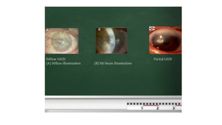
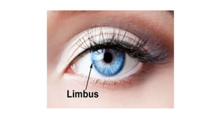
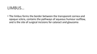
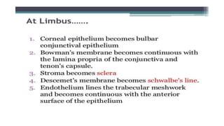
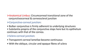
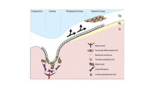
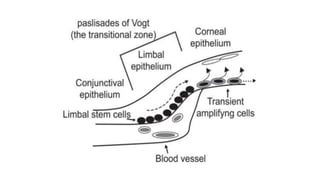
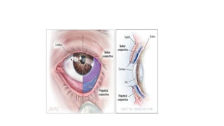
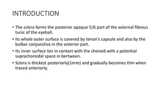
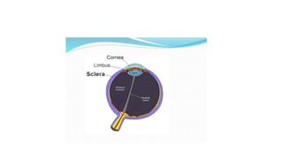
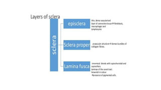
LIMBUS… • The limbus forms the border between the transparent cornea and opaque sclera, contains the pathways of aqueous humour outflow, and is the site of surgical incisions for cataract and glaucoma
2. Anatomical Limbus: Circumcorneal transitional zone of the conjunctivocorneal & corneoscleral junction Conjunctivo-corneal junction: • Bulbar conjunctiva is firmly adherent to underlying structures • Substantia propria of the conjunctiva stops here but its epithelium continues with that of the cornea. Sclero-corneal junction: • Transparent corneal lamellae become continuous • With the oblique, circular and opaque fibres of sclera
3. CONTINUE…. • In the area near limbus, the conjunctiva, tenon’s capsule & the episcleral tissue are fused into a dense tissue which is strongly adherent to corneo scleral junction.It is preferred site for obtaining a firm hold of the eyeball during ocular surgery. • The limbus is a common site for the occurrence of corneal epithelial neoplasm. • The Limbus contains radially oriented fibrovascular ridge known as the palisades of Vogt that may harbour a stem cell population. The palisades of Vogt are more common in the superior and inferior quadrants around the eye
Accommodation/ Accommodation of Eye, Measurement of Accommodation of Eye (hea...Bikash Sapkota
CLICK HERE TO DOWNLOAD FULL PPT ❤❤ https://healthkura.com/measurement-of-accommodation-of-eye/ ❤❤
Dear viewers Check Out my other piece of works at ❤❤❤ https://healthkura.com ❤❤❤
Measurement of Accommodation of eye:
Amplitude, Facility,
Relative Accommodation, Fatigue, Lag,
Dynamic Retinoscopy
Presentation Layout:
-Introduction to accommodation of eye
-Mechanism
-Components
-Measurement of accommodation of eye
- Amplitude
- Facility
- Relative accommodation
- Lag
-Dynamic Retinoscopy
Accommodation
-dioptric adjustment of the crystalline lens of the eye
- to obtain clear vision for a given target of regard
-process by which the refractive power of eye is altered
- to ensure a clear retinal image
For further reading
-Clinical Procedures in Optometry by J.D. Bartlett, J.B. Eskridge, J.F. Amos
-Primary Care Optometry by Theodere Grosvenor
-Borish’s Clinical Refraction by W.J. Benjamin
-Clinical Procedures for Ocular examination by Carlson et al
-American Academy of Ophthalmology
-Optometric Clinical Practice Guideline by American Optometric Association
-Internet
Follow me to get in touch with optometric and ophthalmic updates
Cornea is the clear front surface of the eye. It lies directly in front of the iris and pupil, and it allows light to enter the eye.
Cornea forms the transparent and anterior 1/6th of the external fibrous coat of the globe of the eyeball.
The cornea is the eye's most powerful structure for focusing light that provides approximately 65 to 75 percent of the focusing power of the eye.
The cornea has unmyelinated nerve endings sensitive to touch, temperature and chemicals; a touch of the cornea causes an involuntary reflex to close the eyelid.
This lecture includes anatomy, Physiology of Sclera, if u like it kindly share it with colleagues and like it. I will share more lectures related to eye anatomy and optometry.
Thank You.
This presentation gives a brief idea about angle of anterior chamber along with its structures and diagnostic methods to grade and visualize the structures.
1. Introduction Gross anatomy Layers Blood supply, drainage and nerve supply
2. INTRODUCTION • Sclera forms posterior 5/6th of external tunic , connective tissue coat of eyeball. • it continues with duramater and cornea • Its whole surface covered by tenon’s capsule • Anteriorly covered by- bulbar conjunctiva • Inner surface lies in contact with choroid • With a potential suprachoroidal space in between
3. Equa THICKNESS OF SCLERA
4. • Thickness varies with individual, with age • Thinner- children, elder, F> M • Thickest posteriorly • Gradually becomes thinner when traced anteriorly • Thin at insertion of extraocular muscle
astigmatism
AstigmatismAstigmatism Walter Huang, ODWalter Huang, OD Yuanpei UniversityYuanpei University Department of OptometryDepartment of Optometry
2. DefinitionDefinition When parallel rays of light enter the eyeWhen parallel rays of light enter the eye ((with accommodation relaxedwith accommodation relaxed) and do) and do notnot come to a single point focus on or nearcome to a single point focus on or near the retinathe retina
3. OpticsOptics Power in thePower in the horizontalhorizontal plane projects aplane projects a verticalvertical focal line imagefocal line image Power in thePower in the verticalvertical plane projects aplane projects a horizontalhorizontal focal line imagefocal line image
4. OpticsOptics Refraction of light taking place at a toricRefraction of light taking place at a toric surface: the conoid of Sturmsurface: the conoid of Sturm
TONOMETRY • Tonometry is the procedure performed to determine the intraocular pressure (IOP).
3. CLASSIFICATION TONOMETRY DIRECT INDIRECT Indentation Applanation Manometer
4. APPLANATION Contact Non-contact Goldmann Perkins Air-puff Pulse air
5. INDENTATION TONOMETER • It is based on fundamental fact that plunger will indent a soft eye more than hard eye. • The indentation tonometer in current use is that of Schiotz . • It was devised in 1905 and continued to refine it through 1927.
6. PROCEDURE • Patient should be anaesthetising with 4% lignocaine or 0.5% proparacaine. • with the patient in supine position, looking up at a fixation target while examiners separates the lids and lower the tonometer plate to rest on the cornea so that plunger is free to move. •
. Introduction Biomicroscope derives its name from the fact that it enables the practitioner to observe the living tissue of eye under magnification. It not only provides magnified view of every part of eye but also allows quantitative measurements and photography of every part for documentation.
3. • The lamp facilitates an examination which looks at anterior segment, or frontal structures, of the human eye, which includes the –Eyelid –Cornea –Sclera –Conjunctiva –Iris –Aqueous –Natural crystalline lens and –Anterior vitreous.
4. Important historical landmarks De Wecker 1863 devised a portable ophthalmomicroscope . Albert and Greenough 1891,developed a binocular microscope which provided stereoscopic view. Gullstrand ,1911 introduced the illumination system which had for the first time a slit diapharm in it Therefore Gullstrand is credited with the invention of slit lamp.
The pupil is an opening located in the center of the iris that allows light to enter the retina. • Its function is to control the amount of light entering the eye and it does this via contraction (miosis) and dilation (mydriasis) under the influence of the autonomic nervous system
3. • The iris is a contractile structure, consisting mainly of smooth muscle, surrounding the pupil. Light enters the eye through the pupil, and the iris regulates the amount of light by controlling the size of the pupil.
4. The iris contains two groups of smooth muscles: a circular group called the sphincter pupillae. and a radial group called the dilator pupillae.
5. Parasympathetic pathway • First Order – Retina to Pretectal Nucleus in B/S (at level of Superior colliculus) Second Order – Pretectal nucleus to E/W nucleus (bilateral innervation!) Third Order – E/W nucleus to Ciliary Ganglion Fourth Order – Ciliary Ganglion to Sphincter pupillae (via short ciliary nerves) • • •
The tear film constitutes Three layers :- An outermost lipid (oily) layer An aqueous (watery) layer that makes up 90% of the tear film volume; and A mucin layer that coats the corneal surface.
3. To form smooth optical surface on cornea. To keep the surface of cornea & conjunctiva moist It serve as lubricant It transfer oxygen Provide antibacterial action Wash debris out It provides a pathway for WBC in case of injury
4. Functions of lipid layer Retards evaporation of tear film Prevents the overflow of tears
5. Function of Aqueous Layer Flushes, buffers and lubricates the corneal surface Delivers oxygen and other nutrients to the corneal surface Wash out debris Delivers antibacterial enzymes and antibodies such as lysozyme.
6. Functions of Mucin Layer Spreads tears over corneal surface. Protects the cornea against foreign substances . Makes corneal surface smooth by filling in surface irregularities
Introduction Transparent,avascular,watch-glass like structure. Forms 1/6th part of outer fibrous coat (Sclera) It is the major refracting surface of the eye
3. Dimensions + Avg horizontal dia =11.75 mm (ant surface) + Avg vertical dia = 11 mm (ant surface ) + Avg dia (post surface)= 11.5 mm + Thickness(centre) =0.52mm + Thickness(peripheral) = 0.67mm + Radius of curvature (ant surface) = 7.8mm + Radius of curvature (post surface)= 6.5mm + Refractive power (ant surface) = +48D + Refractive power(post surface)= - 5D + R.I = 1.376
4. Histology + Epithelium + Bowman’s membrane + Stroma + Dua’s layer + Descemet’s membrane + Endothelium
5. Epithelium + Made up of stratified squamous epithelium + Thickness - 50-90 um + 5-6 layers of cells + Regenerative, entire epithelial layer is replaced every 6-8 days + Made up of 3 types of cells - basal,wing, flattened cells + Cells are attached by to each other by means of desmosomes & maculi occludents
6. Bowman’s membrane + Acellular,Non regenerative + Made up of condensed collagen fibrils. + Thickness - 12um + Resistant to infection & injury.
Diagnosis, Management, and Surgery by Adam J. Cohen, Michael Mercandetti & Brian G. Brazzo. The dry eye , a practical approach by Sudi Patel & Kenny J Blades. Jack J Kanski’s clinical ophthalmology Clinical Anatomy of the Eye by Richard S. Snell & Michael A. Lemp.
3. It is concerned with the tear formation & transport. Lacrimal passage includes : Lacrimal gland Conjunctival sac Lacrimal puncta Lacrimal canaliculi Lacrimal sac Nasolacrimal duct
4. The following components of the lacrimal apparatus are discussed : Embryology Osteology Secretory system Excretory system Physiology
5. Ectodermal origin Solid epithelial buds(first 2 months) Supero
Extraocular musles(EOM) They are six in number Four recti: Superior rectus Inferior rectus Medial rectus Lateral rectus Two oblique muscles: Superior oblique Inferior oblique
3. SUPERIOR RECTUS MUSCLE . Origin Superior part of common annular tendon of Zinn Course Passes anterolaterally beneath the levator At 23 degrees with the globe ‘s AP axis Pierces Tenon s capsule Insertion into sclera by flat tendinous 10 mm broad insertion 7.7 mm behind sclero-corneal junction. 42 mm long 9 mm wide
4. Nerve supply Sup division of 3rd N Blood Supply Lateral Muscular br. of Ophthalmic A APPLIED: SR loosely bound to LPS muscle. • During SR resection- eyelid may be pulled forward narr owing palpebral fissure • In hypotropia pseudoptosis may be present Origin of SR and MR are closely attached to the dural sheat h of the optic nerve pain during upward & inward movements of the globe in RETROBULBAR NEURITIS
Diseases of sclera
2. anatomy • Sclera posterior 5/6th opaque part of the external fibrous tunic of the eyeball.
3. • outer surface }covered by Tenon's capsule. • anterior part } covered by bulbar conjunctiva.
4. Its inner surface lies in contact with choroid with a potential suprachoroidal space in between
5. Thickness of sclera. • thinner }children and in females Sclera • thickest} posteriorly (1mm) • gradually becomes thin when traced anteriorly. • thinnest } insertion of extraocular muscles (0.3 mm). • Lamina cribrosa is a sieve-like sclera from which fibres of optic nerve pass.
6. Apertures of sclera • Anterior • Anterior ciliary vessels • Middle • four vortex veins (vena verticosae) • Posterior • Optic nerve • Long & short ciliary nerves
7. Layers of sclera sclera episclera Sclera proper Lamina fusca thin, dense vascularised layer of connective tissue fibroblasts, macrophages and lymphocytes avascular structure dense bundles of collagen fibres. innermost blends with suprachoroidal and supraciliary laminae of the uveal tract. brownish in colour presence of pigmented cells.
Main physiologic function of cornea is to act as a major refracting medium, so that a clear retinal image is formed. • Normal corneal transparency is result of • 1.anatomical factor such as uniform and regular arrangement of corneal epithelium, peculiar arrangement of corneal lamella and corneal vascularity 2.Physiological factor [ie] relative state of corneal dehydration.
3. • Therefore, any process which upsets the anatomy or physiology of cornea will cause LOSS OF TRANSPARENCY to some degree.
4. FACTORS AFFECTING CORNEAL TRANSPARENCY • CORNEAL EPITHELIUM &TEAR FLIM • ARRANGEMENT OF STROMAL LAMELLA • CORNEAL VASCULARIZATION • CORNEAL HYDRATION • CELLULAR FACTORS AFFECTING TRANSPARENCY
CONJUNCTIVA: ANATOMY , PHYSIOLOGY, SYMPTOMATOLOGY AND CLASSIFICATION Pranay Shinde DNB Resident Deen Dayal Upadhyay Hospital,New Delhi
2. ANATOMY It is the mucous membrane covering the under surface of the lids and anterior part of the eyeball upto the cornea.
3. Parts of conjunctiva • Palpebral; covering the lids—firmly adherent. • Forniceal; covering the fornices—loose—thrown into folds. • Bulbar; covering the eyeball—loosely attached except at limbus.
4. Palpebral conjunctiva • Subtarsal sulcus 2mm from posterior edge of the lid margin. • Richly vascular. • Extremely thin. • Strongly bound to the tarsal plate.
5. Palpebral conjunctiva is subdivided into three parts: 1)Marginal 2)Tarsal 3)Orbital
6. Conjunctival fornices • Transitional region between palpebral and bulbar conjunctivae. • Superior fornix 10 mm from limbus. • Inferior fornix 8 mm from limbus. • Lateral fornix 14mm from limbus. • Medially absent. • Ducts of lacrimal glands open into lateral part of superior fornix.
q Colour Vision Deficiency Presented by : Optometrist (intern) Asma Al-Jroudi Saudi Arabia, Riyadh, King Abdulaziz University Hospital 30 Dec 14
2. • What Is Color Vision Deficiency? • Causes Of Color Vision Deficiency • Types Of Color Vision Deficiency • Tretments Of Color Vision Deficiency • Ishihara’s Test • Conclousion
3. What is Colour Blindness? • Color blindness, or color vision deficiency, is the inability or decreased ability to see color, or perceive color differences, under normal lighting conditions. •This condition results from an absence of color- sensitive pigment in the cone cells of the retina, the nerve layer at the back of the eye.
4. What is Colour Blindness? • Cones are the coulored light receptors in back of the eye: Red light receptors, Blue light receptors and Green light receptors. • Colour blindness occurs when one or more of the cone types are defected.
5. Causes of Color Blindness • Genetic: Many more men are affected than women. • Acquired : Chronic illness, Accidents, Medications and Age.
ANATOMY & PHYSIOLOGY Lecturer: Tatyana V. Ryazantseva
2. Outer eye: Eyelids The eyelids fulfill two main functions: protection of the eyeball secretion, distribution and drainage of tears
3. Lid movement The levator extends from an attachment at the orbital apex to attachments at the tarsal plate and skin. ● The lids are securely attached at either end to the bony orbital margin by the medial and lateral palpebral ligaments. Trauma to the medial ligament causes the lid to flop forward and laterally, impairing function and cosmesis.
4. Innervation - Sensory innervation is from the trigeminal (fifth) cranial nerve, via the ophthalmic division (upper lid) and maxillary division (lower lid). - The orbicularis oculi is innervated by the facial (seventh) nerve. - The levator muscle in the upper lid is supplied by the oculomotor (third) nerve.
5. Blood supply and lymphatics The eyelids are supplied by an extensive network of blood vessels which form an anastomosis between branches derived from the external carotid artery via the face and from the internal carotid artery via the orbit.
6. Blood supply and lymphatics Lymphatic fluid drains into the preauricular and submandibular nodes. Preauricular lymphadenopathy is a useful sign of infective eyelid swelling (especially viral).
Anatomy and Physiology of Aqueous Humor Sumit Singh Maharjan
2. Anatomy
3. Angle of anterior chamber
4. Angle of the Anterior chamber
5. Gonioscopic grading of Angle
6. Aqueous Outflow system
7. Trabecular meshwork
8. Functions of Aqueous Humor • Maintenance of Intraocular pressure • Metabolic role cornea lens vitreous and retina • Optical function • Clearing function
9. Physicochemical properties • volume: 0.31ml (0.25ml in Ant. Chamber and 0.06 in post chamber) • Refractive index: 1.336 • Density: slightly greater than water, its viscocity is 1.025-1.040 • Osmotic pressure: slightly hyperosmotic to plasma by 3-5mosm/l • PH: 7.2 • Rate of formation: 2-2.5microliter/min
10. Biochemical composition • Water: 99.9% • Proteins: 5-16mg/100ml • Amino acids: aqueous/plasma concentration varies from 0.08-3.14 • Non colloidal constituents: conc. of ascorbate, pyruvate, lactate in higher am
Vitreous humour
1. Vitreous Humour
2. General features Vitreous humour is an inert ,transparent , colourless, jellylike, hydrophilic gel that serves the optical functions and also acts as important supporting structures for the eyeball. The vitreous cavity is bounded by anteriorly by the lens and ciliary body and posteriorly by the retina Its weighs nearly 4g Vitreous is an extacellular material composed of approximately 99 per cent water
3. Structure The vitreous body is the largest and simplest connective tissue present as a single piece in the human body Divided into three parts- 1. The hyaloid layer or membrane 2. The cortical vitreous and 3. The medullary vitreous
Vitamins all
1. Vitamins. Definition - Organic compound required in small amounts. Vitamin A Vitamin B1, B2, B3, B5, B6, B7, B9, B12 Vitamin D Vitamin E Vitamin K A few wordsabout each.
2. Sourcesin diet - Many plants(photoreceptors), also meat, especially liver. Fat soluble, so you can get too much, or too littleif absorption isaproblem. Vitamin A - Retinol Retinol (vitamin A) Someuses: Vision (11-cis-retinol bound to rhodopsin detectslight in our eyes). Regulating genetranscription (retinoic acid receptorson cell nuclei arepart of a system for regulating transcription of mRNAsfor anumber of genes).
Tear film
1. TEAR FILM
2. The outer most layer of the cornea. It is the exposed part of the eyeball. FUNCTION It provide smooth optical surface It serves to keep the surface of cornea and conjunctiva moist. It serves as a lubricant for the preocular surface and lids It transfer oxygen from the air to the cornea Prevent infection due to the presence of antibacterial substance like lysozymes,and other protein. It wash away debris and irritants Provides pathway to WBC in case of injury.
3. LAYERS OF TEAR FILM It consist of three layers: 1.Lipid layer 2.Aqueous layer 3.Mucoid layer 1.LIPID LAYER
Synthesis
1. Synthesis of Fatty acids
2. Fatty acids are synthesized mainly by de novo synthetic pathway operating in the cytoplasm . It is called as extramitochondrial or cytoplasmic fatty acid synthase system. The major fatty acids synthesised de novo is palmitic acid, the 16C saturated fatty acids. The process occurs in liver ,adipose tissue ,kidney and mammary glands. Fatty acids synthase (FAS) complex : This system exists as multi- enzyme complex .This enzyme form a dimer with identical subunits. Each subunits of the complex is organised into 3 domains with 7 enzyme.
Retina
1. RETINA
2. Photoreceptor cells • These are the primary neurons in the visual pathway • Lie at the outer edge of the retina • The photoreceptor cells differentiate longitudinally into four major regions: 1. the inner segment containing the metabolic apparatus 2. the outer segment containing the visual pigment for the conversion of light into neuroelectrical energy 3.a region containing the cell nucleus 4. a synaptic
More from C L GUPTA EYE INSTITUTE MORADABAD UTTER PRADESH (20)
3. LIMBUS…
• The limbus forms the border between the transparent cornea and
opaque sclera, contains the pathways of aqueous humour outflow,
and is the site of surgical incisions for cataract and glaucoma
4.
5. Anatomical Limbus: Circumcorneal transitional zone of the
conjunctivocorneal & corneoscleral junction
Conjunctivo-corneal junction:
• Bulbar conjunctiva is firmly adherent to underlying structures
• Substantia propria of the conjunctiva stops here but its epithelium
continues with that of the cornea.
Sclero-corneal junction:
• Transparent corneal lamellae become continuous
• With the oblique, circular and opaque fibres of sclera
6.
7.
8.
9.
10.
11.
12. CONTINUE….
• In the area near limbus, the conjunctiva, tenon’s capsule & the
episcleral tissue are fused into a dense tissue which is strongly
adherent to corneo scleral junction.It is preferred site for obtaining a
firm hold of the eyeball during ocular surgery.
• The limbus is a common site for the occurrence of corneal epithelial
neoplasm.
• The Limbus contains radially oriented fibrovascular ridge known as
the palisades of Vogt that may harbour a stem cell population. The
palisades of Vogt are more common in the superior and inferior
quadrants around the eye
13. CONTINUE…
• Limbal epithelial stem cells reside in the basal layer of the epithelium,
which undulates at the limbus.
• Daughter transient amplifying cells (TACs) divide and migrate towards
the central cornea to replenish the epithelium, which rests on
Bowmans layer .
• The stroma of the limbal epithelial stem cell niche is populated with
fibroblasts and melanocytes and also has a blood supply.
14.
15.
16.
17. • LESC deficiency can occur as a result of primary or acquired insults.
Partial or full LESC deficiency leads to deleterious effects on corneal
wound healing and surface integrity .
• Deficiency can arise following injuries including chemical or thermal
burns and through diseases such as aniridia and Stevens Johnson
syndrome .
• As a result of LESC deficiency conjunctivalisation , neovascularization ,
chronic inflammation, recurrent erosions, ulceration and stromal
scarring can occur causing painful vision loss .
18. • Long term restoration of visual function requires renewal of the
corneal epithelium, through replacement of the stem cell population
has traditionally been achieved by grafting limbal auto- or allografts .
• Recently it has been demonstrated that other stem cell populations
including human embryonic stem cell and hair follicle stem cell can be
driven towards a corneal epithelial-like phenotype.
• These exciting data may lead to alternative therapeutic strategies in
the future for patients blinded by ocular surface disease cause by
failure of LESC function.
22. INTRODUCTION
• The sclera forms the posterior opaque 5/6 part of the external fibrous
tunic of the eyeball.
• Its whole outer surface is covered by tenon’s capsule and also by the
bulbar conjunctiva in the anterior part.
• Its inner surface lies in contact with the choroid with a potential
suprachoroidal space in bertween.
• Sclera is thickest posteriorly(1mm) and gradually becomes thin when
traced anteriorly.
23.
24. Special regions of sclera
Scleral sulcus
• It is an indentation(furrow) on the inner surface of the anterior most point
of the sclera near the limbus.
Sclera spur
• It is a circular flang of the anterior most part of the sclera which lie deep to
schlemm’s canal.
• It appears wedge-shaped in section.
Lamina cribrosa
• It is a sieve-like sclera from which the fibres of the optic nerve pass.
25. Scleral apertures(emissaria): Sclera has three sets of apertures
1. Posterior aperture : situated around the optic nerve.
2. Middle apertures: situated 4-7mm posterior to the equator.
3. Anterior aperture: situated 3-4mm away from the limbus
28. Episcleral tissue
• It is a thin, dense vascularised layer of the connective tissue which
covers the sclera proper.
• Anteriorly it becomes continuous with the tenon’s capsule
29.
30. Sclera proper
• It is an avascular structure which consist of dense bundles of
collagens fibres crossing each other in all direction.
• Variability in collagen fiber diameter, interlacing in bundles of
collagen, and relative deficiency in water-binding substances accounts
for the scleral dull-white color.
• This arrangement makes the sclera opaque in contrast to cornea.
• Mucopolysaccharides are present in the interfibrillar space of the
collagen fibre.
• Few fibroblasts are also present in this layer.
31. Lamina fusca
• It is the innermost part of sclera which blends with suprachoroidal
and supraciliary lamina of the uveal tract.
• It is brownish in colour owing to the presence of pigment cells.
32. Blood supply
• The episclera receives its blood supply from the anterior ciliary arteries,
anterior to the insertions of the rectus muscles and the long and short
posterior ciliary arteries.
Nerve Supply
• The sclera is supplied by the branches from the long ciliary nerves
anteriorly and short ciliary nerves behind the equator. Rich in nerve supply
so causes pain- inflammation, stretching due to oedema and movement of
eye
33. Function
• Protects intraocular components from trauma, light, and mechanical
displacement
• Withstands the considerable expansive force generated by the
intraocular pressure maintaining the shape of the globe
• Provides attachment sites for the extraocular muscles.
34. • Inflammations of sclera
1. Episcleritis (superficial)
2. Scleritis(deep)