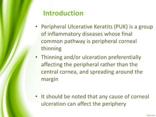
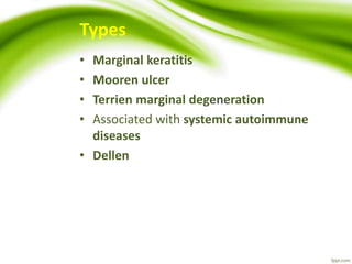
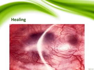
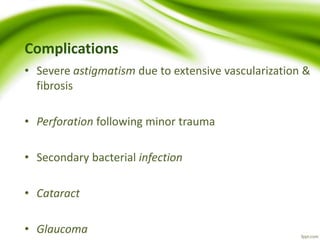
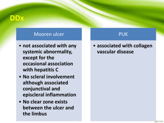
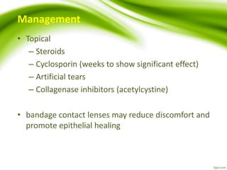
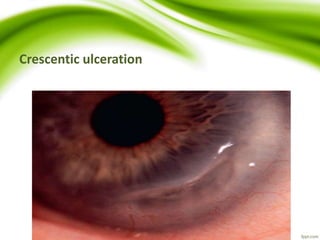
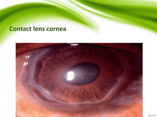
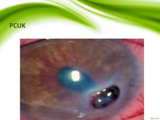
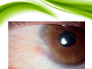
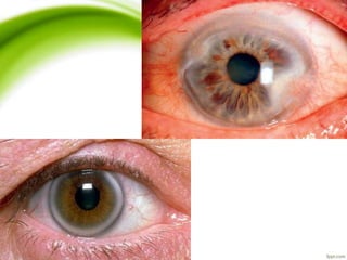
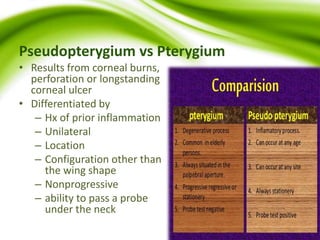
Peripheral Ulcerative Keratitis
Dr Md Ferdous Islam
Department of Ophthalmology
CMH,Dhaka
Introduction
• Peripheral Ulcerative Keratits (PUK) is a group
of inflammatory diseases whose final
common pathway is peripheral corneal
thinning
• Thinning and/or ulceration preferentially
affecting the peripheral rather than the
central cornea, and spreading around the
margin
• It should be noted that any cause of corneal
ulceration can affect the periphery
Types
• Marginal keratitis
• Mooren ulcer
• Terrien marginal degeneration
• Associated with systemic autoimmune
diseases
• Dellen
Mooren Ulcer
• Rare autoimmune disease
• Characterized by
– progressive,
– peripheral,
– circumferential,
– stromal corneal ulceration
– with later central spread
Forms unilateral
• Older patient
• Equal sex distribution
• Slowly progressive
• Responds well to
medical therapy
bilateral
• younger patients
• Indian males
• more aggressive
• likely to need
systemic
immunosuppression
• poorer prognosis
• associated with
severe pain
Symptoms and signs
 Peripheral ulceration
 involving the superficial one-third of the stroma,
 variable epithelial loss,
 Several distinct foci and subsequently coalesce
 Undermined and infiltrated leading edge is characteristic
 Limbitis may be present but no scleritis
 Progressive circumferential and central stromal thinning
 Vascularization involving the bed of the ulcer up to its leading edge but
not beyond
 The healing stage is characterized by thinning, vascularization and
scarring
Local peripheral ulceration
Undermining
Circumferential
Healing
Complications
• Severe astigmatism due to extensive vascularization &
fibrosis
• Perforation following minor trauma
• Secondary bacterial infection
• Cataract
• Glaucoma
DDx
Mooren ulcer
• not associated with any
systemic abnormality,
except for the
occasional association
with hepatitis C
• No scleral involvement
although associated
conjunctival and
episcleral inflammation
• No clear zone exists
between the ulcer and
the limbus
PUK
• associated with collagen
vascular disease
Management
• Topical
– Steroids
– Cyclosporin (weeks to show significant effect)
– Artificial tears
– Collagenase inhibitors (acetylcystine)
• bandage contact lenses may reduce discomfort and
promote epithelial healing
• Conjunctival resection
– may be combined with excision of necrotic tissue
– performed if there is no response to topical steroids
– The excised area should extend 4 mm back from the
limbus and 2 mm beyond the circumferential margins
– Keratoepithelioplasty (suturing of a donor corneal
lenticule onto the scleral bed) may be combined to
produce a physical barrier against conjunctival
regrowth and further melting
– Steroids are continued postoperatively
Systemic
• Immunosuppression should be instituted earlier for
– bilateral disease
– advanced involvement at presentation
• Systemic collagenase inhibitors such as doxycycline
• Lamellar keratectomy involving dissection of the
residual central island in advanced disease may
remove the stimulus for further inflammation
Peripheral Ulcerative Keratitis Associated With
Systemic Autoimmune Disease
• Destructive inflammation of the peripheral cornea
associated with corneal epithelial sloughing and
keratolysis
• The mechanism includes immune complex
deposition in peripheral cornea, episcleral and
conjunctival capillary occlusion with secondary
cytokine release and inflammatory cell recruitment,
the upregulation of collagenases and reduced activity
of their inhibitors
Systemic associations
• Rheumatoid arthritis (most common)
– PUK is bilateral in 30% and tends to occur in advanced RA
• Wegener granulomatosis (2nd most common)
– In contrast to RA ocular complications are the initial presentation in
50%
• Other conditions include polyarteritis nodosa, relapsing
polychondritis ,SLE , Churg – Strauss ,Microscopic Polyangiitis,
Inflammatory Bowel Disease
Signs
 Crescentic ulceration
 with an epithelial defect, thinning and stromal infiltration at
the limbus
 Spread is circumferential and occasionally central
 in contrast to Mooren ulcer, extension into the sclera may
occur
 Limbitis, episcleritis or scleritis are usually present
 Contact lens cornea
 Perforation
 Rheumatoid paracentral ulcerative keratitis (PCUK)
 punched-out more centrally located lesion with little infiltrate
in a quiet eye
 Perforation can occur rapidly
Crescentic ulceration
Contact lens cornea
PCUK
Management
 Principally with systemic immunosuppression in
collaboration with a rheumatologist
 Topical
 Artificial tears (preservative-free)
 Antibiotics as prophylaxis
 Steroids may worsen thinning so are generally avoided
 Systemic
 Steroids (via pulsed IV administration) are used to control
acute disease, with immunosuppressive therapy and
biological blockers for longer-term management
 Tetracycline for its anticollagenase effect
 Surgical management is generally as for Mooren ulcer,
including conjunctival excision if medical treatment is
ineffective
Terrien Marginal Degeneration
• Idiopathic thinning of the peripheral cornea
• Young adult to elderly patients
• Uncommon
• 75% males
• Usually bilateral
Symptoms
• Asymptomatic
• gradual visual deterioration can occur due to
astigmatism
• A few patients experience episodic pain and
inflammation
Signs
 Fine yellow–white refractile stromal opacities, frequently
associated with mild superficial vascularization,
 usually start superiorly,
 spread circumferentially
 separated from the limbus by a clear zone
 no epithelial defect, and on cursory examination the condition
may resemble arcus senilis
 Slowly progressive circumferential thinning
 results in a peripheral gutter, the outer slope of which shelves
gradually, while the central part rises sharply. A band of lipid is
commonly present at the central edge
 Perforation is rare but may be spontaneous or follow blunt
trauma
 Pseudopterygia sometimes develop
Pseudopterygium
Pseudopterygium vs Pterygium
• Results from corneal burns,
perforation or longstanding
corneal ulcer
• Differentiated by
– Hx of prior inflammation
– Unilateral
– Location
– Configuration other than
the wing shape
– Nonprogressive
– ability to pass a probe
under the neck
Management
• Safety spectacles if thinning is significant
• Contact lenses for astigmatism. Scleral or soft lenses
with rigid gas permeable ‘piggybacking’
• Surgery – crescentic or annular excision of the gutter
with lamellar or full-thickness transplantation
Marginal Keratitis
• Caused by hypersensitivity reaction against
Staphylococcal exotoxin and cell wall proteins with
deposition of Ag-Ab complex in peripheral cornea
• Leisons are culture negative but S. aureus can be
isolated from lid margin
Signs and symptoms
• Chronic Blepheritis
• Subepithelial marginal infiltrates seperated from
limbus by a clear zone
• Conjunctival hyperemia
• Coalescence and circumferential spread
• Little or no AC reaction
• Resolution usually occurs in 1-4 wks, occasionally
there is residual superficial scarring
Treatment
• Weak topical steroid
• May be combined with a topical antibiotic
• Tetracycline orally
• For children , breastfeeding and pregnancy
erythromycin
• Treatment of blepheritis
Dellen
• Localized corneal disturbance associated with
drying of a focal area
• Usually associated with an adjacent elevated
lesion as pinguecula or large subconjunctival
haemorrhage that impairs physiological
lubrication
Ref
• Kanski’s Clinical Ophthalmology A Systemic Approach
by Brad Bowling. 8th Edition
• American Academy of Ophthalmology
Thank You

Peripheral Ulcerative Keratitis.Dr Ferdous

  • 1.
    Peripheral Ulcerative Keratitis DrMd Ferdous Islam Department of Ophthalmology CMH,Dhaka
  • 2.
    Introduction • Peripheral UlcerativeKeratits (PUK) is a group of inflammatory diseases whose final common pathway is peripheral corneal thinning • Thinning and/or ulceration preferentially affecting the peripheral rather than the central cornea, and spreading around the margin • It should be noted that any cause of corneal ulceration can affect the periphery
  • 3.
    Types • Marginal keratitis •Mooren ulcer • Terrien marginal degeneration • Associated with systemic autoimmune diseases • Dellen
  • 4.
    Mooren Ulcer • Rareautoimmune disease • Characterized by – progressive, – peripheral, – circumferential, – stromal corneal ulceration – with later central spread
  • 5.
    Forms unilateral • Olderpatient • Equal sex distribution • Slowly progressive • Responds well to medical therapy bilateral • younger patients • Indian males • more aggressive • likely to need systemic immunosuppression • poorer prognosis • associated with severe pain
  • 6.
    Symptoms and signs Peripheral ulceration  involving the superficial one-third of the stroma,  variable epithelial loss,  Several distinct foci and subsequently coalesce  Undermined and infiltrated leading edge is characteristic  Limbitis may be present but no scleritis  Progressive circumferential and central stromal thinning  Vascularization involving the bed of the ulcer up to its leading edge but not beyond  The healing stage is characterized by thinning, vascularization and scarring
  • 7.
  • 8.
  • 9.
  • 10.
  • 11.
    Complications • Severe astigmatismdue to extensive vascularization & fibrosis • Perforation following minor trauma • Secondary bacterial infection • Cataract • Glaucoma
  • 12.
    DDx Mooren ulcer • notassociated with any systemic abnormality, except for the occasional association with hepatitis C • No scleral involvement although associated conjunctival and episcleral inflammation • No clear zone exists between the ulcer and the limbus PUK • associated with collagen vascular disease
  • 13.
    Management • Topical – Steroids –Cyclosporin (weeks to show significant effect) – Artificial tears – Collagenase inhibitors (acetylcystine) • bandage contact lenses may reduce discomfort and promote epithelial healing
  • 14.
    • Conjunctival resection –may be combined with excision of necrotic tissue – performed if there is no response to topical steroids – The excised area should extend 4 mm back from the limbus and 2 mm beyond the circumferential margins – Keratoepithelioplasty (suturing of a donor corneal lenticule onto the scleral bed) may be combined to produce a physical barrier against conjunctival regrowth and further melting – Steroids are continued postoperatively
  • 15.
    Systemic • Immunosuppression shouldbe instituted earlier for – bilateral disease – advanced involvement at presentation • Systemic collagenase inhibitors such as doxycycline • Lamellar keratectomy involving dissection of the residual central island in advanced disease may remove the stimulus for further inflammation
  • 16.
    Peripheral Ulcerative KeratitisAssociated With Systemic Autoimmune Disease • Destructive inflammation of the peripheral cornea associated with corneal epithelial sloughing and keratolysis • The mechanism includes immune complex deposition in peripheral cornea, episcleral and conjunctival capillary occlusion with secondary cytokine release and inflammatory cell recruitment, the upregulation of collagenases and reduced activity of their inhibitors
  • 17.
    Systemic associations • Rheumatoidarthritis (most common) – PUK is bilateral in 30% and tends to occur in advanced RA • Wegener granulomatosis (2nd most common) – In contrast to RA ocular complications are the initial presentation in 50% • Other conditions include polyarteritis nodosa, relapsing polychondritis ,SLE , Churg – Strauss ,Microscopic Polyangiitis, Inflammatory Bowel Disease
  • 18.
    Signs  Crescentic ulceration with an epithelial defect, thinning and stromal infiltration at the limbus  Spread is circumferential and occasionally central  in contrast to Mooren ulcer, extension into the sclera may occur  Limbitis, episcleritis or scleritis are usually present  Contact lens cornea  Perforation  Rheumatoid paracentral ulcerative keratitis (PCUK)  punched-out more centrally located lesion with little infiltrate in a quiet eye  Perforation can occur rapidly
  • 19.
  • 20.
  • 21.
  • 22.
    Management  Principally withsystemic immunosuppression in collaboration with a rheumatologist  Topical  Artificial tears (preservative-free)  Antibiotics as prophylaxis  Steroids may worsen thinning so are generally avoided  Systemic  Steroids (via pulsed IV administration) are used to control acute disease, with immunosuppressive therapy and biological blockers for longer-term management  Tetracycline for its anticollagenase effect  Surgical management is generally as for Mooren ulcer, including conjunctival excision if medical treatment is ineffective
  • 24.
    Terrien Marginal Degeneration •Idiopathic thinning of the peripheral cornea • Young adult to elderly patients • Uncommon • 75% males • Usually bilateral
  • 25.
    Symptoms • Asymptomatic • gradualvisual deterioration can occur due to astigmatism • A few patients experience episodic pain and inflammation
  • 26.
    Signs  Fine yellow–whiterefractile stromal opacities, frequently associated with mild superficial vascularization,  usually start superiorly,  spread circumferentially  separated from the limbus by a clear zone  no epithelial defect, and on cursory examination the condition may resemble arcus senilis  Slowly progressive circumferential thinning  results in a peripheral gutter, the outer slope of which shelves gradually, while the central part rises sharply. A band of lipid is commonly present at the central edge  Perforation is rare but may be spontaneous or follow blunt trauma  Pseudopterygia sometimes develop
  • 29.
  • 30.
    Pseudopterygium vs Pterygium •Results from corneal burns, perforation or longstanding corneal ulcer • Differentiated by – Hx of prior inflammation – Unilateral – Location – Configuration other than the wing shape – Nonprogressive – ability to pass a probe under the neck
  • 31.
    Management • Safety spectaclesif thinning is significant • Contact lenses for astigmatism. Scleral or soft lenses with rigid gas permeable ‘piggybacking’ • Surgery – crescentic or annular excision of the gutter with lamellar or full-thickness transplantation
  • 32.
    Marginal Keratitis • Causedby hypersensitivity reaction against Staphylococcal exotoxin and cell wall proteins with deposition of Ag-Ab complex in peripheral cornea • Leisons are culture negative but S. aureus can be isolated from lid margin
  • 33.
    Signs and symptoms •Chronic Blepheritis • Subepithelial marginal infiltrates seperated from limbus by a clear zone • Conjunctival hyperemia • Coalescence and circumferential spread • Little or no AC reaction • Resolution usually occurs in 1-4 wks, occasionally there is residual superficial scarring
  • 34.
    Treatment • Weak topicalsteroid • May be combined with a topical antibiotic • Tetracycline orally • For children , breastfeeding and pregnancy erythromycin • Treatment of blepheritis
  • 35.
    Dellen • Localized cornealdisturbance associated with drying of a focal area • Usually associated with an adjacent elevated lesion as pinguecula or large subconjunctival haemorrhage that impairs physiological lubrication
  • 37.
    Ref • Kanski’s ClinicalOphthalmology A Systemic Approach by Brad Bowling. 8th Edition • American Academy of Ophthalmology
  • 38.