Download to read offline


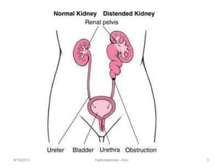
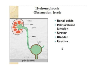


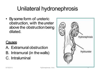
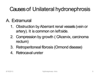

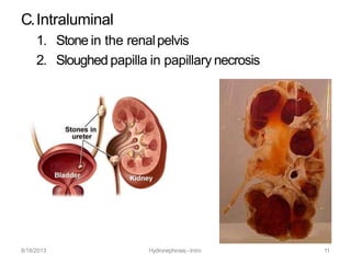


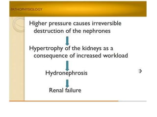


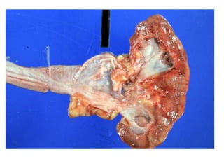


Hydronephrosis is the dilatation of the renal pelvis or calyces, which can be associated with obstruction. It has various causes including extramural obstruction from aberrant vessels or tumors, intramural obstruction from congenital issues or tumors, and intraluminal obstruction from stones or papillary necrosis. Bilateral hydronephrosis can result from urethral obstruction or issues affecting both kidneys. Imaging studies are used to diagnose and characterize hydronephrosis. Long term obstruction can lead to thinning of the renal parenchyma and loss of kidney function.


