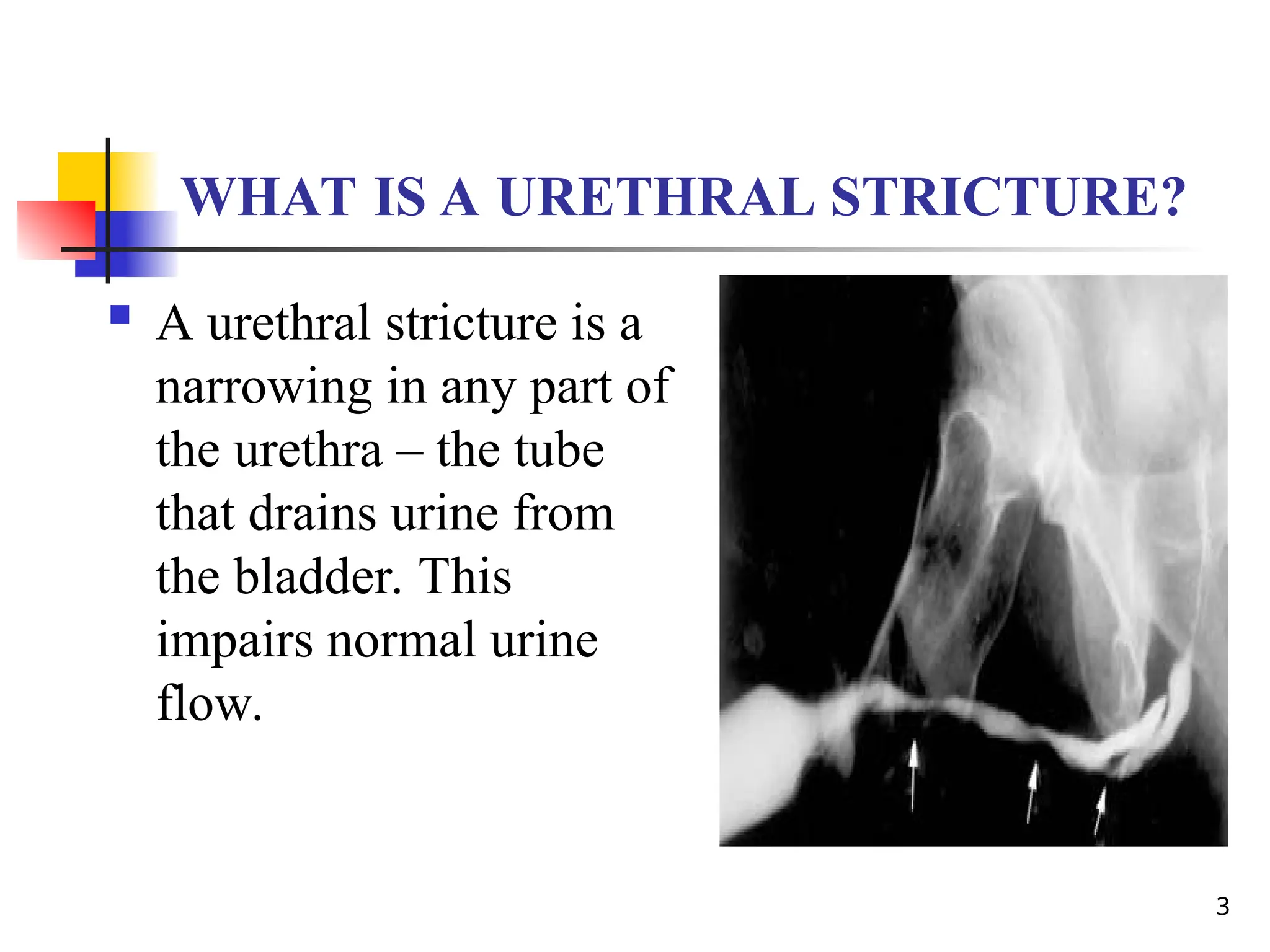
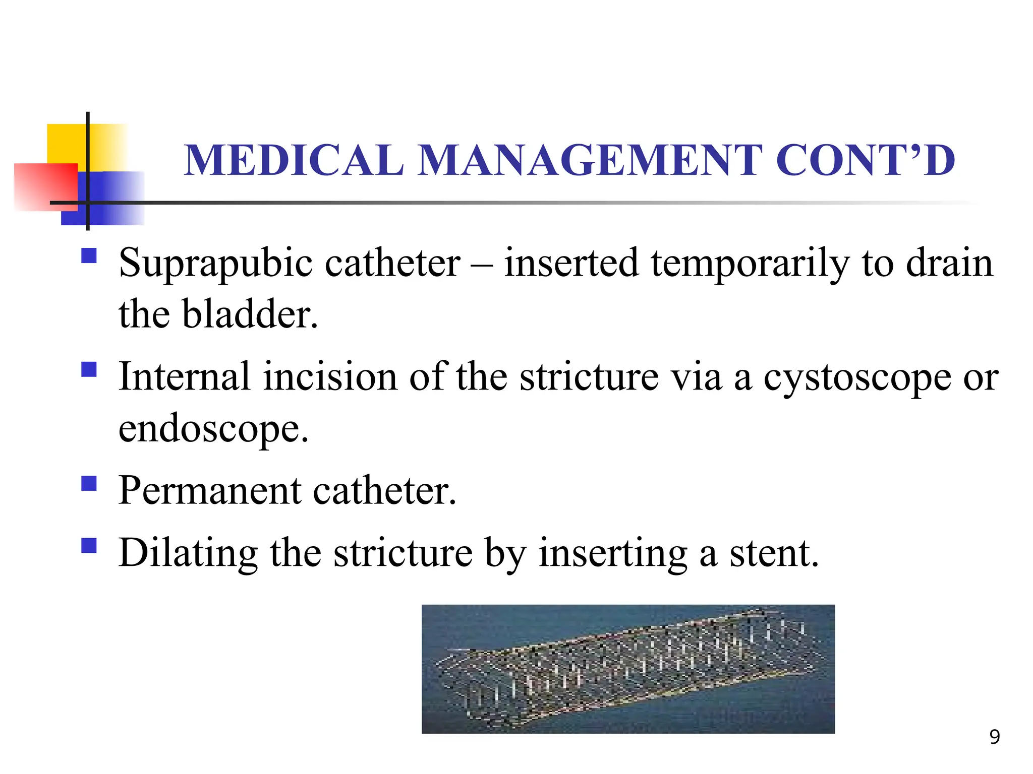
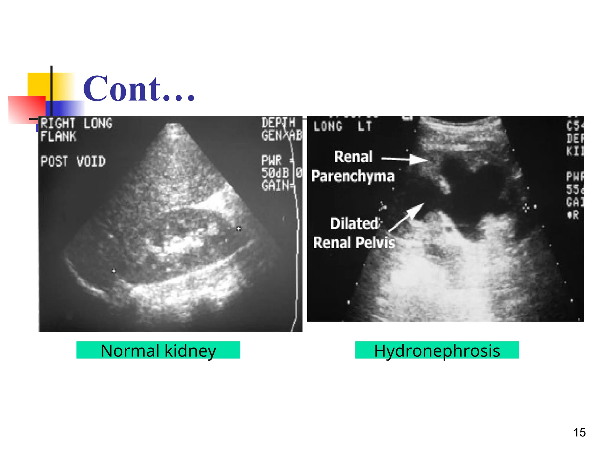
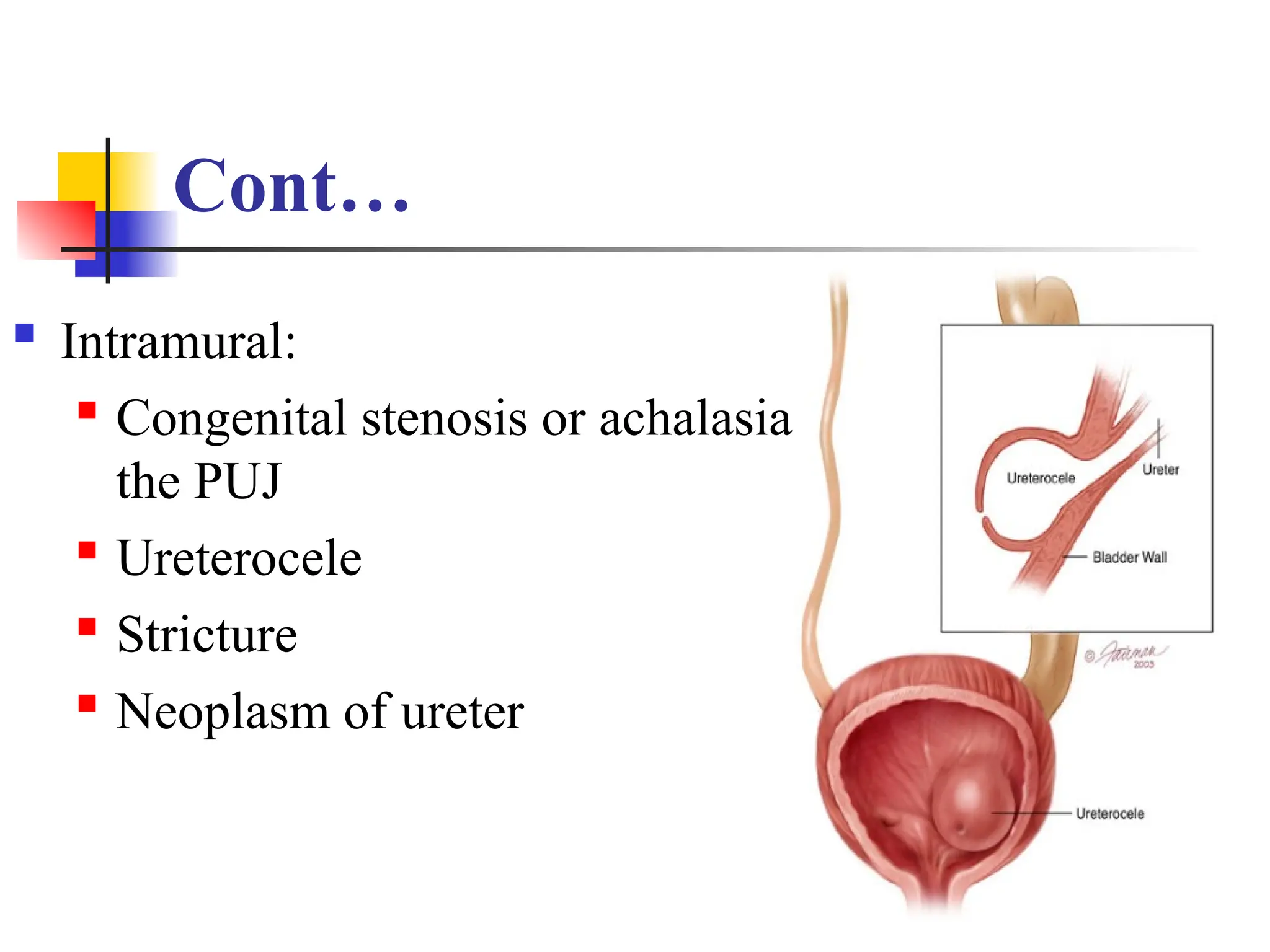
URETHRAL STRICTURES
Prepared by
Ms. Maira Shaheen
LEARNING OBJECTIVES
By the end of the session learners will be able to:
Discuss the causes, Pathophysiology and
manifestation of the following Urethral strictures and
Hydronephrosis
Discuss Assessment, Diagnostic Test, Medical,
Surgical and Nursing Management of Urethral
strictures and Hydronephrosis
2
WHAT IS A URETHRAL STRICTURE?
 A urethral stricture is a
narrowing in any part of
the urethra – the tube
that drains urine from
the bladder. This
impairs normal urine
flow.
3
ETIOLOGY
 Urethral injury, disease, catheterization or surgery
that results in inflammation or scar tissue.
 Urethral infections, such as gonorrhea.
 A tumor in the urethra (rarely)
 A complication of radiation treatment of the pelvis
 May be congenital or acquired.
4
RISK FACTORS
 Increased risk is associated with men who have a
history of sexually transmitted disease, repeated
episodes of urethritis, and benign prostatic
hyperplasia. There is also increased risk of
stricture after an injury or trauma to the pelvic
region. Any instrumentation entering the urethra
increases the chance of developing urethral
strictures.
5
SIGNS AND SYMPTOMS
 Dysuria.
 Hematuria.
 Weak stream.
 Splaying of the urine stream.
 Nocturia.
 Incomplete emptying.
 Pain with bladder distention.
 Urinary tract infection (fever and malaise may be
present with a urinary tract infection.
6
DIAGNOSTICS
 Voiding cystourethrography (pictured)
 Cystoscopy
7
MEDICAL MANAGEMENT
 Surgery to correct
The condition – open
Urethroplasty.
8
MEDICAL MANAGEMENT CONT’D
 Suprapubic catheter – inserted temporarily to drain
the bladder.
 Internal incision of the stricture via a cystoscope or
endoscope.
 Permanent catheter.
 Dilating the stricture by inserting a stent.
9
NURSING INTERVENTIONS
 Adequate hydration – to decrease discomfort
when voiding
 Monitor urine output.
 Mild analgesics.
 Sitz baths – to encourage voiding.
 If urethroplasty is performed – splint the catheter
to support the catheter line. Do not cause tension
on the catheter.
10
PROGNOSIS
 Without appropriate treatment, the stricture will
almost always recur. But, after surgical correction
or dilation the prognosis is favorable.
11
HYDRONEPHROSIS
Prepared by
Ms. Maira
HYDRONEPHROSIS
 Dilatation of renal pelvis & calyces with
accompanying destruction of the kidney
parenchyma
 Usually due to partial obstruction to the outflow of
urine
13
Hydronephrosis
14
Cont…
15
Normal kidney Hydronephrosis
Normal Intravenous Pyelogram (IVP)
16
ETIOLOGY
 Primary or secondary
 Primary: idiopathic
 Secondary: maybe unilateral or bilateral
17
CAUSES OF UNILATERAL
HYDORNEPHROSIS
 Maybe extramural, intramural or Intraluminal
 Extramural:

Pressure on the ureter by loaded sigmoid colon, gravid
uterus, uterine & ovarian tumors

Involvement of the ureter by malignant neoplasm outside it
e.g. carcinoma of the cervix, uterus, colon, rectum, prostate

Aberrant renal vessels

Idiopathic retroperitoneal fibrosis
18
Cont…
 Intramural:
 Congenital stenosis or achalasia of
the PUJ
 Ureterocele
 Stricture
 Neoplasm of ureter
19
Cont…
 Intraluminal:
 Calculus
 Congenital folds at the upper end
of the ureter
20
Causes of bilateral hydornephrosis
 Due to pathologies in the urethra or the urinary bladder
 Causes in the urethra:

Pin – hole meatus
 Congenital valves

BPH & carcinoma of prostate

Stricture
 Carcinoma of cervix & uterus
21
Cont…
 Due to pathologies in the urethra or the urinary
bladder
 Causes in the bladder:
 Calculus
 Neoplasms
 Sphincter dysfunction
22
23
24
PATHOPHYSIOLOGY
 Dilatation of the renal pelvis & calyces
 Types of hydronephrosis:
 Pelvic type
 Renal type
 Pelvirenal type: most common type, both the
pelvis & calyces are equally dilated
25
26
CLINICAL FEATURES
 May depend on unilateral, intermittent or bilateral
hydronephrosis
 Unilateral hydronephrosis:
 Dull ache & sense of weight on the affected
side of the loin
 Causes of the hydronephrosis
27
Cont…
 Intermittent hydronephrosis:
 Dietl’s crisis
 Bilateral hydronephrosis:
 Features of the causes
 Bilateral renal swelling
28
SPECIAL INVESTIGATIONS
 Straight abdomen of the X- ray
 Excretory urography
 Retrograde urography
 Ultrasound
29
TREATMENT
Secondary hydronephrosis: treatment of the cause
 Primary hydronephrosis:
 Pyeloplasty:

Anderson- Hynes

Culp

Foley
 Nephrectomy
30
31
32
33
PRINCIPLES OF PYELOPLASTY
 To reduce the size of the renal pelvis
 Excision of the UPJ
 The ureter is attached to the most dependent part
of the pelvis
34
35
extraperitoneal flank
approach, bed of the twelfth
rib
Cont…
36
37
REFERENCE
 Smeltzer, S. C., Bare, B. G., Hinkle, J. L., &
Cheever, K. H. (2010). Brunner and
Suddarth’s textbook of medical-surgical nursing
(12th ed.). Philadelphia:Lippincott Williams &
Wilkins.
38

Lect 37 urethral-strictures and hydronephrosis.ppt

  • 1.
  • 2.
    LEARNING OBJECTIVES By theend of the session learners will be able to: Discuss the causes, Pathophysiology and manifestation of the following Urethral strictures and Hydronephrosis Discuss Assessment, Diagnostic Test, Medical, Surgical and Nursing Management of Urethral strictures and Hydronephrosis 2
  • 3.
    WHAT IS AURETHRAL STRICTURE?  A urethral stricture is a narrowing in any part of the urethra – the tube that drains urine from the bladder. This impairs normal urine flow. 3
  • 4.
    ETIOLOGY  Urethral injury,disease, catheterization or surgery that results in inflammation or scar tissue.  Urethral infections, such as gonorrhea.  A tumor in the urethra (rarely)  A complication of radiation treatment of the pelvis  May be congenital or acquired. 4
  • 5.
    RISK FACTORS  Increasedrisk is associated with men who have a history of sexually transmitted disease, repeated episodes of urethritis, and benign prostatic hyperplasia. There is also increased risk of stricture after an injury or trauma to the pelvic region. Any instrumentation entering the urethra increases the chance of developing urethral strictures. 5
  • 6.
    SIGNS AND SYMPTOMS Dysuria.  Hematuria.  Weak stream.  Splaying of the urine stream.  Nocturia.  Incomplete emptying.  Pain with bladder distention.  Urinary tract infection (fever and malaise may be present with a urinary tract infection. 6
  • 7.
    DIAGNOSTICS  Voiding cystourethrography(pictured)  Cystoscopy 7
  • 8.
    MEDICAL MANAGEMENT  Surgeryto correct The condition – open Urethroplasty. 8
  • 9.
    MEDICAL MANAGEMENT CONT’D Suprapubic catheter – inserted temporarily to drain the bladder.  Internal incision of the stricture via a cystoscope or endoscope.  Permanent catheter.  Dilating the stricture by inserting a stent. 9
  • 10.
    NURSING INTERVENTIONS  Adequatehydration – to decrease discomfort when voiding  Monitor urine output.  Mild analgesics.  Sitz baths – to encourage voiding.  If urethroplasty is performed – splint the catheter to support the catheter line. Do not cause tension on the catheter. 10
  • 11.
    PROGNOSIS  Without appropriatetreatment, the stricture will almost always recur. But, after surgical correction or dilation the prognosis is favorable. 11
  • 12.
  • 13.
    HYDRONEPHROSIS  Dilatation ofrenal pelvis & calyces with accompanying destruction of the kidney parenchyma  Usually due to partial obstruction to the outflow of urine 13
  • 14.
  • 15.
  • 16.
  • 17.
    ETIOLOGY  Primary orsecondary  Primary: idiopathic  Secondary: maybe unilateral or bilateral 17
  • 18.
    CAUSES OF UNILATERAL HYDORNEPHROSIS Maybe extramural, intramural or Intraluminal  Extramural:  Pressure on the ureter by loaded sigmoid colon, gravid uterus, uterine & ovarian tumors  Involvement of the ureter by malignant neoplasm outside it e.g. carcinoma of the cervix, uterus, colon, rectum, prostate  Aberrant renal vessels  Idiopathic retroperitoneal fibrosis 18
  • 19.
    Cont…  Intramural:  Congenitalstenosis or achalasia of the PUJ  Ureterocele  Stricture  Neoplasm of ureter 19
  • 20.
    Cont…  Intraluminal:  Calculus Congenital folds at the upper end of the ureter 20
  • 21.
    Causes of bilateralhydornephrosis  Due to pathologies in the urethra or the urinary bladder  Causes in the urethra:  Pin – hole meatus  Congenital valves  BPH & carcinoma of prostate  Stricture  Carcinoma of cervix & uterus 21
  • 22.
    Cont…  Due topathologies in the urethra or the urinary bladder  Causes in the bladder:  Calculus  Neoplasms  Sphincter dysfunction 22
  • 23.
  • 24.
  • 25.
    PATHOPHYSIOLOGY  Dilatation ofthe renal pelvis & calyces  Types of hydronephrosis:  Pelvic type  Renal type  Pelvirenal type: most common type, both the pelvis & calyces are equally dilated 25
  • 26.
  • 27.
    CLINICAL FEATURES  Maydepend on unilateral, intermittent or bilateral hydronephrosis  Unilateral hydronephrosis:  Dull ache & sense of weight on the affected side of the loin  Causes of the hydronephrosis 27
  • 28.
    Cont…  Intermittent hydronephrosis: Dietl’s crisis  Bilateral hydronephrosis:  Features of the causes  Bilateral renal swelling 28
  • 29.
    SPECIAL INVESTIGATIONS  Straightabdomen of the X- ray  Excretory urography  Retrograde urography  Ultrasound 29
  • 30.
    TREATMENT Secondary hydronephrosis: treatmentof the cause  Primary hydronephrosis:  Pyeloplasty:  Anderson- Hynes  Culp  Foley  Nephrectomy 30
  • 31.
  • 32.
  • 33.
  • 34.
    PRINCIPLES OF PYELOPLASTY To reduce the size of the renal pelvis  Excision of the UPJ  The ureter is attached to the most dependent part of the pelvis 34
  • 35.
  • 36.
  • 37.
  • 38.
    REFERENCE  Smeltzer, S.C., Bare, B. G., Hinkle, J. L., & Cheever, K. H. (2010). Brunner and Suddarth’s textbook of medical-surgical nursing (12th ed.). Philadelphia:Lippincott Williams & Wilkins. 38