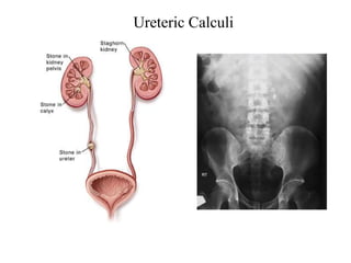
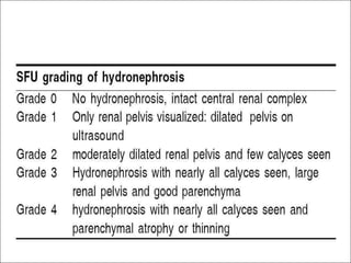
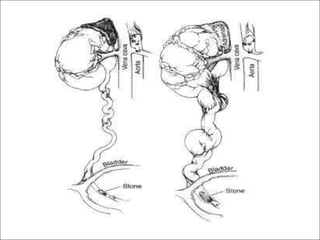
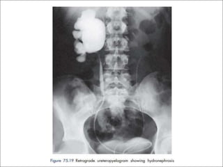
Hydronephrosis
Dr.Akmal
Associate Professor
Department of General Surgery
AVMC & H
• Hydronephrosis is an aseptic dilatation of the kidney caused by
obstruction.
• Dilation of renal pelvis and calycesassociated with
progressive atrophy of the kidney.
• Urine outflow obstruction.
• Obstructive uropathy indicates impedance of urinary flow
anywhere along the urinary tract and damage to renal parenchyma
due to obstruction at any site.
Definition
HYDRONEPHROSIS: CAUSES
Causes of Unilateral Ureteric Obstruction
Extramural obstruction:
Adjacent structures neoplasm like those of cervix, prostate, rectum,
colon or caecum
Idiopathic retroperitoneal fibrosis
Retrocaval ureter
• Intramural obstruction:
Congenital stenosis, physiological narrowing of the pelviureteric
junction leading to pelviureteric junction obstruction
Ureterocele and congenital small ureteric orifice
Ureterocele
Cobra head appearance of
ureterocele
CAUSESOF UNILATERALURETERIC
OBSTRUCTION
• Intramural obstruction:
 Inflammatory stricture following removal of ureteric
calculus, repair of a damaged ureter or tuberculous infection
 Neoplasm of the ureter or bladder cancer involving the ureteric
orifice
• Intraluminal obstruction:
 Calculus in the pelvis or ureter
 Sloughed papilla in papillary necrosis(more commonly in
• diabetics, analgesic abusers and sickle cell disease)
Ureteric Calculi
BILA
TERALHYDRONEPHROSIS
It iscommonly result of urethral obstruction.
Causes:
Congenital
 Posterior urethral valves
 Urethral atresia
Acquired
 Benign prostatic enlargement
 Carcinoma of the prostate
 Postoperative bladder neck scarring
 Urethral stricture
 Phimosis
Posterior Urethral Valve
PA
THOPHYSIOLOGY
• Even with complete obstruction, glomerular filtration persists for
some time
• Thefiltrate diffuses back to the interstitium an perirenal
spaces
• Affected calyces and pelvis becomes dilated
• High pressure transmitted through collecting ducts:
Cortex: Renal atrophy
Medulla: Renal vasculature compression with diminished
inner medullar blood flow
s
ac
• Thekidney becomes athin walled, lobulated, fluid filled
PATHOPHYSIOLOGY
• Effective hydroureteronephrosis on renal function depends on
whether it is totally or partially obstructive and unilateral or
bilateral
• Effects of obstruction of the kidney are time dependent.
Within several hours, changes are evident but:
1–2 week:glomerular destruction, tubular atrophy, and
interstitial fibrosis occur
By6–8 week: irreversible damage occurs
CLINICALFEA
TURES
Unilateral Hydronephrosis ( commonly by idiopathic
pelvicureteric junction obstruction or calculus)
• More common in women and on right side
• May remain silent for long periods, being apparent in course of imaging
studies (about 3%of population)
• Mild pain or dull aching in loin (dragging heaviness worsened by
excessivefluid intake)
• Kidney may be palpable
• Intermittent Hydronephrosis (Dietl’s crisis)
CLINICALFEA
TURES
Bilateral Hydronephrosis:
Dysuria,
 Loin pain
 Features of bladder outlet obstruction: Polyuria, Nocturia,
Hesitancy
 Kidneys usually not palpable
 Inability to concentrate urine which may be associated with distal
tubular acidosis, chronic tubulointerstitial nephritis, renal and renal
salt wasting
CLINICALFEATURES
• Complete Bilateral Obstruction:
Rapid onset oliguria or anuria that is incompatible with
survival until obstruction is relieved.After relief, post
obstructive diuresis with large amount of sodium chloride
• Ureters and pelvis dilatation in pregnancy:
Up to 20th week .Backto normal within 12 weeks of delivery
Effect of high progesterone on smooth muscles
POSSIBLEEXAMINATION
FINDINGS
• General condition: pain or localized symptoms
• Abdominal, flank, or pelvic mass
• Flank tenderness can occur along with acute obstruction
and with calculi or infection
• V
aginal exam– Ureteral prolapse
• Digital rectal exam– Enlarged prostate, nodularity suggestive of
prostate cancer
INVESTIGATIONS
• Ultrasound scanning:
least invasive, regularly used for pelviureteric junction
obstruction
• Intravenous Pyelogram:
Significant function in obstructed kidney
Contrast fills the obstructed system down to blockage
Cantake follow up films 36 hours after the injection of
contrast
INVESTIGATIONS
Isotope renography
 Best to confirm obstructive dilatation of collectingsystem
 Technetium 99m-labelled DTP
A (diethylenetriaminepenta- acetic
acid or MAG-3) injected intravenously and tracked using gamma
camera
 99mTc-DTP
Astays in renal pelvis in obstructed site
Isotope renogram series shows a late accumulation and
persistence of radioactivity in the left kidney
INVESTIGATIONS
Whitaker test
 Percutaneous puncture made in kidney, fluid is infused at
constant rate with monitoring intrapelvic pressure,
 Abnormal rise in intrapelvic pressure confirms obstruction
Retrograde Pyelography
 Confirms site of obstruction
 Done immediately before corrective surgery
TREATMENT
• Hydronephrosis is not a specific diagnosis but a finding or sign
• Management is highly dependent on underlying condition
and the timing (acute vs. chronic)
• Urgent decompression isneeded with:
– Severe pain
– Active urinary tract infection and acute kidney insufficiency
–Retrograde ureteral stent or percutaneous nephrostomy
can provide equally effective drainage
MEDICALTHERAPY
• Patients with infection and hydronephrosis require antibiotic
therapy and drainage
• Renal failure and electrolyte abnormalities should be
corrected in conjunction with drainage
• Along with these, catheter drainage may be required as well
TREATMENT
• Indications for surgery:
Bouts of renal pain
Increasing Hydronephrosis
Evidence of parenchymal damage and infection
• Mild casesfollowed by serial ultrasound scans and operated upon
if dilatation is increasing
• Nephrectomy considered only when kidney largely
destroyed
ANDERSONS-HYNES
PYELOPLASTY
• Upper third of ureter and renal pelvis is mobilized
• Renal vein can be divided but artery should be preserved
• Anastomosis formed in front of artery
• A nephrostomy tube or ureteric stent protects the
anastomosis
• Laparoscopic pyeloplasty, a minimal access procedure is
becoming increasingly popular
ENDOSCOPICPYELOPLASTY
• Disruption of pelviureteric junction by aballoon passed up
the ureter and distended under radiographic control
• Long term efficacy still need to be proved
THANK YOU

Hydronephrosis.pptx

  • 1.
  • 2.
    • Hydronephrosis isan aseptic dilatation of the kidney caused by obstruction. • Dilation of renal pelvis and calycesassociated with progressive atrophy of the kidney. • Urine outflow obstruction. • Obstructive uropathy indicates impedance of urinary flow anywhere along the urinary tract and damage to renal parenchyma due to obstruction at any site. Definition
  • 3.
    HYDRONEPHROSIS: CAUSES Causes ofUnilateral Ureteric Obstruction Extramural obstruction: Adjacent structures neoplasm like those of cervix, prostate, rectum, colon or caecum Idiopathic retroperitoneal fibrosis Retrocaval ureter • Intramural obstruction: Congenital stenosis, physiological narrowing of the pelviureteric junction leading to pelviureteric junction obstruction Ureterocele and congenital small ureteric orifice
  • 4.
  • 5.
    CAUSESOF UNILATERALURETERIC OBSTRUCTION • Intramuralobstruction:  Inflammatory stricture following removal of ureteric calculus, repair of a damaged ureter or tuberculous infection  Neoplasm of the ureter or bladder cancer involving the ureteric orifice • Intraluminal obstruction:  Calculus in the pelvis or ureter  Sloughed papilla in papillary necrosis(more commonly in • diabetics, analgesic abusers and sickle cell disease)
  • 6.
  • 7.
    BILA TERALHYDRONEPHROSIS It iscommonly resultof urethral obstruction. Causes: Congenital  Posterior urethral valves  Urethral atresia Acquired  Benign prostatic enlargement  Carcinoma of the prostate  Postoperative bladder neck scarring  Urethral stricture  Phimosis
  • 8.
  • 11.
    PA THOPHYSIOLOGY • Even withcomplete obstruction, glomerular filtration persists for some time • Thefiltrate diffuses back to the interstitium an perirenal spaces • Affected calyces and pelvis becomes dilated • High pressure transmitted through collecting ducts: Cortex: Renal atrophy Medulla: Renal vasculature compression with diminished inner medullar blood flow s ac • Thekidney becomes athin walled, lobulated, fluid filled
  • 12.
    PATHOPHYSIOLOGY • Effective hydroureteronephrosison renal function depends on whether it is totally or partially obstructive and unilateral or bilateral • Effects of obstruction of the kidney are time dependent. Within several hours, changes are evident but: 1–2 week:glomerular destruction, tubular atrophy, and interstitial fibrosis occur By6–8 week: irreversible damage occurs
  • 14.
    CLINICALFEA TURES Unilateral Hydronephrosis (commonly by idiopathic pelvicureteric junction obstruction or calculus) • More common in women and on right side • May remain silent for long periods, being apparent in course of imaging studies (about 3%of population) • Mild pain or dull aching in loin (dragging heaviness worsened by excessivefluid intake) • Kidney may be palpable • Intermittent Hydronephrosis (Dietl’s crisis)
  • 15.
    CLINICALFEA TURES Bilateral Hydronephrosis: Dysuria,  Loinpain  Features of bladder outlet obstruction: Polyuria, Nocturia, Hesitancy  Kidneys usually not palpable  Inability to concentrate urine which may be associated with distal tubular acidosis, chronic tubulointerstitial nephritis, renal and renal salt wasting
  • 16.
    CLINICALFEATURES • Complete BilateralObstruction: Rapid onset oliguria or anuria that is incompatible with survival until obstruction is relieved.After relief, post obstructive diuresis with large amount of sodium chloride • Ureters and pelvis dilatation in pregnancy: Up to 20th week .Backto normal within 12 weeks of delivery Effect of high progesterone on smooth muscles
  • 17.
    POSSIBLEEXAMINATION FINDINGS • General condition:pain or localized symptoms • Abdominal, flank, or pelvic mass • Flank tenderness can occur along with acute obstruction and with calculi or infection • V aginal exam– Ureteral prolapse • Digital rectal exam– Enlarged prostate, nodularity suggestive of prostate cancer
  • 18.
    INVESTIGATIONS • Ultrasound scanning: leastinvasive, regularly used for pelviureteric junction obstruction • Intravenous Pyelogram: Significant function in obstructed kidney Contrast fills the obstructed system down to blockage Cantake follow up films 36 hours after the injection of contrast
  • 19.
    INVESTIGATIONS Isotope renography  Bestto confirm obstructive dilatation of collectingsystem  Technetium 99m-labelled DTP A (diethylenetriaminepenta- acetic acid or MAG-3) injected intravenously and tracked using gamma camera  99mTc-DTP Astays in renal pelvis in obstructed site
  • 20.
    Isotope renogram seriesshows a late accumulation and persistence of radioactivity in the left kidney
  • 21.
    INVESTIGATIONS Whitaker test  Percutaneouspuncture made in kidney, fluid is infused at constant rate with monitoring intrapelvic pressure,  Abnormal rise in intrapelvic pressure confirms obstruction Retrograde Pyelography  Confirms site of obstruction  Done immediately before corrective surgery
  • 23.
    TREATMENT • Hydronephrosis isnot a specific diagnosis but a finding or sign • Management is highly dependent on underlying condition and the timing (acute vs. chronic) • Urgent decompression isneeded with: – Severe pain – Active urinary tract infection and acute kidney insufficiency –Retrograde ureteral stent or percutaneous nephrostomy can provide equally effective drainage
  • 24.
    MEDICALTHERAPY • Patients withinfection and hydronephrosis require antibiotic therapy and drainage • Renal failure and electrolyte abnormalities should be corrected in conjunction with drainage • Along with these, catheter drainage may be required as well
  • 25.
    TREATMENT • Indications forsurgery: Bouts of renal pain Increasing Hydronephrosis Evidence of parenchymal damage and infection • Mild casesfollowed by serial ultrasound scans and operated upon if dilatation is increasing • Nephrectomy considered only when kidney largely destroyed
  • 26.
    ANDERSONS-HYNES PYELOPLASTY • Upper thirdof ureter and renal pelvis is mobilized • Renal vein can be divided but artery should be preserved • Anastomosis formed in front of artery • A nephrostomy tube or ureteric stent protects the anastomosis • Laparoscopic pyeloplasty, a minimal access procedure is becoming increasingly popular
  • 28.
    ENDOSCOPICPYELOPLASTY • Disruption ofpelviureteric junction by aballoon passed up the ureter and distended under radiographic control • Long term efficacy still need to be proved
  • 29.