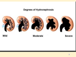
Dr Vijay Kumar
Associate Professor
HYDRONEPHROSIS
1
Hydronephrosis.
• Hydronephrosis - dilation of
the renal pelvis and calyces
with urine, associated with
progressive atrophy of the
kidney due to obstruction to
the outflow of urine
2
3
Types of Obstruction
• SUDDEN / INSIDIOUS
• PARTIAL / COMPLETE
• UNILATERAL / BILATERAL
4
Epidemiology
• Autopsy 3.1%
• Differences based on sex did not become
apparent until age 20 years.
• At age 20-60 years, hydronephrosis was
more common in women, due to pregnancy
and gynecologic malignancy.
• In men, prostatic diseases - cause of the rise
in prevalence > 60 years.
.5
6
Types of Lesions
Causative lesions may be:-
• Intrinsic to the urinary tract or
• Extrinsic lesions - compress ureter
7
Obstructive lesions of the urinary tract
8
Etiology
• 1. Congenital anomalies
• 2. Urinary calculi
• 3. Benign prostatic hypertrophy
• 4. Tumors
• 5. Inflammation
• 6. Sloughed papillae or blood clots
• 7. Pregnancy
• 8. Uterine prolapse and cystocele
• 9. Functional disorders
9
Etiology
• Congenital
– urethral valves or stricture
– stenosis at the UVJ or PUJ → majority of cases
in children.
• Calculi are most common in young adults
• Prostatic hypertrophy or carcinoma
retroperitoneal or pelvic neoplasms, and
calculi → in older patients.
10
Physiological
• Hydronephrosis or hydroureter is normal pregnancy
• More prominent on the right
• These changes can be visualized on USG in 2nd
trimester
• Resolve 6-12 weeks postpartum
11
12
13
Acute Vs. Chronic
• Acute hydronephrosis , when corrected,
allows full recovery of renal function.
• Chronic hydronephrosis - loss of function is
irreversible
• If acute unilateral obstruction is corrected
within 2 weeks, full recovery of renal
function is possible
• After 6 weeks of obstruction, function is
irreversibly lost
14
When the obstruction is SUDDEN & COMPLETE
GFR IS REDUCED
• It leads to
• MILD DILATION OF PELVIS and CALYCES
and sometimes to
• ATROPHY OF THE RENAL PARENCHYMA
15
When the obstruction is
• SUBTOTAL or INTERMITTENT
→GFR IS NOT SUPPRESSED and
• PROGRESSIVE DILATION
ENSUES
• Kidney may be slightly to massively
ENLARGED, depending on the
degree and the duration 16
• In far-advanced cases the kidney may
become transformed into
• A THIN-WALLED CYSTIC
STRUCTURE with a diameter 15 - 20 cm
• with striking PARENCHYMAL
ATROPHY
• TOTAL OBLITERATION OF THE
PYRAMIDS, and
• THINNING OF THE CORTEX
17
Complications
Urinary stasis may result in
• Infection
• Renal scarring
• Calculus formation
• Sepsis
• Renal failure
• Hypertension
18
Clinical Features
I. Acute obstruction –
• PAIN d/t distention of the renal
capsule
• The site of obstruction -
– Upper ureteral or renal pelvic lesions
lead to flank pain or tenderness
– Lower ureteral obstruction causes pain
that may radiate to the ipsilateral
testicle or labia.
19
Clinical Features
• II Unilateral HDN remain SILENT FOR
LONG PERIODS, since the opposite kidney
can maintain adequate renal function.
• Sometimes detected in routine USG or IVP
20
Clinical Features
III. In B/L partial obstruction the earliest
manifestation is inability to concentrate
the urine, reflected by
1. POLYURIA
2. NOCTURIA.
21
Clinical Features
Other symptoms
• Anuria
• Hematuria
• Fever
22
Clinical Features:Physical Exam
• Kidney lump
• With B/L hydronephrosis, lower limb
edema may occur.
• Renal angle tenderness +
• Distended bladder
23
Diagnostic Studies
• Laboratory Tests
• Imaging
– X-ray + Contrast studies.
– USG
– CT
– MRI
– Nuclear Scan
• Other Tests
24
Laboratory Studies
• Urinalysis-
– Pyuria -means- infection.
– Microscopic hematuria → presence of a stone
or tumor.
• Leukocytosis -acute infection.
• Serum Creatinine levels
• Hypercalcemia can be a life-threatening
condition.
25
Imaging studies
• Renal ultrasonography is the test of choice
• Intravenous Urography
• CT Indications :
– Ultrasonography results are equivocal
– The kidneys cannot be well visualized
– The cause of the obstruction cannot be identified.
26
DTPA renal scan - kidney function and blood flow
Technetium-99m (Tc-99m) labeled DTPA(Diethylenetriaminepentaacetic
acid) is injected into the bloodstream, and its clearance from the kidneys
→ renal function. 27
D
T
P
A
Imaging in children
• Antenatal hydronephrosis -visible in USG
usually occurs in the II trimester
• Postnatal USG
• A voiding cystourethrography (VCUG) -
detect VUR and, in boys, evaluate the
posterior urethra.
• Magnetic resonance urography (MRU)
28
Differential Diagnosis
Other problems to consider in the differential
diagnosis include the following:
• Peripelvic cyst
• Congenital megacalyces
• Calyceal diverticula
• Capacious extrarenal pelvis
• Pyelonephritis
29
Non Operative Therapy
• Pain control
• Treatment or prevention of infection
• Oral alkalinization therapy for uric acid
stones and
• Steroid therapy for retroperitoneal fibrosis.
30
Minimally invasive Therapy
• Extracorporeal shockwave lithotripsy
• Urethral Catheterisation.
• SPC
• Ureteral catheterisation +- stent
• Nephrostomy
• Post. Urethral Valve fulguration.
31
Operative Therapy:Indications
• Any signs of infection Fever, leukocytosis
• The potential for loss of renal function also
adds to the urgency (eg, hydronephrosis or
hydroureter bilaterally or in a solitary
kidney)
• Refractory pain in a patient with an
obstructing ureteral calculus necessitates
intervention
• Intractable nausea and vomiting.
32
33

hydronephrosis-221122055345-ef1e2953.pptx

  • 1.
    Dr Vijay Kumar AssociateProfessor HYDRONEPHROSIS 1
  • 2.
    Hydronephrosis. • Hydronephrosis -dilation of the renal pelvis and calyces with urine, associated with progressive atrophy of the kidney due to obstruction to the outflow of urine 2
  • 3.
  • 4.
    Types of Obstruction •SUDDEN / INSIDIOUS • PARTIAL / COMPLETE • UNILATERAL / BILATERAL 4
  • 5.
    Epidemiology • Autopsy 3.1% •Differences based on sex did not become apparent until age 20 years. • At age 20-60 years, hydronephrosis was more common in women, due to pregnancy and gynecologic malignancy. • In men, prostatic diseases - cause of the rise in prevalence > 60 years. .5
  • 6.
  • 7.
    Types of Lesions Causativelesions may be:- • Intrinsic to the urinary tract or • Extrinsic lesions - compress ureter 7
  • 8.
    Obstructive lesions ofthe urinary tract 8
  • 9.
    Etiology • 1. Congenitalanomalies • 2. Urinary calculi • 3. Benign prostatic hypertrophy • 4. Tumors • 5. Inflammation • 6. Sloughed papillae or blood clots • 7. Pregnancy • 8. Uterine prolapse and cystocele • 9. Functional disorders 9
  • 10.
    Etiology • Congenital – urethralvalves or stricture – stenosis at the UVJ or PUJ → majority of cases in children. • Calculi are most common in young adults • Prostatic hypertrophy or carcinoma retroperitoneal or pelvic neoplasms, and calculi → in older patients. 10
  • 11.
    Physiological • Hydronephrosis orhydroureter is normal pregnancy • More prominent on the right • These changes can be visualized on USG in 2nd trimester • Resolve 6-12 weeks postpartum 11
  • 12.
  • 13.
  • 14.
    Acute Vs. Chronic •Acute hydronephrosis , when corrected, allows full recovery of renal function. • Chronic hydronephrosis - loss of function is irreversible • If acute unilateral obstruction is corrected within 2 weeks, full recovery of renal function is possible • After 6 weeks of obstruction, function is irreversibly lost 14
  • 15.
    When the obstructionis SUDDEN & COMPLETE GFR IS REDUCED • It leads to • MILD DILATION OF PELVIS and CALYCES and sometimes to • ATROPHY OF THE RENAL PARENCHYMA 15
  • 16.
    When the obstructionis • SUBTOTAL or INTERMITTENT →GFR IS NOT SUPPRESSED and • PROGRESSIVE DILATION ENSUES • Kidney may be slightly to massively ENLARGED, depending on the degree and the duration 16
  • 17.
    • In far-advancedcases the kidney may become transformed into • A THIN-WALLED CYSTIC STRUCTURE with a diameter 15 - 20 cm • with striking PARENCHYMAL ATROPHY • TOTAL OBLITERATION OF THE PYRAMIDS, and • THINNING OF THE CORTEX 17
  • 18.
    Complications Urinary stasis mayresult in • Infection • Renal scarring • Calculus formation • Sepsis • Renal failure • Hypertension 18
  • 19.
    Clinical Features I. Acuteobstruction – • PAIN d/t distention of the renal capsule • The site of obstruction - – Upper ureteral or renal pelvic lesions lead to flank pain or tenderness – Lower ureteral obstruction causes pain that may radiate to the ipsilateral testicle or labia. 19
  • 20.
    Clinical Features • IIUnilateral HDN remain SILENT FOR LONG PERIODS, since the opposite kidney can maintain adequate renal function. • Sometimes detected in routine USG or IVP 20
  • 21.
    Clinical Features III. InB/L partial obstruction the earliest manifestation is inability to concentrate the urine, reflected by 1. POLYURIA 2. NOCTURIA. 21
  • 22.
    Clinical Features Other symptoms •Anuria • Hematuria • Fever 22
  • 23.
    Clinical Features:Physical Exam •Kidney lump • With B/L hydronephrosis, lower limb edema may occur. • Renal angle tenderness + • Distended bladder 23
  • 24.
    Diagnostic Studies • LaboratoryTests • Imaging – X-ray + Contrast studies. – USG – CT – MRI – Nuclear Scan • Other Tests 24
  • 25.
    Laboratory Studies • Urinalysis- –Pyuria -means- infection. – Microscopic hematuria → presence of a stone or tumor. • Leukocytosis -acute infection. • Serum Creatinine levels • Hypercalcemia can be a life-threatening condition. 25
  • 26.
    Imaging studies • Renalultrasonography is the test of choice • Intravenous Urography • CT Indications : – Ultrasonography results are equivocal – The kidneys cannot be well visualized – The cause of the obstruction cannot be identified. 26
  • 27.
    DTPA renal scan- kidney function and blood flow Technetium-99m (Tc-99m) labeled DTPA(Diethylenetriaminepentaacetic acid) is injected into the bloodstream, and its clearance from the kidneys → renal function. 27 D T P A
  • 28.
    Imaging in children •Antenatal hydronephrosis -visible in USG usually occurs in the II trimester • Postnatal USG • A voiding cystourethrography (VCUG) - detect VUR and, in boys, evaluate the posterior urethra. • Magnetic resonance urography (MRU) 28
  • 29.
    Differential Diagnosis Other problemsto consider in the differential diagnosis include the following: • Peripelvic cyst • Congenital megacalyces • Calyceal diverticula • Capacious extrarenal pelvis • Pyelonephritis 29
  • 30.
    Non Operative Therapy •Pain control • Treatment or prevention of infection • Oral alkalinization therapy for uric acid stones and • Steroid therapy for retroperitoneal fibrosis. 30
  • 31.
    Minimally invasive Therapy •Extracorporeal shockwave lithotripsy • Urethral Catheterisation. • SPC • Ureteral catheterisation +- stent • Nephrostomy • Post. Urethral Valve fulguration. 31
  • 32.
    Operative Therapy:Indications • Anysigns of infection Fever, leukocytosis • The potential for loss of renal function also adds to the urgency (eg, hydronephrosis or hydroureter bilaterally or in a solitary kidney) • Refractory pain in a patient with an obstructing ureteral calculus necessitates intervention • Intractable nausea and vomiting. 32
  • 33.