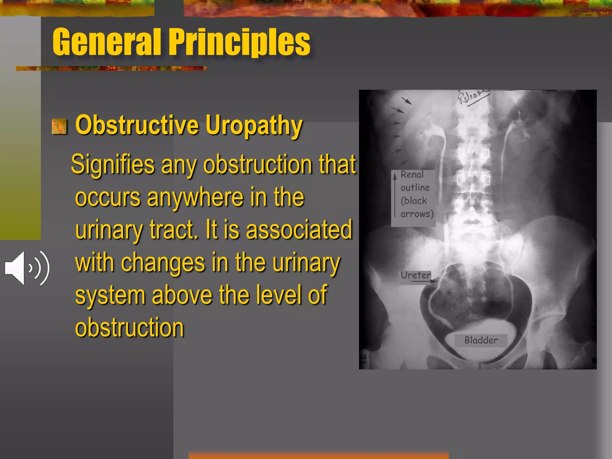
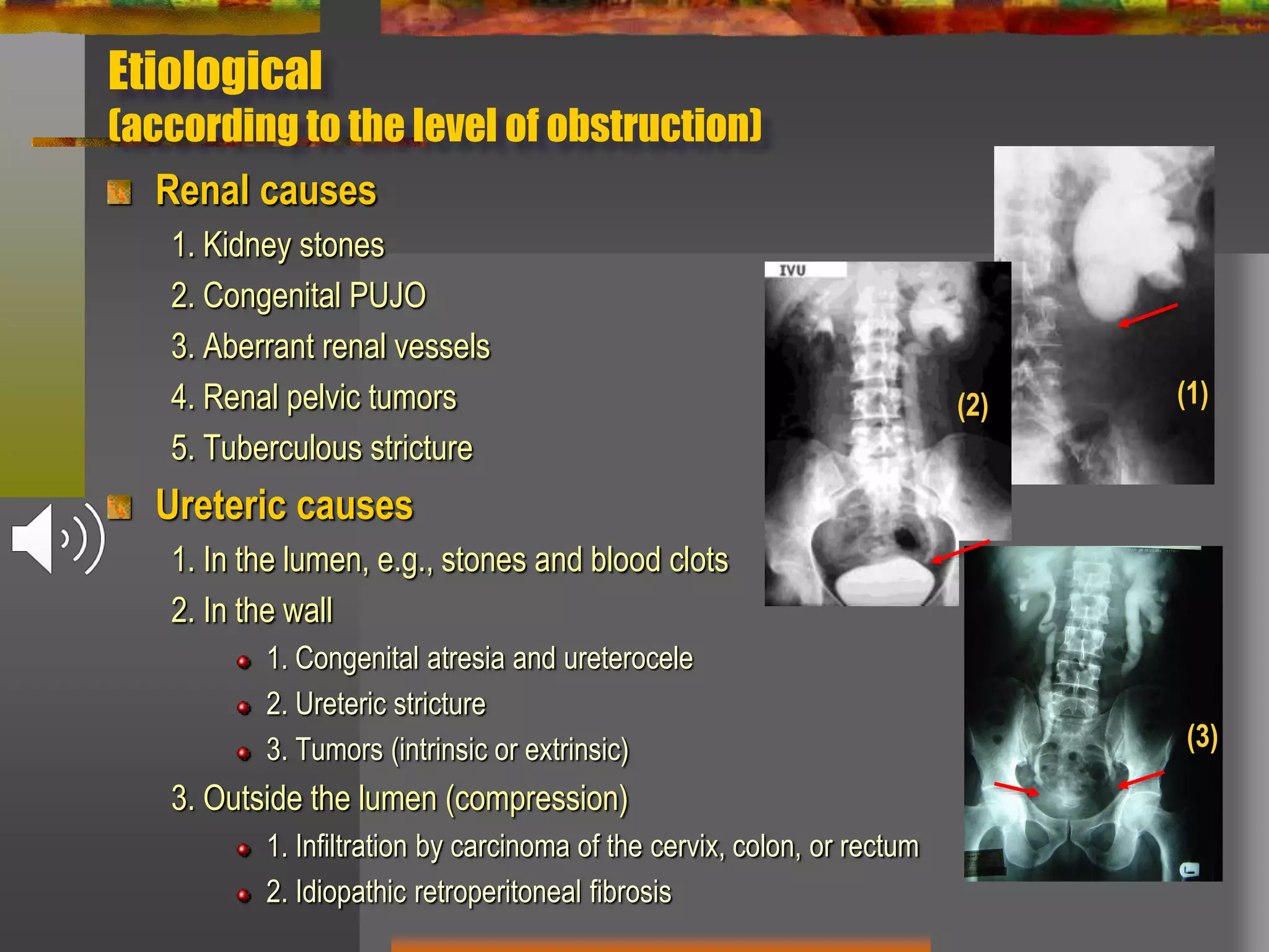
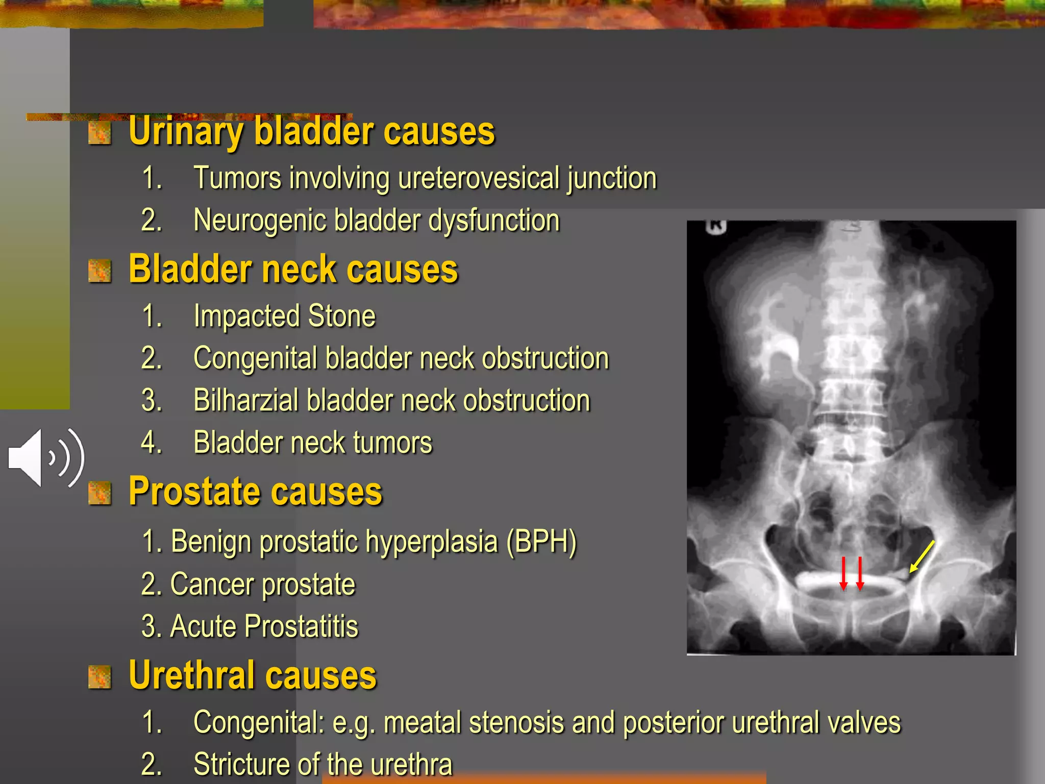
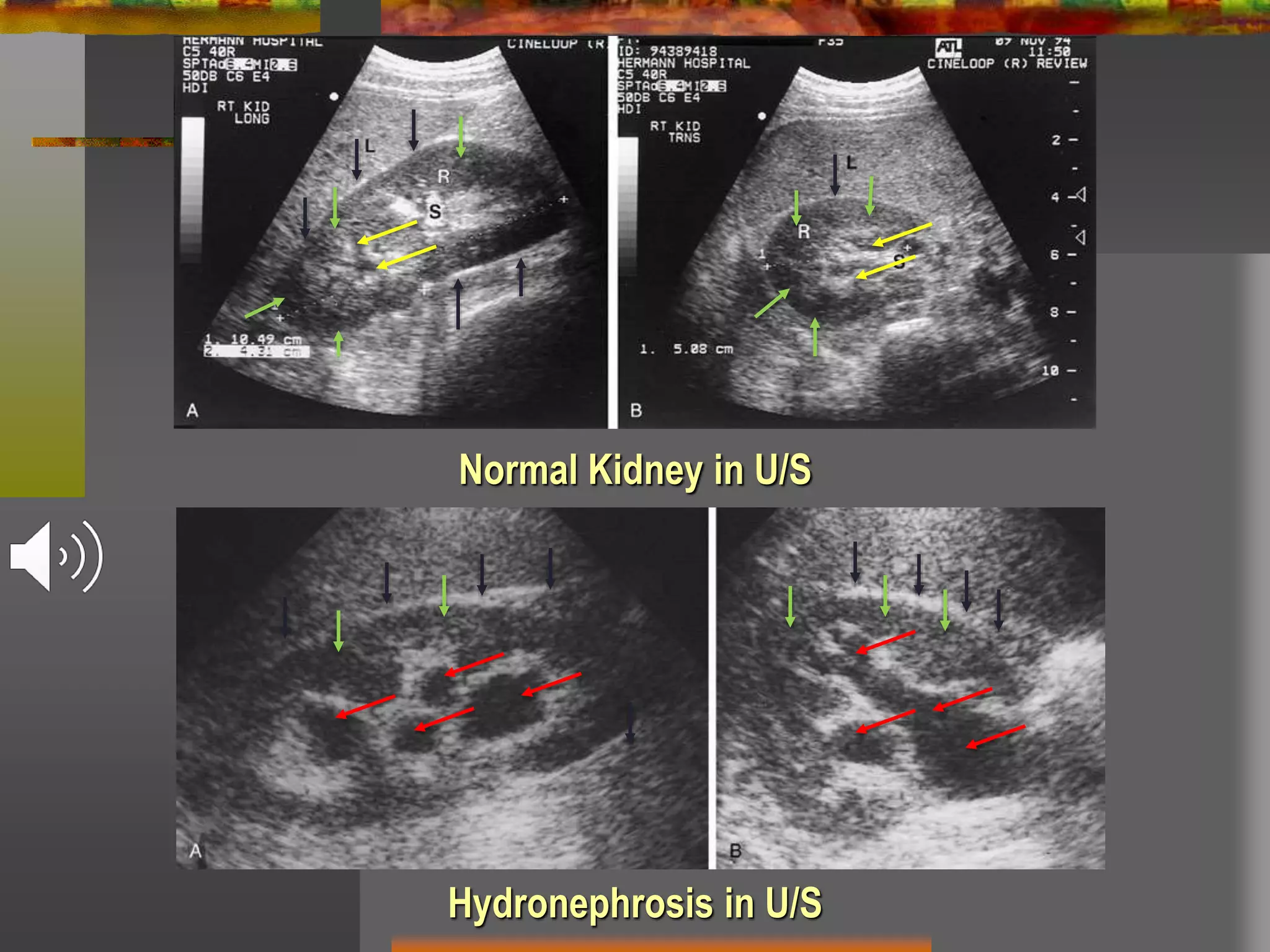
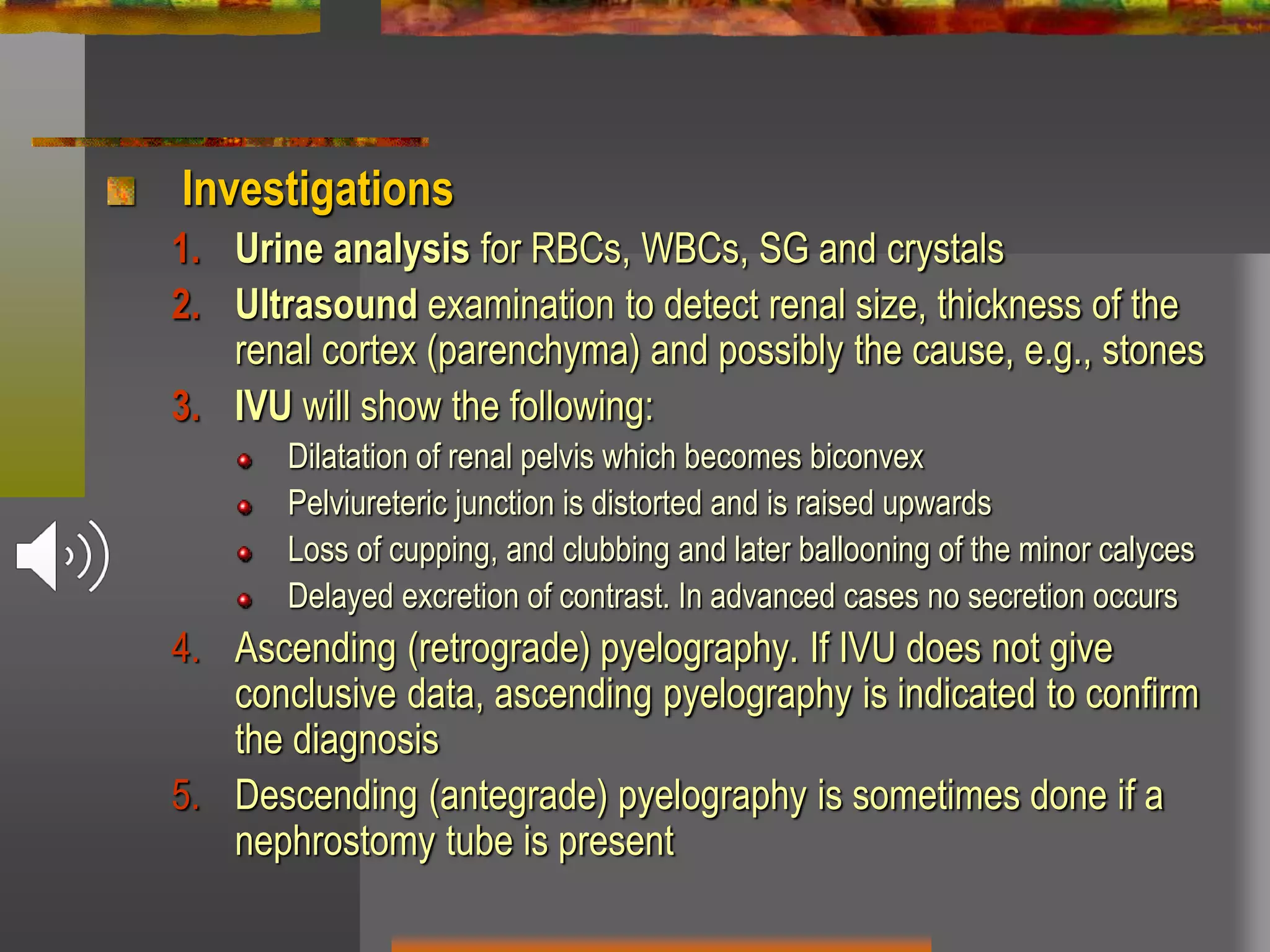
SlideShare a Scribd company logo
Obstructive Uropathy
By
Ayman Rashed, MD
Lecturer of Urology
General Principles
Obstructive Uropathy
Signifies any obstruction that
occurs anywhere in the
urinary tract. It is associated
with changes in the urinary
system above the level of
obstruction
Classification
Obstruction can be divided according to:
Acute or chronic obstruction
Partial or complete obstruction
Unilateral or bilateral obstruction
Congenital or acquired obstruction
Extrinsic or intrinsic obstruction
Etiological (according to the cause of obstruction)
Etiological
(according to the level of obstruction)
Renal causes
1. Kidney stones
2. Congenital PUJO
3. Aberrant renal vessels
4. Renal pelvic tumors
5. Tuberculous stricture
Ureteric causes
1. In the lumen, e.g., stones and blood clots
2. In the wall
1. Congenital atresia and ureterocele
2. Ureteric stricture
3. Tumors (intrinsic or extrinsic)
3. Outside the lumen (compression)
1. Infiltration by carcinoma of the cervix, colon, or rectum
2. Idiopathic retroperitoneal fibrosis
(1)(2)
(3)
Urinary bladder causes
1. Tumors involving ureterovesical junction
2. Neurogenic bladder dysfunction
Bladder neck causes
1. Impacted Stone
2. Congenital bladder neck obstruction
3. Bilharzial bladder neck obstruction
4. Bladder neck tumors
Prostate causes
1. Benign prostatic hyperplasia (BPH)
2. Cancer prostate
3. Acute Prostatitis
Urethral causes
1. Congenital: e.g. meatal stenosis and posterior urethral valves
2. Stricture of the urethra
Pathology
Urethra
Increased hydrostatic pressure leads to urethral dilatation above
the obstruction
Urinary bladder
Early, (Compensated Phase): Hypertrophy of the detrusor
muscle --- bladder Trabeculation --- Saccules (mucosa
protrudes between muscle fibers) --- Diverticulae develop when
mucosa bulges outside bladder wall
Later, (Decompensated Phase): the bladder dilates more and
its wall thins out --- Atony of the bladder --- Urinary retention
Ureter
Early there is muscular hypertrophy of the wall, later atony occurs.
The ureter becomes dilated (hydroureter), elongated and tortuous
Kidney
• Morphological changes: Normally, the pressure within the
renal pelvis is close to zero. With obstruction this pressure
increases and pelvicalyceal system dilates leading to
hydronephrosis
1. In early stages, Renal pelvic musculature undergoes
compensatory hypertrophy. Later, muscles become stretched and
atonic (decompensated)
2. Calyces are the first to suffer from obstruction. The normal
concave appearance (cupping) of minor calyces , becomes flat
then rounded (clubbing) and lastly ballooned
3. Renal parenchyma starts to thin out as it suffers from ischaemic
atrophy as it is compressed between renal capsule externally, and
the centrally increasing intra-pelvic pressure
4. At a later stage, the kidney is completely destroyed and appears
as a thin-walled sac filled with clear fluid or, if infected, with pus
IVP: Bilateral Hydro-uretero-nephrosis
Due to Bilateral Ureteric Stricture
Functional changes
1. Intra-pelvic pressure continues to rise till it overcomes
the glomerular filtration pressure, where urine secretion
stops
2. Glomerular filtration rate and the renal plasma flow are
reduced and renal concentrating power is gradually lost
4. The contralateral normal kidney undergoes
compensatory hypertrophy in order to maintain an
overall normal renal function
5. In neglected bilateral obstruction, renal failure will occur
With relief of obstruction
Early treatment leads to remarkable improvement of morphology
and function
Delay in treatment results in some degree of irreversible renal
damage
Factors affecting morphological and functional changes
1. Level of obstruction (proximal or distal “uni or bilateral”)
2. Degree of obstruction (complete or incomplete)
3. Duration of obstruction. Long-standing partial or intermittent
obstruction tends to produce significant proximal dilatation. Acute
obstruction, on the other hand, tends to produce mild dilatation
but more rapid deterioration of function
4. Infection on top of obstruction
Complications
1. Stasis means infection --- faster deterioration of renal
function
2. Stone formation
3. Rupture of hydronephrotic kidney (rare, with trauma)
4. Renal failure in bilateral cases
Urgent Acute Obstructive Conditions
Acute Urinary Retention
Calcular Anuria
Bilateral ligation of both ureters
Hydronephrosis
Definition
Hydronephrosis is chronic aseptic distension of the renal pelvis
and calyces due to obstruction of the urinary tract
Aetiology
1. All causes of chronic obstructive uropathy
2. Pregnancy: especially on the right side. Dilatation occurs during
the first few weeks and reaches maximum between fifth and sixth
month. It is due to atony of ureteric musculature due to high
progesterone level. Also, the fetal head may compress both
ureters. Hydronephrosis of pregnancy subsides within 2-12 weeks
after delivery
3. Vesicoureteric reflux
Normal IVU
Bilateral Hydronephrosis
due to Pregnancy near term
Carcinoma of the colon invading the right ureter
An antegrade pyeloureterogram shows a ureteral stent
Hydronephrosis in U/S
Normal Kidney in U/S
Clinical features
1. Pain: There is usually slight dull aching pain or a
sense of discomfort in the loin
2. Swelling: In late stages a cystic swelling in renal angle
3. Polyuria of low specific gravity
4. Manifestations of renal failure, if present
Complications: As mentioned in general principles
Differential diagnosis:
Polycystic kidney
Hypernephroma
Investigations
1. Urine analysis for RBCs, WBCs, SG and crystals
2. Ultrasound examination to detect renal size, thickness of the
renal cortex (parenchyma) and possibly the cause, e.g., stones
3. IVU will show the following:
Dilatation of renal pelvis which becomes biconvex
Pelviureteric junction is distorted and is raised upwards
Loss of cupping, and clubbing and later ballooning of the minor calyces
Delayed excretion of contrast. In advanced cases no secretion occurs
4. Ascending (retrograde) pyelography. If IVU does not give
conclusive data, ascending pyelography is indicated to confirm
the diagnosis
5. Descending (antegrade) pyelography is sometimes done if a
nephrostomy tube is present
Normal Bilateral Ascending (retrograde) Pyelography
Treatment
Removing cause of obstruction
Nephrectomy in selected cases
The kidney is non functioning with infusion urography
Sonography showed very thin parenchyma
Renal scan shows zero function
The opposite kidney is normal
In compromised cases with bad other kidney, a
percutaneous nephrostomy may be left for long time till
obstruction is treated
In advanced cases of PUJO --- Pyeloplasty to provide
adequate drainage of the lowest part of the pelvis
Ureteric Stricture
Aetiology
1. Congenital: PUJO or UVJO (pin-hole meatus)
2. Traumatic: Following ureteric injuries e.g. Iatrogenic
3. Inflammatory
4. Bilharzial stricture: commonly affects the intramural and lower
part of the ureter. It may be bilateral
5. Tuberculous stricture: affects the whole ureter and is usually
multiple and bilateral
6. Long-standing stone may lead to stricture
7. Neoplastic: Carcinoma of the ureter (intrinsic) or more often,
invasion of ureter (extrinsic) by a malignant growth as carcinoma
of the uterus, colon, or rectum
Clinical features
1. Renal pain (dull aching) due to distension of the renal pelvis
2. Progressive hydronephrosis
3. Recurrent attacks of pyelonephritis
4. In Egypt, bilateral hydronephrosis is a common sequel of
bilateral Bilharzial ureteric stricture. These patients may present
by chronic renal failure
Investigations
1. Blood urea, serum creatinine and serum electrolytes
2. U/S will demonstrate hydronephrosis
3. IVU will determine site of the stricture and degree of functional
capacity of both kidneys
4. Retrograde and Antegrade pyelography
Treatment (mainly surgical)
Stricture of upper 2/3 (above the level of ischial spine)
Excision and end-to-end anastomosis: Both ends of the ureter
are trimmed obliquely, spatulated and then anastomosed by
interrupted sutures of 4-0 Vicryl over a ureteric stent which is
removed later by a cystoscopic forceps
Stricture of lower 1/3
Ureterovesical reimplantation: The ureter is divided above the
stricture and then a new ureterovesical anastomosis is established..
Bladder (Boari's) flap: for long stricture. A flap can be prepared
from the bladder wall, transformed into tube and anastomosed to
ureter
Stricture of long segment of ureter:
Ileal loop replacement
Pre- and Post- Dismembered Pyeloplasty for PUJO
Retroperitoneal Fibrosis
Types
Primary (idiopathic) retroperitoneal fibrosis
Unknown aetiology but may be drug-induced, e.g., methysergide that is used for
prophylaxis against migraine
The patient may presents acutely with anuria. Ureteric catheterization is urgently
done to relieve obstruction. Unlike calcular anuria, the catheters pass up to the
kidneys easily.
ESR, urea and creatinine are usually elevated.
IVU and/or ascending pyelography demonstrates that both ureters are pulled
medially towards the mid line.
CT shows a retroperitoneal fibrous plaque in which both ureters and great
abdominal vessels (aorta an IVC) are embedded. Both kidneys show
hydronephrotic changes.
Definitive treatment is Surgical Ureterolysis i.e. release of ureters from fibrous
tissue, then wrapping them in pedicled omentum inside the peritoneal cavity to
avoid re-obstruction. Postoperative corticosteroid therapy is believed to reduce
the possibility of recurrence
Secondary retroperitoneal fibrosis
Diffuse infiltrating retroperitoneal malignancy
Previous leakage of an aortic aneurysm
Retroperitoneal Fibrosis
Obstructive Uropathy "online"

More Related Content

What's hot

Vesicouretric reflux
Vesicouretric refluxVesicouretric reflux
Vesicouretric reflux
sanyal1981
 
Urinary tract injury (kidney injury)
Urinary tract injury (kidney injury)Urinary tract injury (kidney injury)
Urinary tract injury (kidney injury)
sunil kumar daha
 
Obstructive Uropathy.pptx
Obstructive Uropathy.pptxObstructive Uropathy.pptx
Obstructive Uropathy.pptx
AishaAkram13
 
Obstructive uropathy+urolithias
Obstructive uropathy+urolithiasObstructive uropathy+urolithias
Obstructive uropathy+urolithias
Soumya Ranjan Parida
 
The Kidney: OBSTRUCTIVE UROPATHY
The Kidney: OBSTRUCTIVE UROPATHYThe Kidney: OBSTRUCTIVE UROPATHY
The Kidney: OBSTRUCTIVE UROPATHY
Dr. Roopam Jain
 
Post obstructive diuresis
Post obstructive diuresisPost obstructive diuresis
Post obstructive diuresis
Garima Aggarwal
 
Posterior urethral valves- Pediatric Surgery
Posterior urethral valves- Pediatric SurgeryPosterior urethral valves- Pediatric Surgery
Posterior urethral valves- Pediatric Surgery
Selvaraj Balasubramani
 
Hiv associated nephropathy (hivan)
Hiv associated nephropathy (hivan)Hiv associated nephropathy (hivan)
Hiv associated nephropathy (hivan)
carol dzorani
 
congenital anomalies of renal system
congenital anomalies of renal systemcongenital anomalies of renal system
congenital anomalies of renal systemRia Saira
 
Urinary Tract Obstruction
Urinary Tract ObstructionUrinary Tract Obstruction
Urinary Tract ObstructionBayu_F_Wibowo
 
Urinary tract obstrution
Urinary tract obstrutionUrinary tract obstrution
Urinary tract obstrution
DR MUKESH SAH
 
Evaluation of hematuria
Evaluation of hematuria Evaluation of hematuria
Evaluation of hematuria
SomendraBansal
 
PCNL - the Perfect Puncture
PCNL - the Perfect PuncturePCNL - the Perfect Puncture
PCNL - the Perfect Puncture
Siewhong Ho
 
Genito urinary tuberculosis
Genito urinary tuberculosisGenito urinary tuberculosis
Genito urinary tuberculosis
Annie Agarwal
 
Puj obstruction
Puj obstructionPuj obstruction
Puj obstruction
DRTALALBALLOUT
 
Anuria & acute retention
Anuria & acute retentionAnuria & acute retention
Anuria & acute retention
Mohammed Abd El Wadood
 
Uretheral stricture
Uretheral strictureUretheral stricture
Uretheral stricture
Sumer Yadav
 
Renal trauma
Renal traumaRenal trauma
Renal trauma
Jibran Mohsin
 
Urological emergencies
Urological emergenciesUrological emergencies
Urological emergencies
Ahmed Eliwa
 
Renal Artery Stenosis
Renal Artery StenosisRenal Artery Stenosis
Renal Artery Stenosis
KhalafAlGhamdi
 

What's hot (20)

Vesicouretric reflux
Vesicouretric refluxVesicouretric reflux
Vesicouretric reflux
 
Urinary tract injury (kidney injury)
Urinary tract injury (kidney injury)Urinary tract injury (kidney injury)
Urinary tract injury (kidney injury)
 
Obstructive Uropathy.pptx
Obstructive Uropathy.pptxObstructive Uropathy.pptx
Obstructive Uropathy.pptx
 
Obstructive uropathy+urolithias
Obstructive uropathy+urolithiasObstructive uropathy+urolithias
Obstructive uropathy+urolithias
 
The Kidney: OBSTRUCTIVE UROPATHY
The Kidney: OBSTRUCTIVE UROPATHYThe Kidney: OBSTRUCTIVE UROPATHY
The Kidney: OBSTRUCTIVE UROPATHY
 
Post obstructive diuresis
Post obstructive diuresisPost obstructive diuresis
Post obstructive diuresis
 
Posterior urethral valves- Pediatric Surgery
Posterior urethral valves- Pediatric SurgeryPosterior urethral valves- Pediatric Surgery
Posterior urethral valves- Pediatric Surgery
 
Hiv associated nephropathy (hivan)
Hiv associated nephropathy (hivan)Hiv associated nephropathy (hivan)
Hiv associated nephropathy (hivan)
 
congenital anomalies of renal system
congenital anomalies of renal systemcongenital anomalies of renal system
congenital anomalies of renal system
 
Urinary Tract Obstruction
Urinary Tract ObstructionUrinary Tract Obstruction
Urinary Tract Obstruction
 
Urinary tract obstrution
Urinary tract obstrutionUrinary tract obstrution
Urinary tract obstrution
 
Evaluation of hematuria
Evaluation of hematuria Evaluation of hematuria
Evaluation of hematuria
 
PCNL - the Perfect Puncture
PCNL - the Perfect PuncturePCNL - the Perfect Puncture
PCNL - the Perfect Puncture
 
Genito urinary tuberculosis
Genito urinary tuberculosisGenito urinary tuberculosis
Genito urinary tuberculosis
 
Puj obstruction
Puj obstructionPuj obstruction
Puj obstruction
 
Anuria & acute retention
Anuria & acute retentionAnuria & acute retention
Anuria & acute retention
 
Uretheral stricture
Uretheral strictureUretheral stricture
Uretheral stricture
 
Renal trauma
Renal traumaRenal trauma
Renal trauma
 
Urological emergencies
Urological emergenciesUrological emergencies
Urological emergencies
 
Renal Artery Stenosis
Renal Artery StenosisRenal Artery Stenosis
Renal Artery Stenosis
 

Similar to Obstructive Uropathy "online"

Posterior urethral valve
Posterior urethral valvePosterior urethral valve
Posterior urethral valve
Makafui Yigah
 
Renal pathology lecture 2&3. Infection of upper and lower urinary tract sufia...
Renal pathology lecture 2&3. Infection of upper and lower urinary tract sufia...Renal pathology lecture 2&3. Infection of upper and lower urinary tract sufia...
Renal pathology lecture 2&3. Infection of upper and lower urinary tract sufia...
Sufia Husain
 
hydronephrosis.pptx
hydronephrosis.pptxhydronephrosis.pptx
hydronephrosis.pptx
Ray Victor
 
Common urological emergencies
Common urological emergencies   Common urological emergencies
Common urological emergencies
Uthamalingam Murali
 
ACUTE AND CHRONIC URINARY RETENTION.pptx
ACUTE AND CHRONIC URINARY RETENTION.pptxACUTE AND CHRONIC URINARY RETENTION.pptx
ACUTE AND CHRONIC URINARY RETENTION.pptx
DrAmitt Mishra
 
Management of pelviureteric junction obstruction onyeze copy
Management of pelviureteric junction obstruction onyeze   copyManagement of pelviureteric junction obstruction onyeze   copy
Management of pelviureteric junction obstruction onyeze copy
Chigozie Onyeze
 
Ureteropelvic junction obstruction in children
Ureteropelvic junction obstruction in childrenUreteropelvic junction obstruction in children
Ureteropelvic junction obstruction in children
MUSTAFA MAJID
 
Mcu rgu ppt
Mcu rgu pptMcu rgu ppt
Mcu rgu ppt
Naba Kumar Barman
 
Hydronephrosis and Pyonephrosis
Hydronephrosis and PyonephrosisHydronephrosis and Pyonephrosis
Hydronephrosis and Pyonephrosis
Dr. Aryan (Anish Dhakal)
 
Radiology 5th year, 4th lecture (Dr. Nasrin Alatrushi)
Radiology 5th year, 4th lecture (Dr. Nasrin Alatrushi)Radiology 5th year, 4th lecture (Dr. Nasrin Alatrushi)
Radiology 5th year, 4th lecture (Dr. Nasrin Alatrushi)
College of Medicine, Sulaymaniyah
 
Hydronephrosis.pptx
Hydronephrosis.pptxHydronephrosis.pptx
Hydronephrosis.pptx
Sreevani49
 
Posterior Urethral Valves
Posterior Urethral ValvesPosterior Urethral Valves
Posterior Urethral Valves
Efosa Aimien
 
Hydronephrosis
HydronephrosisHydronephrosis
Hydronephrosis
Yogesh Dengale
 
Hydronephrosis
HydronephrosisHydronephrosis
Hydronephrosis
DRTALALBALLOUT
 
Renal disorders
Renal disordersRenal disorders
Renal disorders
Zuhair Mustafa
 
Acute Urinary Retention.pptx
Acute Urinary Retention.pptxAcute Urinary Retention.pptx
Acute Urinary Retention.pptx
AgnimaAnne
 
Urinary Outflow Obstruction
Urinary Outflow ObstructionUrinary Outflow Obstruction
Urinary Outflow ObstructionDr Harim Mohsin
 
Hydronephrosis.pptx
Hydronephrosis.pptxHydronephrosis.pptx
Hydronephrosis.pptx
Pradeep Pande
 
Retention of urine
Retention of urineRetention of urine
Retention of urinePrabha Om
 
Ureteral stricture
Ureteral strictureUreteral stricture
Ureteral stricture
sonia dagar
 

Similar to Obstructive Uropathy "online" (20)

Posterior urethral valve
Posterior urethral valvePosterior urethral valve
Posterior urethral valve
 
Renal pathology lecture 2&3. Infection of upper and lower urinary tract sufia...
Renal pathology lecture 2&3. Infection of upper and lower urinary tract sufia...Renal pathology lecture 2&3. Infection of upper and lower urinary tract sufia...
Renal pathology lecture 2&3. Infection of upper and lower urinary tract sufia...
 
hydronephrosis.pptx
hydronephrosis.pptxhydronephrosis.pptx
hydronephrosis.pptx
 
Common urological emergencies
Common urological emergencies   Common urological emergencies
Common urological emergencies
 
ACUTE AND CHRONIC URINARY RETENTION.pptx
ACUTE AND CHRONIC URINARY RETENTION.pptxACUTE AND CHRONIC URINARY RETENTION.pptx
ACUTE AND CHRONIC URINARY RETENTION.pptx
 
Management of pelviureteric junction obstruction onyeze copy
Management of pelviureteric junction obstruction onyeze   copyManagement of pelviureteric junction obstruction onyeze   copy
Management of pelviureteric junction obstruction onyeze copy
 
Ureteropelvic junction obstruction in children
Ureteropelvic junction obstruction in childrenUreteropelvic junction obstruction in children
Ureteropelvic junction obstruction in children
 
Mcu rgu ppt
Mcu rgu pptMcu rgu ppt
Mcu rgu ppt
 
Hydronephrosis and Pyonephrosis
Hydronephrosis and PyonephrosisHydronephrosis and Pyonephrosis
Hydronephrosis and Pyonephrosis
 
Radiology 5th year, 4th lecture (Dr. Nasrin Alatrushi)
Radiology 5th year, 4th lecture (Dr. Nasrin Alatrushi)Radiology 5th year, 4th lecture (Dr. Nasrin Alatrushi)
Radiology 5th year, 4th lecture (Dr. Nasrin Alatrushi)
 
Hydronephrosis.pptx
Hydronephrosis.pptxHydronephrosis.pptx
Hydronephrosis.pptx
 
Posterior Urethral Valves
Posterior Urethral ValvesPosterior Urethral Valves
Posterior Urethral Valves
 
Hydronephrosis
HydronephrosisHydronephrosis
Hydronephrosis
 
Hydronephrosis
HydronephrosisHydronephrosis
Hydronephrosis
 
Renal disorders
Renal disordersRenal disorders
Renal disorders
 
Acute Urinary Retention.pptx
Acute Urinary Retention.pptxAcute Urinary Retention.pptx
Acute Urinary Retention.pptx
 
Urinary Outflow Obstruction
Urinary Outflow ObstructionUrinary Outflow Obstruction
Urinary Outflow Obstruction
 
Hydronephrosis.pptx
Hydronephrosis.pptxHydronephrosis.pptx
Hydronephrosis.pptx
 
Retention of urine
Retention of urineRetention of urine
Retention of urine
 
Ureteral stricture
Ureteral strictureUreteral stricture
Ureteral stricture
 

Recently uploaded

Charaka Samhita Sutra sthana Chapter 15 Upakalpaniyaadhyaya
Charaka Samhita Sutra sthana Chapter 15 UpakalpaniyaadhyayaCharaka Samhita Sutra sthana Chapter 15 Upakalpaniyaadhyaya
Charaka Samhita Sutra sthana Chapter 15 Upakalpaniyaadhyaya
Dr KHALID B.M
 
Physiology of Chemical Sensation of smell.pdf
Physiology of Chemical Sensation of smell.pdfPhysiology of Chemical Sensation of smell.pdf
Physiology of Chemical Sensation of smell.pdf
MedicoseAcademics
 
Lung Cancer: Artificial Intelligence, Synergetics, Complex System Analysis, S...
Lung Cancer: Artificial Intelligence, Synergetics, Complex System Analysis, S...Lung Cancer: Artificial Intelligence, Synergetics, Complex System Analysis, S...
Lung Cancer: Artificial Intelligence, Synergetics, Complex System Analysis, S...
Oleg Kshivets
 
Alcohol_Dr. Jeenal Mistry MD Pharmacology.pdf
Alcohol_Dr. Jeenal Mistry MD Pharmacology.pdfAlcohol_Dr. Jeenal Mistry MD Pharmacology.pdf
Alcohol_Dr. Jeenal Mistry MD Pharmacology.pdf
Dr Jeenal Mistry
 
Physiology of Special Chemical Sensation of Taste
Physiology of Special Chemical Sensation of TastePhysiology of Special Chemical Sensation of Taste
Physiology of Special Chemical Sensation of Taste
MedicoseAcademics
 
THOA 2.ppt Human Organ Transplantation Act
THOA 2.ppt Human Organ Transplantation ActTHOA 2.ppt Human Organ Transplantation Act
THOA 2.ppt Human Organ Transplantation Act
DrSathishMS1
 
Phone Us ❤85270-49040❤ #ℂall #gIRLS In Surat By Surat @ℂall @Girls Hotel With...
Phone Us ❤85270-49040❤ #ℂall #gIRLS In Surat By Surat @ℂall @Girls Hotel With...Phone Us ❤85270-49040❤ #ℂall #gIRLS In Surat By Surat @ℂall @Girls Hotel With...
Phone Us ❤85270-49040❤ #ℂall #gIRLS In Surat By Surat @ℂall @Girls Hotel With...
Savita Shen $i11
 
POST OPERATIVE OLIGURIA and its management
POST OPERATIVE OLIGURIA and its managementPOST OPERATIVE OLIGURIA and its management
POST OPERATIVE OLIGURIA and its management
touseefaziz1
 
Are There Any Natural Remedies To Treat Syphilis.pdf
Are There Any Natural Remedies To Treat Syphilis.pdfAre There Any Natural Remedies To Treat Syphilis.pdf
Are There Any Natural Remedies To Treat Syphilis.pdf
Little Cross Family Clinic
 
Antiulcer drugs Advance Pharmacology .pptx
Antiulcer drugs Advance Pharmacology .pptxAntiulcer drugs Advance Pharmacology .pptx
Antiulcer drugs Advance Pharmacology .pptx
Rohit chaurpagar
 
ANATOMY AND PHYSIOLOGY OF URINARY SYSTEM.pptx
ANATOMY AND PHYSIOLOGY OF URINARY SYSTEM.pptxANATOMY AND PHYSIOLOGY OF URINARY SYSTEM.pptx
ANATOMY AND PHYSIOLOGY OF URINARY SYSTEM.pptx
Swetaba Besh
 
Superficial & Deep Fascia of the NECK.pptx
Superficial & Deep Fascia of the NECK.pptxSuperficial & Deep Fascia of the NECK.pptx
Superficial & Deep Fascia of the NECK.pptx
Dr. Rabia Inam Gandapore
 
Ophthalmology Clinical Tests for OSCE exam
Ophthalmology Clinical Tests for OSCE examOphthalmology Clinical Tests for OSCE exam
Ophthalmology Clinical Tests for OSCE exam
KafrELShiekh University
 
heat stroke and heat exhaustion in children
heat stroke and heat exhaustion in childrenheat stroke and heat exhaustion in children
heat stroke and heat exhaustion in children
SumeraAhmad5
 
Cervical & Brachial Plexus By Dr. RIG.pptx
Cervical & Brachial Plexus By Dr. RIG.pptxCervical & Brachial Plexus By Dr. RIG.pptx
Cervical & Brachial Plexus By Dr. RIG.pptx
Dr. Rabia Inam Gandapore
 
Prix Galien International 2024 Forum Program
Prix Galien International 2024 Forum ProgramPrix Galien International 2024 Forum Program
Prix Galien International 2024 Forum Program
Levi Shapiro
 
HOT NEW PRODUCT! BIG SALES FAST SHIPPING NOW FROM CHINA!! EU KU DB BK substit...
HOT NEW PRODUCT! BIG SALES FAST SHIPPING NOW FROM CHINA!! EU KU DB BK substit...HOT NEW PRODUCT! BIG SALES FAST SHIPPING NOW FROM CHINA!! EU KU DB BK substit...
HOT NEW PRODUCT! BIG SALES FAST SHIPPING NOW FROM CHINA!! EU KU DB BK substit...
GL Anaacs
 
24 Upakrama.pptx class ppt useful in all
24 Upakrama.pptx class ppt useful in all24 Upakrama.pptx class ppt useful in all
24 Upakrama.pptx class ppt useful in all
DrSathishMS1
 
TEST BANK for Operations Management, 14th Edition by William J. Stevenson, Ve...
TEST BANK for Operations Management, 14th Edition by William J. Stevenson, Ve...TEST BANK for Operations Management, 14th Edition by William J. Stevenson, Ve...
TEST BANK for Operations Management, 14th Edition by William J. Stevenson, Ve...
kevinkariuki227
 
BENIGN PROSTATIC HYPERPLASIA.BPH. BPHpdf
BENIGN PROSTATIC HYPERPLASIA.BPH. BPHpdfBENIGN PROSTATIC HYPERPLASIA.BPH. BPHpdf
BENIGN PROSTATIC HYPERPLASIA.BPH. BPHpdf
DR SETH JOTHAM
 

Recently uploaded (20)

Charaka Samhita Sutra sthana Chapter 15 Upakalpaniyaadhyaya
Charaka Samhita Sutra sthana Chapter 15 UpakalpaniyaadhyayaCharaka Samhita Sutra sthana Chapter 15 Upakalpaniyaadhyaya
Charaka Samhita Sutra sthana Chapter 15 Upakalpaniyaadhyaya
 
Physiology of Chemical Sensation of smell.pdf
Physiology of Chemical Sensation of smell.pdfPhysiology of Chemical Sensation of smell.pdf
Physiology of Chemical Sensation of smell.pdf
 
Lung Cancer: Artificial Intelligence, Synergetics, Complex System Analysis, S...
Lung Cancer: Artificial Intelligence, Synergetics, Complex System Analysis, S...Lung Cancer: Artificial Intelligence, Synergetics, Complex System Analysis, S...
Lung Cancer: Artificial Intelligence, Synergetics, Complex System Analysis, S...
 
Alcohol_Dr. Jeenal Mistry MD Pharmacology.pdf
Alcohol_Dr. Jeenal Mistry MD Pharmacology.pdfAlcohol_Dr. Jeenal Mistry MD Pharmacology.pdf
Alcohol_Dr. Jeenal Mistry MD Pharmacology.pdf
 
Physiology of Special Chemical Sensation of Taste
Physiology of Special Chemical Sensation of TastePhysiology of Special Chemical Sensation of Taste
Physiology of Special Chemical Sensation of Taste
 
THOA 2.ppt Human Organ Transplantation Act
THOA 2.ppt Human Organ Transplantation ActTHOA 2.ppt Human Organ Transplantation Act
THOA 2.ppt Human Organ Transplantation Act
 
Phone Us ❤85270-49040❤ #ℂall #gIRLS In Surat By Surat @ℂall @Girls Hotel With...
Phone Us ❤85270-49040❤ #ℂall #gIRLS In Surat By Surat @ℂall @Girls Hotel With...Phone Us ❤85270-49040❤ #ℂall #gIRLS In Surat By Surat @ℂall @Girls Hotel With...
Phone Us ❤85270-49040❤ #ℂall #gIRLS In Surat By Surat @ℂall @Girls Hotel With...
 
POST OPERATIVE OLIGURIA and its management
POST OPERATIVE OLIGURIA and its managementPOST OPERATIVE OLIGURIA and its management
POST OPERATIVE OLIGURIA and its management
 
Are There Any Natural Remedies To Treat Syphilis.pdf
Are There Any Natural Remedies To Treat Syphilis.pdfAre There Any Natural Remedies To Treat Syphilis.pdf
Are There Any Natural Remedies To Treat Syphilis.pdf
 
Antiulcer drugs Advance Pharmacology .pptx
Antiulcer drugs Advance Pharmacology .pptxAntiulcer drugs Advance Pharmacology .pptx
Antiulcer drugs Advance Pharmacology .pptx
 
ANATOMY AND PHYSIOLOGY OF URINARY SYSTEM.pptx
ANATOMY AND PHYSIOLOGY OF URINARY SYSTEM.pptxANATOMY AND PHYSIOLOGY OF URINARY SYSTEM.pptx
ANATOMY AND PHYSIOLOGY OF URINARY SYSTEM.pptx
 
Superficial & Deep Fascia of the NECK.pptx
Superficial & Deep Fascia of the NECK.pptxSuperficial & Deep Fascia of the NECK.pptx
Superficial & Deep Fascia of the NECK.pptx
 
Ophthalmology Clinical Tests for OSCE exam
Ophthalmology Clinical Tests for OSCE examOphthalmology Clinical Tests for OSCE exam
Ophthalmology Clinical Tests for OSCE exam
 
heat stroke and heat exhaustion in children
heat stroke and heat exhaustion in childrenheat stroke and heat exhaustion in children
heat stroke and heat exhaustion in children
 
Cervical & Brachial Plexus By Dr. RIG.pptx
Cervical & Brachial Plexus By Dr. RIG.pptxCervical & Brachial Plexus By Dr. RIG.pptx
Cervical & Brachial Plexus By Dr. RIG.pptx
 
Prix Galien International 2024 Forum Program
Prix Galien International 2024 Forum ProgramPrix Galien International 2024 Forum Program
Prix Galien International 2024 Forum Program
 
HOT NEW PRODUCT! BIG SALES FAST SHIPPING NOW FROM CHINA!! EU KU DB BK substit...
HOT NEW PRODUCT! BIG SALES FAST SHIPPING NOW FROM CHINA!! EU KU DB BK substit...HOT NEW PRODUCT! BIG SALES FAST SHIPPING NOW FROM CHINA!! EU KU DB BK substit...
HOT NEW PRODUCT! BIG SALES FAST SHIPPING NOW FROM CHINA!! EU KU DB BK substit...
 
24 Upakrama.pptx class ppt useful in all
24 Upakrama.pptx class ppt useful in all24 Upakrama.pptx class ppt useful in all
24 Upakrama.pptx class ppt useful in all
 
TEST BANK for Operations Management, 14th Edition by William J. Stevenson, Ve...
TEST BANK for Operations Management, 14th Edition by William J. Stevenson, Ve...TEST BANK for Operations Management, 14th Edition by William J. Stevenson, Ve...
TEST BANK for Operations Management, 14th Edition by William J. Stevenson, Ve...
 
BENIGN PROSTATIC HYPERPLASIA.BPH. BPHpdf
BENIGN PROSTATIC HYPERPLASIA.BPH. BPHpdfBENIGN PROSTATIC HYPERPLASIA.BPH. BPHpdf
BENIGN PROSTATIC HYPERPLASIA.BPH. BPHpdf
 

Obstructive Uropathy "online"

  • 1. Obstructive Uropathy By Ayman Rashed, MD Lecturer of Urology
  • 2. General Principles Obstructive Uropathy Signifies any obstruction that occurs anywhere in the urinary tract. It is associated with changes in the urinary system above the level of obstruction
  • 3. Classification Obstruction can be divided according to: Acute or chronic obstruction Partial or complete obstruction Unilateral or bilateral obstruction Congenital or acquired obstruction Extrinsic or intrinsic obstruction Etiological (according to the cause of obstruction)
  • 4. Etiological (according to the level of obstruction) Renal causes 1. Kidney stones 2. Congenital PUJO 3. Aberrant renal vessels 4. Renal pelvic tumors 5. Tuberculous stricture Ureteric causes 1. In the lumen, e.g., stones and blood clots 2. In the wall 1. Congenital atresia and ureterocele 2. Ureteric stricture 3. Tumors (intrinsic or extrinsic) 3. Outside the lumen (compression) 1. Infiltration by carcinoma of the cervix, colon, or rectum 2. Idiopathic retroperitoneal fibrosis (1)(2) (3)
  • 5. Urinary bladder causes 1. Tumors involving ureterovesical junction 2. Neurogenic bladder dysfunction Bladder neck causes 1. Impacted Stone 2. Congenital bladder neck obstruction 3. Bilharzial bladder neck obstruction 4. Bladder neck tumors Prostate causes 1. Benign prostatic hyperplasia (BPH) 2. Cancer prostate 3. Acute Prostatitis Urethral causes 1. Congenital: e.g. meatal stenosis and posterior urethral valves 2. Stricture of the urethra
  • 6. Pathology Urethra Increased hydrostatic pressure leads to urethral dilatation above the obstruction Urinary bladder Early, (Compensated Phase): Hypertrophy of the detrusor muscle --- bladder Trabeculation --- Saccules (mucosa protrudes between muscle fibers) --- Diverticulae develop when mucosa bulges outside bladder wall Later, (Decompensated Phase): the bladder dilates more and its wall thins out --- Atony of the bladder --- Urinary retention Ureter Early there is muscular hypertrophy of the wall, later atony occurs. The ureter becomes dilated (hydroureter), elongated and tortuous
  • 7. Kidney • Morphological changes: Normally, the pressure within the renal pelvis is close to zero. With obstruction this pressure increases and pelvicalyceal system dilates leading to hydronephrosis 1. In early stages, Renal pelvic musculature undergoes compensatory hypertrophy. Later, muscles become stretched and atonic (decompensated) 2. Calyces are the first to suffer from obstruction. The normal concave appearance (cupping) of minor calyces , becomes flat then rounded (clubbing) and lastly ballooned 3. Renal parenchyma starts to thin out as it suffers from ischaemic atrophy as it is compressed between renal capsule externally, and the centrally increasing intra-pelvic pressure 4. At a later stage, the kidney is completely destroyed and appears as a thin-walled sac filled with clear fluid or, if infected, with pus
  • 8. IVP: Bilateral Hydro-uretero-nephrosis Due to Bilateral Ureteric Stricture
  • 9. Functional changes 1. Intra-pelvic pressure continues to rise till it overcomes the glomerular filtration pressure, where urine secretion stops 2. Glomerular filtration rate and the renal plasma flow are reduced and renal concentrating power is gradually lost 4. The contralateral normal kidney undergoes compensatory hypertrophy in order to maintain an overall normal renal function 5. In neglected bilateral obstruction, renal failure will occur
  • 10. With relief of obstruction Early treatment leads to remarkable improvement of morphology and function Delay in treatment results in some degree of irreversible renal damage Factors affecting morphological and functional changes 1. Level of obstruction (proximal or distal “uni or bilateral”) 2. Degree of obstruction (complete or incomplete) 3. Duration of obstruction. Long-standing partial or intermittent obstruction tends to produce significant proximal dilatation. Acute obstruction, on the other hand, tends to produce mild dilatation but more rapid deterioration of function 4. Infection on top of obstruction
  • 11. Complications 1. Stasis means infection --- faster deterioration of renal function 2. Stone formation 3. Rupture of hydronephrotic kidney (rare, with trauma) 4. Renal failure in bilateral cases Urgent Acute Obstructive Conditions Acute Urinary Retention Calcular Anuria Bilateral ligation of both ureters
  • 12. Hydronephrosis Definition Hydronephrosis is chronic aseptic distension of the renal pelvis and calyces due to obstruction of the urinary tract Aetiology 1. All causes of chronic obstructive uropathy 2. Pregnancy: especially on the right side. Dilatation occurs during the first few weeks and reaches maximum between fifth and sixth month. It is due to atony of ureteric musculature due to high progesterone level. Also, the fetal head may compress both ureters. Hydronephrosis of pregnancy subsides within 2-12 weeks after delivery 3. Vesicoureteric reflux
  • 14. Bilateral Hydronephrosis due to Pregnancy near term
  • 15. Carcinoma of the colon invading the right ureter An antegrade pyeloureterogram shows a ureteral stent
  • 17. Clinical features 1. Pain: There is usually slight dull aching pain or a sense of discomfort in the loin 2. Swelling: In late stages a cystic swelling in renal angle 3. Polyuria of low specific gravity 4. Manifestations of renal failure, if present Complications: As mentioned in general principles Differential diagnosis: Polycystic kidney Hypernephroma
  • 18. Investigations 1. Urine analysis for RBCs, WBCs, SG and crystals 2. Ultrasound examination to detect renal size, thickness of the renal cortex (parenchyma) and possibly the cause, e.g., stones 3. IVU will show the following: Dilatation of renal pelvis which becomes biconvex Pelviureteric junction is distorted and is raised upwards Loss of cupping, and clubbing and later ballooning of the minor calyces Delayed excretion of contrast. In advanced cases no secretion occurs 4. Ascending (retrograde) pyelography. If IVU does not give conclusive data, ascending pyelography is indicated to confirm the diagnosis 5. Descending (antegrade) pyelography is sometimes done if a nephrostomy tube is present
  • 19. Normal Bilateral Ascending (retrograde) Pyelography
  • 20. Treatment Removing cause of obstruction Nephrectomy in selected cases The kidney is non functioning with infusion urography Sonography showed very thin parenchyma Renal scan shows zero function The opposite kidney is normal In compromised cases with bad other kidney, a percutaneous nephrostomy may be left for long time till obstruction is treated In advanced cases of PUJO --- Pyeloplasty to provide adequate drainage of the lowest part of the pelvis
  • 21. Ureteric Stricture Aetiology 1. Congenital: PUJO or UVJO (pin-hole meatus) 2. Traumatic: Following ureteric injuries e.g. Iatrogenic 3. Inflammatory 4. Bilharzial stricture: commonly affects the intramural and lower part of the ureter. It may be bilateral 5. Tuberculous stricture: affects the whole ureter and is usually multiple and bilateral 6. Long-standing stone may lead to stricture 7. Neoplastic: Carcinoma of the ureter (intrinsic) or more often, invasion of ureter (extrinsic) by a malignant growth as carcinoma of the uterus, colon, or rectum
  • 22. Clinical features 1. Renal pain (dull aching) due to distension of the renal pelvis 2. Progressive hydronephrosis 3. Recurrent attacks of pyelonephritis 4. In Egypt, bilateral hydronephrosis is a common sequel of bilateral Bilharzial ureteric stricture. These patients may present by chronic renal failure Investigations 1. Blood urea, serum creatinine and serum electrolytes 2. U/S will demonstrate hydronephrosis 3. IVU will determine site of the stricture and degree of functional capacity of both kidneys 4. Retrograde and Antegrade pyelography
  • 23. Treatment (mainly surgical) Stricture of upper 2/3 (above the level of ischial spine) Excision and end-to-end anastomosis: Both ends of the ureter are trimmed obliquely, spatulated and then anastomosed by interrupted sutures of 4-0 Vicryl over a ureteric stent which is removed later by a cystoscopic forceps Stricture of lower 1/3 Ureterovesical reimplantation: The ureter is divided above the stricture and then a new ureterovesical anastomosis is established.. Bladder (Boari's) flap: for long stricture. A flap can be prepared from the bladder wall, transformed into tube and anastomosed to ureter Stricture of long segment of ureter: Ileal loop replacement
  • 24. Pre- and Post- Dismembered Pyeloplasty for PUJO
  • 25. Retroperitoneal Fibrosis Types Primary (idiopathic) retroperitoneal fibrosis Unknown aetiology but may be drug-induced, e.g., methysergide that is used for prophylaxis against migraine The patient may presents acutely with anuria. Ureteric catheterization is urgently done to relieve obstruction. Unlike calcular anuria, the catheters pass up to the kidneys easily. ESR, urea and creatinine are usually elevated. IVU and/or ascending pyelography demonstrates that both ureters are pulled medially towards the mid line. CT shows a retroperitoneal fibrous plaque in which both ureters and great abdominal vessels (aorta an IVC) are embedded. Both kidneys show hydronephrotic changes. Definitive treatment is Surgical Ureterolysis i.e. release of ureters from fibrous tissue, then wrapping them in pedicled omentum inside the peritoneal cavity to avoid re-obstruction. Postoperative corticosteroid therapy is believed to reduce the possibility of recurrence Secondary retroperitoneal fibrosis Diffuse infiltrating retroperitoneal malignancy Previous leakage of an aortic aneurysm

Editor's Notes

  1. This is Dr. Ayman Rashed, Urology department. and this is the first online narrated Urologic lecture for students of 6th Grade at faculty of medicine, October 6th University. today’s lecture “ Obstructive Urology” is the keynote of Urology by which you can understand most of Urology, that is why I prefer to start with
  2. what is meant by Obstructive Uropathy? this is to signify any obstruction that occurs anywhere in the urinary tract from the kidneys above through ureters down to the urinary bladder and finally out of the body through the urethra. It is associated with changes in the urinary system above the level of obstruction for example dilatation. the picture in this slide denoting IVP of normal appearance of the urinary system. you can see obviously the collecting system with cupping, normal renal pelvis on both sides, normal caliber of ureters and normal filling of the bladder. by the way this is incomplete film because the urethra couldn’t be shown in this film
  3. the obstruction can be classified according to the following Is it Acute or chronic Partial or complete Unilateral or bilateral Congenital or acquired Extrinsic or intrinsic Etiological (according to the cause of obstruction)
  4. according to etiology will further subdivided according to the level of obstruction. so, at the renal level etiology may be due to: Renal stones, Congenital PUJO, Aberrant renal vessels, Renal pelvic tumors, Tuberculous stricture. at this level you will note dilatation of the collecting system what we call Hydronephrosis as appeared in IVP film No. (1) where the obstruction at the level of renal pelvis red arrow mostly due to congenital pelvi-ureteric junction obstruction (PUJ) when the ureter is obstructed at any level it will cause dilatation including the ureter above the obstruction and the kidney or the collection system what we call hydrouretronephrosis. This may be due to causes inside the the ureteric lumen like stones as in film No. (2) red arrow or blood clots or causes in the wall of the ureter as ureteric stricture as in film No. (3) showing bilateral hydrouretronephrosis due to bilateral ureteric strictures (red arrows, atresia, uretrocele
  5. at the urinary bladder level the causes of obstruction may include : Impacted stone at bladder neck, Tumors involving ureterovesical junction or Neurogenic bladder dysfunction. at the level of the prostate: BPH Cancer prostate or even Acute prostatitis can cause obstruction. as you see in the picture this an IVP film denoting a filling defect due to enlarged prostate (red arrows) note the smooth edge of the filling defect of the prostate that elevating the bladder upward causing outlet obstruction and the diverticulum on the left side of the bladder (yellow arrow) due to increased pressure inside the bladder. finally at the level of the urethra the obstruction may be due to Congenital: e.g. meatal stenosis and posterior urethral valves or due to Stricture of the urethra
  6. Now what are the pathologies resulted as a consequence of the obstruction as regard to the urethra it is just a dilatation above the level of obstruction. As regard to the urinary bladder: the pathology occurred subdivided to Early (Compensated Phase): where Hypertrophy of the detrusor muscle --- bladder Trabeculation --- Saccules (mucosa protrudes between muscle fibers) --- Diverticulae develop when mucosa bulges outside bladder wall as we just have seen in the previous slide IVP film. then Later (Decompensated Phase): when the bladder couldn’t contract anymore against the obstruction the bladder dilates more and its wall thins out --- Atony of the bladder --- Urinary retention As regard to the Ureters. they take the same way as the Bladder
  7. The renal pathology will suffer from obstruction Morphologically and functionally the morphologic changes will occur due to elevated hydrostatic pressure inside the collecting system (from normally zero) leading to hydronephrosis. the severity of the changes depends upon many factors that we will discuss later. So, in early stages the renal pelvis musculature undergoes compensatory hypertrophy. Later, muscles become stretched and atonic (decompensated) The Calyces are the first to suffer from obstruction. The normal concave appearance (cupping) of minor calyces , becomes flat then rounded (clubbing) and lastly ballooned Renal parenchyma starts to thin out as it suffers from ischaemic atrophy as it is compressed between renal capsule externally, and the centrally increasing intra-pelvic pressure At a later stage, the kidney is completely destroyed and appears as a thin-walled sac filled with clear fluid or, if infected, with pus
  8. this is an IVP film: an example for hydrouerteronephrosis due to bilateral lower ureteric stricture (red arrows). Note the clubbing of the calyces more evident on Lt side (yellow arrows)
  9. the functional changes include stopping of urine secretion due to increased intra-pelvic pressure which at some point will overcome the glomular filtration pressure this will subsequently reduce the filtration rate and reduce the concentrating power of the kidney. the contralateral normal kidney undergoes compensatory hypertrophy in order to maintain an overall normal renal function. In neglected bilateral obstruction, renal failure will occur
  10. Early treatment leads to remarkable improvement of morphology and function, while Delay in treatment results in some degree of irreversible renal damage. the factors affecting the degree of renal changes include 1. Level of obstruction (proximal or distal “uni or bilateral”) 2. Degree of obstruction 3. Duration of obstruction. Long-standing partial or intermittent obstruction tends to produce significant proximal dilatation. Acute obstruction, on the other hand, tends to produce mild dilatation but more rapid deterioration of function 4. Infection on top of obstruction
  11. the complications of urinary obstruction include 1. Stasis means infection --- faster deterioration of renal function 2. Stone formation 3. Rupture of hydronephrotic kidney (rare, with trauma) 4. Renal failure in bilateral cases and the emergency conditions are Acute Urinary Retention where the urine couldn’t expelled out of the bladder Calcular Anuria where the obstruction above the level of the bladder and the urine cannot reach down to the bladder Bilateral ligation of both ureters is another example of anuria
  12. Hydronephrosis as a separate entity. means chronic aseptic distension of the renal pelvis and calyces due to obstruction of the urinary tract, so it is a manifestation of a hidden disease and a diagnosis Aetiology 1. All causes of chronic obstructive uropathy commonly due to stones within the urinary tract 2. Pregnancy: especially on the right side. Dilatation occurs during the first few weeks and reaches maximum between fifth and sixth month. It is due to atony of ureteric musculature due to high progesterone level. Also, the fetal head may compress both ureters. Hydronephrosis of pregnancy subsides within 2-12 weeks after delivery 3. Vesicoureteric reflux
  13. This is again normal IVP showing normal urinary system
  14. this is an IVP done by mistake elsewhere for a pregnant woman near full term. the fetal sac shadow appeared to bilaterally obstructing the urinary system (compression from outside) resulting in bilateral Hydroureteronephrosis. of course it is a temporary condition which relieved after labor.
  15. this is an antegrade pyelography study showing the nephrostomy tube (red arrows) inside the kidney and the contrast media was injected through this tube to assess the morphology of the Rt. kidney which is obstructed by cancer colon invading the ureter. in such cases we try to maintain the kidney function and patency of the tract by inserting a ureteric stent (yellow arrows) or by directing the urine directly outside by nephrostomy tube (red arrow)
  16. here in you can see normal Renal U/S above and hydronephrosis in the below picture. Note the difference between normal calyces (yellow arrows) and dilated calyces (red arrows). Also note, renal capsule (black arrows) and Renal parenchyma (green arrows)
  17. clinical features include: dull aching pain due to stretch of renal capsule, swelling can be palpated in late stages as a cystic swelling in renal angle. lab investigations may reveal polyuria with low specific gravity due to loss of concentration power and elevated serum urea and creatinine in bilateral renal hydronephrosis cases denoting renal failure Dx include polycystic kidney and hypernephroma both can be investigated using U/S and CT
  18. these are the investigations that should be done to reveal the etiology of Hydronephrosis
  19. this a Retrograde or ascending pyelography study showing the cystoscope (red arrows) introduced through the urethra inside the bladder and two ureteric catheters (yellow arrows) advanced in both ureters though which contrast media is injected to delineate the urinary tract. this is a morphologic study by which you can study the anatomy of urinary tract but not functional study as you inject the contrast media not secreted by the kidney unlike in IVP which assess morphologic and functional parameters of the urinary tract
  20. of course treatment will depend on treatment of the cause for example removing obstructing stones. however in some cases with end stage non functioning kidney the nephrectomy may be the treatment of choice. In compromised cases with bad other kidney, a percutaneous nephrostomy may be left for long time till obstruction is treated In advanced cases of PUJO --- Pyeloplasty to provide adequate drainage of the lowest part of the pelvis
  21. Stricture is also another separate entity and also is a manifestation for hidden etiology which may be one of the following Congenital: PUJO or UVJO (pin-hole meatus) Traumatic: Following ureteric injuries e.g. Iatrogenic Inflammatory Bilharzial stricture: commonly affects the intramural and lower part of the ureter. It may be bilateral Tuberculous stricture: affects the whole ureter and is usually multiple and bilateral Long-standing stone may lead to stricture Neoplastic: Carcinoma of the ureter (intrinsic) or more often, invasion of ureter (extrinsic) by a malignant growth as carcinoma of the uterus, colon, or rectum
  22. this is an example for PUJ obstruction on Lt picture and after excision of the strictured part of the ureter (red arrow) and repair on the rt picture
  23. here in you can see ascending pyelography showing straightening of the Lt ureter and its shifting toward spinal column also hydronephrosis is noted these features denoting retroperitoneal fibrosis