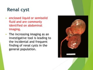
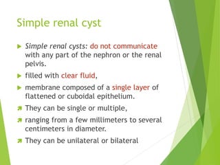
Simple Renal Cysts
by:-
Hasanain g. Khudhair (hasanain.ghaleb@gmail.com)
Afnan Adil Abbas
Zahraa Ahmed Abdulkadhim
Kerbala university / college of medicine
department of surgery
20-11-2017
Renal cyst
 enclosed liquid or semisolid
fluid and are commonly
identified on abdominal
imaging.
 The increasing imaging as an
investigative tool is leading to
the incidental and frequent
finding of renal cysts in the
general population.
Simple renal cyst
 Simple renal cysts: do not communicate
with any part of the nephron or the renal
pelvis.
 filled with clear fluid,
 membrane composed of a single layer of
flattened or cuboidal epithelium.
 They can be single or multiple,
 ranging from a few millimeters to several
centimeters in diameter.
 They can be unilateral or bilateral
morphological
description, Stage I renal
cyst (Bosniak
Classification)
Simple vs nonsimple ?!?!
 The presence of a solitary or multiple
renal cysts has been generally considered
benign in the absence of a family history
of renal cystic disease or evidence of
chronic kidney disease.
 a number of recent studies have
questioned this consensus by reported
associations with the development of
hypertension or malignant change. ???
 radiologists (to indicate non-complex
lesions)
 nephrologists (to indicate age-related
non-hereditary lesions)
 We propose that the term ‘simple’ be
replaced with the morphological
description, Stage I renal cyst (Bosniak
Classification)
Complicated CYSTS
Complicated cysts are
cysts that do not meet
the criteria of simple
cysts and thus require
further workup.
Prevalence
 Increases with age,
 the precise prevalence depending on the
method of diagnosis.
 On CT, 20% of adults have renal cysts by
age 40y and 33% by the age of 60.1 At
post-mortem, 50% of subjects aged >50y
have simple cysts. ( age )
 Cysts do not usually increase in size with
age, but may increase in number.
 Males and females are affected equally.
Pathogenesis
 diverticulae in the distal convoluted
tubule or collecting ducts due to
weakening of the tubular basement
membrane precipitated by obstruction-
related back pressure or age.
 This theoryhas since been refuted.
Pathogenesis
 The currently accepted hypothesis is that
renal ischaemia or injury prompts an
aberrant hypertrophic response leading to
cyst growth and leads to further nephron
loss due to compensatory hyperfiltration.
 The risk of exposure to subclinical renal
injury likely increases with age and
ageing is clearly associated with both a
decline in glomerular filtration rate and
an increasing prevalence of renal cysts.
The most important
principle in appropriately
managing Stage I renal
cysts is to establish the
correct diagnosis.
Presentation
 Simple cysts are most commonly
diagnosed as an incidental finding
following a renal ultrasound scan (USS) or
CT performed for other purposes.
 The majority are asymptomatic;
 very large cysts may present as an
abdominal mass or cause dull flank or
back pain.
 Acute severe loin pain may follow
bleeding into a cyst (causing sudden
distension of the wall).
Presentation
 Rupture (spontaneous or following renal
trauma) is rare.
 Rupture into the pelvicalyceal system can
produce haematuria.
 Infected cysts (rare) present with flank
pain and fever.
 Very occasionally, large cysts can cause
obstruction and hydronephrosis.
Differential diagnosis
Renal cell carcinoma (4–7% of RCC are
cystic).
Early autosomal dominant polycystic kidney
disease (ADPKD—diffuse, multiple or
bilateral cysts, associated with hepatic
cysts.
Complex renal cysts (i.e. those which
contain blood, pus or calcification).
Differential diagnosis
Renal cell carcinoma (4–7% of RCC are
cystic).
Early autosomal dominant polycystic kidney
disease (ADPKD—diffuse, multiple or
bilateral cysts, associated with hepatic
cysts.
Complex renal cysts (i.e. those which
contain blood, pus or calcification).
Investigation
 Ultrasound
 Computerized tomography (CT) scan
 contrast-enhanced ultrasound (CEUS),
alternative modality to CT avoids the use
of potentially nephrotoxic contrast agents
and ionizing radiation
 IVU
Radiographic features , US:
Anechoic
Enhanced through-transmission
Sharply marginated,
smooth walls
Radiographic features , CT:
Smooth cyst wall
Sharp demarcation
Homogenous Water
density (< 10-15 HU)
No significant enhancement
after IV contrast (<5HU)
Cyst wall too thin to be seen by CT
Radiographic features , IVP:
Lucent defect
Cortical bulge
Round indentations on
collecting system
Be Careful:
True renal cysts should always
be differentiated from
hydronephrosis, calyceal
diverticulum,
Differentiate renal cyst from
hypoechoic renal artery
aneurysm using color Doppler
US and Angiography
Hydronephrosis:
Calyceal
Diverticulum
Renal Artery Aneurysm
Biopsy
 Image-guided cyst aspiration or biopsy
can be used to help diagnose
indeterminate cysts and prevent
unnecessary surgery.
The most important
principle in appropriately
managing Stage I renal
cysts is to establish the
correct diagnosis.
Treatment
 A simple cyst (type I: round or spherical,
smooth wall, distinct outline, and no
internal echoes) requires no further
investigation, no treatment, and no
follow-up.
Treatment
 In the rare situation where the cyst is
thought to be the cause of symptoms
(e.g. back or flank pain), treatment
options include percutaneous aspiration
and injection of sclerosing agent or open
or laparoscopic surgical excision of the
cyst wall.
 In the rare event of cyst infection,
percutaneous drainage and antibiotics are
indicated.
Treatment
 Cysts with features on USS suggesting
possible malignancy (calcification,
septation, irregular margins) should be
investigated by CT with contrast.
A 42-year-old female with back pain, hematuria, and a
renal mass discovered by lumbar spine MR.
hyperdense (55
HU) 3 cm mass.
enhance to 88 HU after IV
contrast
A 42-year-old female with back pain, hematuria, and a
renal mass discovered by lumbar spine MR.
hyperdense (55
HU) 3 cm mass.
enhance to 88 HU after IV
contrast
Renal cell carcinoma
Thank
you
Thank
you

Simple renal cysts

  • 1.
    Simple Renal Cysts by:- Hasanaing. Khudhair (hasanain.ghaleb@gmail.com) Afnan Adil Abbas Zahraa Ahmed Abdulkadhim Kerbala university / college of medicine department of surgery 20-11-2017
  • 2.
    Renal cyst  enclosedliquid or semisolid fluid and are commonly identified on abdominal imaging.  The increasing imaging as an investigative tool is leading to the incidental and frequent finding of renal cysts in the general population.
  • 3.
    Simple renal cyst Simple renal cysts: do not communicate with any part of the nephron or the renal pelvis.  filled with clear fluid,  membrane composed of a single layer of flattened or cuboidal epithelium.  They can be single or multiple,  ranging from a few millimeters to several centimeters in diameter.  They can be unilateral or bilateral
  • 4.
    Simple renal cyst Simple renal cysts: do not communicate with any part of the nephron or the renal pelvis.  filled with clear fluid,  membrane composed of a single layer of flattened or cuboidal epithelium.  They can be single or multiple,  ranging from a few millimeters to several centimeters in diameter.  They can be unilateral or bilateral morphological description, Stage I renal cyst (Bosniak Classification)
  • 6.
    Simple vs nonsimple?!?!  The presence of a solitary or multiple renal cysts has been generally considered benign in the absence of a family history of renal cystic disease or evidence of chronic kidney disease.  a number of recent studies have questioned this consensus by reported associations with the development of hypertension or malignant change. ???
  • 7.
     radiologists (toindicate non-complex lesions)  nephrologists (to indicate age-related non-hereditary lesions)  We propose that the term ‘simple’ be replaced with the morphological description, Stage I renal cyst (Bosniak Classification)
  • 8.
    Complicated CYSTS Complicated cystsare cysts that do not meet the criteria of simple cysts and thus require further workup.
  • 9.
    Prevalence  Increases withage,  the precise prevalence depending on the method of diagnosis.  On CT, 20% of adults have renal cysts by age 40y and 33% by the age of 60.1 At post-mortem, 50% of subjects aged >50y have simple cysts. ( age )  Cysts do not usually increase in size with age, but may increase in number.  Males and females are affected equally.
  • 11.
    Pathogenesis  diverticulae inthe distal convoluted tubule or collecting ducts due to weakening of the tubular basement membrane precipitated by obstruction- related back pressure or age.  This theoryhas since been refuted.
  • 12.
    Pathogenesis  The currentlyaccepted hypothesis is that renal ischaemia or injury prompts an aberrant hypertrophic response leading to cyst growth and leads to further nephron loss due to compensatory hyperfiltration.  The risk of exposure to subclinical renal injury likely increases with age and ageing is clearly associated with both a decline in glomerular filtration rate and an increasing prevalence of renal cysts.
  • 13.
    The most important principlein appropriately managing Stage I renal cysts is to establish the correct diagnosis.
  • 14.
    Presentation  Simple cystsare most commonly diagnosed as an incidental finding following a renal ultrasound scan (USS) or CT performed for other purposes.  The majority are asymptomatic;  very large cysts may present as an abdominal mass or cause dull flank or back pain.  Acute severe loin pain may follow bleeding into a cyst (causing sudden distension of the wall).
  • 15.
    Presentation  Rupture (spontaneousor following renal trauma) is rare.  Rupture into the pelvicalyceal system can produce haematuria.  Infected cysts (rare) present with flank pain and fever.  Very occasionally, large cysts can cause obstruction and hydronephrosis.
  • 16.
    Differential diagnosis Renal cellcarcinoma (4–7% of RCC are cystic). Early autosomal dominant polycystic kidney disease (ADPKD—diffuse, multiple or bilateral cysts, associated with hepatic cysts. Complex renal cysts (i.e. those which contain blood, pus or calcification).
  • 17.
    Differential diagnosis Renal cellcarcinoma (4–7% of RCC are cystic). Early autosomal dominant polycystic kidney disease (ADPKD—diffuse, multiple or bilateral cysts, associated with hepatic cysts. Complex renal cysts (i.e. those which contain blood, pus or calcification).
  • 18.
    Investigation  Ultrasound  Computerizedtomography (CT) scan  contrast-enhanced ultrasound (CEUS), alternative modality to CT avoids the use of potentially nephrotoxic contrast agents and ionizing radiation  IVU
  • 19.
    Radiographic features ,US: Anechoic Enhanced through-transmission Sharply marginated, smooth walls
  • 20.
    Radiographic features ,CT: Smooth cyst wall Sharp demarcation Homogenous Water density (< 10-15 HU) No significant enhancement after IV contrast (<5HU) Cyst wall too thin to be seen by CT
  • 21.
    Radiographic features ,IVP: Lucent defect Cortical bulge Round indentations on collecting system
  • 22.
    Be Careful: True renalcysts should always be differentiated from hydronephrosis, calyceal diverticulum, Differentiate renal cyst from hypoechoic renal artery aneurysm using color Doppler US and Angiography
  • 23.
  • 24.
  • 25.
  • 26.
    Biopsy  Image-guided cystaspiration or biopsy can be used to help diagnose indeterminate cysts and prevent unnecessary surgery.
  • 27.
    The most important principlein appropriately managing Stage I renal cysts is to establish the correct diagnosis.
  • 28.
    Treatment  A simplecyst (type I: round or spherical, smooth wall, distinct outline, and no internal echoes) requires no further investigation, no treatment, and no follow-up.
  • 29.
    Treatment  In therare situation where the cyst is thought to be the cause of symptoms (e.g. back or flank pain), treatment options include percutaneous aspiration and injection of sclerosing agent or open or laparoscopic surgical excision of the cyst wall.  In the rare event of cyst infection, percutaneous drainage and antibiotics are indicated.
  • 30.
    Treatment  Cysts withfeatures on USS suggesting possible malignancy (calcification, septation, irregular margins) should be investigated by CT with contrast.
  • 31.
    A 42-year-old femalewith back pain, hematuria, and a renal mass discovered by lumbar spine MR. hyperdense (55 HU) 3 cm mass. enhance to 88 HU after IV contrast
  • 32.
    A 42-year-old femalewith back pain, hematuria, and a renal mass discovered by lumbar spine MR. hyperdense (55 HU) 3 cm mass. enhance to 88 HU after IV contrast Renal cell carcinoma
  • 33.