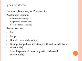
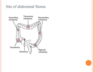
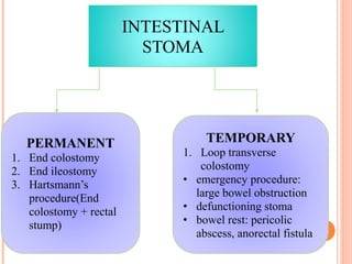
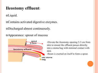
Definition
Greek: ‘mouth’
Stoma = an artificial opening in the abdominal wall, which
connects a hollow viscus(bowel, urinary tract) to the outside
environment/ to divert faeces or urine to the exterior which is
collected in an external appliance.
Natural openings: Nostrils, mouth, anus.
Intestinal stoma = opening of the intestinal tract onto
abdominal wall
Viscus: large interior organ in any of the great body cavities, e
specially those in the abdomen.
STOMA
Types of stoma
•Duration (Temporary or Permanent )
•Anatomical location:
•CNS: ventriculostomy
•Respiratory: tracheostomy
•GIT: ileostomy, colostomy
•Reconstruction:
 End
 Loop
 Double Barrel(Mickulicz)
 Bishop-Koop(distal ileostomy with end to side ileas
anatomosis)
 Santulli(proximal ileostomy with end-to-side
anastomosis)
Permanent stoma
● Necessary when there is no
distal bowel segment
remaining after resection or
when for some reason the
bowel cannot be re-joined
● Usually below the belt line
● Permanent colostomy: left
iliac fossa (LIF)
● Permanent ileostomy: right
iliac fossa(RIF)
Temporary stoma
• Relieve complete distal
large bowel obstruction
causing proximal
dilatation
Indication of Stoma
1. Feeding
– Percutaneous endoscopic gastrostmoy (PEG)
2. Lavage
– Appendectomy
3. Decompression
4. Diversion
– Protection/defunction of distal bowel anastomosis
● Previous contaminated bowel
● Iliorectal anastomosis
– Urinary diversion following cytectomy
5. Exteriorization
– Perforated or contaminated bowel (distal abscess or fistula)
– Permanent stoma (APR of rectum)
Site of abdominal Stoma
●Preparation of patient undergoing Stoma
1. Psychosocial and physical preparation
2. Explanation if indication and complication
3. Request help of Clinical Nurse Specialist in Stoma care
pre-operatively, who will mark the site.
4. Marking the stoma site (Pt standing up)
– Pt able to see the stoma well
– 5 cm from the umbilicus (spino-umbilical line away from all bony
prominence)
– Away from scar & skin creases
– Away from bony points or waistline of clothes
– Easily accessible to Pt (not under a large fold of fat)
5. The stoma within rectus abdominis sheath
●Examination of Stoma
● Inspection
1. Site
2. Types of stoma
3. Surrounding skin
4. Covering of surrounding skin
5. Loop
6. Stoma functioning
7. Stoma discharge
– Colour
– Type
– Amaount
● Palpation
1. General abdominal
palpation
2. Stoma?
● Percussion
– Shifting dullness
● Auscultation
– Bowel sound
● End examination
– PR exam
●Complication of Stoma
● General(d/t u/l dz)
– Stoma diarrhea
● Water&electrolyte
imbalance
● Hypokalemia
– Nutritional disorders
– Stones
● Gallstone
● Renal stone
– Psychosexual
– Residual disease
● Crohn’s disease
● Parastomal fistula
● Specific
– Skin excoriation
– Prolapse/gangrene/ necrosis
of distal end
– Bleeding
– Retraction
– Parastomal hernia
– Fistula formation
– Stenosis of orifice
● Cause constipation
1.Local: skin excoriation, dermatitis, candidiatis, ischaemia,
2.Structural: retracted, prolapse, stenosis, parastomal hernia
(support corset)
3.Systemic:dehydration,electrolyte imbalance, malabsorption
INTESTINAL
STOMA
PERMANENT
1. End colostomy
2. End ileostomy
3. Hartsmann’s
procedure(End
colostomy + rectal
stump)
TEMPORARY
1. Loop transverse
colostomy
• emergency procedure:
large bowel obstruction
• defunctioning stoma
• bowel rest: pericolic
abscess, anorectal fistula
ILEOSTOMY
essential in the management of neonates with certain
types of distal intestinal obstruction
e.g: long segment Hirschsprung disease, complex
meconium ileus, gastroschisis with atresia
Ileostomies are commonly placed to divert bowel
contents in neonatal necrotizing enterocolitis, ulcerative
colitis, familial polyposis
Ileostomy effluent:
Liquid.
Contains activated digestive enzymes.
Discharged almost continuously.
Appearance: sprout of mucosa
-Elevate the ileostomy opening 2-3 cm from
skin to ensure the effluent passes directly
into a stoma bag with minimal contact with
skin.
-Ileum is exerted on itself to form a spout.
End Ileostomy
Loop Ileostomy
Colostomy
A colostomy is an artificial opening made in the large
bowel to divert faeces and flatus to the exterior, where it
can be collected in an external appliance
Indication :
 Imperforate anus,
 Hirschsprung disease,
 Abdomino-perineal resection of a low rectal
 anal canal tumour
 diverticular disease.
Colostomy
By anatomy :
•Transverse colostomy
•Descending colostomy
•Sigmoid colostomy
By function :
•Decompressing
•Diversion
By construction :
• End
• Loop
• Double barrel
COLOSTOMY
●Type:
 Temporary (loop colostomy)
 Permanent (end colostomy)
●Indications:
 Diverticular disease
 Colorectal cancer
●Appearance:
 Flush with the skin (#)
 Mucosa sutured to skin
●Location:
 Permanent at LIF
Temporary at LIF or right hypochondrium
●Effluent:
intermittent and solid
Colostomies are sutured flush with skin.
Allowed to pout slightly to prevent retraction after weight gain
End Colostomy
Double-barrel colostomy
When creating a double-barrel colostomy, the surgeon divides
the bowel completely.(2 stoma besides each other and separate
from each other)
Each opening is brought to the surface as a separate stoma
Proximal-end = end stoma (secrets stool), needs a drainage
bag.
Distal-end= mucous fistula (secretes mucus)
Temporary stoma
Hartmann’s Procedure
 Surgical diversion of urinary system
 Done for baldder Ca, urinary incontinence and neuropathic
bladders
Formation of urostomy
Needs ileal conduit, a segment of viable ileum mad like a tube where 1 end is open
(used as stoma) and another end is closed( used as reserve).
Ureters are implanted into this isolated segment of small bowel tube
The open-end of conduit is everted to create a similar spout as ileostomy and allows
diversion of urine from kidneys to outside the abdomen and collected by stoma bag
Urostomy
Loop stoma – temporary stoma
VASCULAR COMPROMISE
● Ischaemia due to operative tissue trauma
● Intestinal necrosis due to ligation of arterial
supply/inadequate collateral arterial circulation
● Venous outflow obstruction > venous
congestion >necrosis of stoma
Stoma care
Parents, as well as older children, must be carefully taught and reassured before leaving the
hospital and on subsequent follow-up visits.
Properly fitted appliances should remain in situ for several days (change every 3 days).
There are two basic types of pediatric appliances:
the one-piece pouching system in which the adhesive skin barrier is already attached
to the pouch
the two-piece system in which the adhesive skin barrier is separate from the pouch.
Candidiasis remains a common problem in the parastomal skin, and local antifungal
medication should be used at the earliest sign of irritation.
With skin excoriation, the area is exposed to air and a synthetic barrier is applied. A
hairdryer can be useful.
application of silver nitrate may be necessary to control granulation tissue around the
mucosa-skin interface in the early stages.
Colostomy bags and appliances
Stoma
Stoma

Stoma

  • 1.
    Definition Greek: ‘mouth’ Stoma =an artificial opening in the abdominal wall, which connects a hollow viscus(bowel, urinary tract) to the outside environment/ to divert faeces or urine to the exterior which is collected in an external appliance. Natural openings: Nostrils, mouth, anus. Intestinal stoma = opening of the intestinal tract onto abdominal wall Viscus: large interior organ in any of the great body cavities, e specially those in the abdomen. STOMA
  • 2.
    Types of stoma •Duration(Temporary or Permanent ) •Anatomical location: •CNS: ventriculostomy •Respiratory: tracheostomy •GIT: ileostomy, colostomy •Reconstruction:  End  Loop  Double Barrel(Mickulicz)  Bishop-Koop(distal ileostomy with end to side ileas anatomosis)  Santulli(proximal ileostomy with end-to-side anastomosis)
  • 3.
    Permanent stoma ● Necessarywhen there is no distal bowel segment remaining after resection or when for some reason the bowel cannot be re-joined ● Usually below the belt line ● Permanent colostomy: left iliac fossa (LIF) ● Permanent ileostomy: right iliac fossa(RIF) Temporary stoma • Relieve complete distal large bowel obstruction causing proximal dilatation
  • 4.
    Indication of Stoma 1.Feeding – Percutaneous endoscopic gastrostmoy (PEG) 2. Lavage – Appendectomy 3. Decompression 4. Diversion – Protection/defunction of distal bowel anastomosis ● Previous contaminated bowel ● Iliorectal anastomosis – Urinary diversion following cytectomy 5. Exteriorization – Perforated or contaminated bowel (distal abscess or fistula) – Permanent stoma (APR of rectum)
  • 5.
  • 6.
    ●Preparation of patientundergoing Stoma 1. Psychosocial and physical preparation 2. Explanation if indication and complication 3. Request help of Clinical Nurse Specialist in Stoma care pre-operatively, who will mark the site. 4. Marking the stoma site (Pt standing up) – Pt able to see the stoma well – 5 cm from the umbilicus (spino-umbilical line away from all bony prominence) – Away from scar & skin creases – Away from bony points or waistline of clothes – Easily accessible to Pt (not under a large fold of fat) 5. The stoma within rectus abdominis sheath
  • 7.
    ●Examination of Stoma ●Inspection 1. Site 2. Types of stoma 3. Surrounding skin 4. Covering of surrounding skin 5. Loop 6. Stoma functioning 7. Stoma discharge – Colour – Type – Amaount ● Palpation 1. General abdominal palpation 2. Stoma? ● Percussion – Shifting dullness ● Auscultation – Bowel sound ● End examination – PR exam
  • 8.
    ●Complication of Stoma ●General(d/t u/l dz) – Stoma diarrhea ● Water&electrolyte imbalance ● Hypokalemia – Nutritional disorders – Stones ● Gallstone ● Renal stone – Psychosexual – Residual disease ● Crohn’s disease ● Parastomal fistula ● Specific – Skin excoriation – Prolapse/gangrene/ necrosis of distal end – Bleeding – Retraction – Parastomal hernia – Fistula formation – Stenosis of orifice ● Cause constipation 1.Local: skin excoriation, dermatitis, candidiatis, ischaemia, 2.Structural: retracted, prolapse, stenosis, parastomal hernia (support corset) 3.Systemic:dehydration,electrolyte imbalance, malabsorption
  • 9.
    INTESTINAL STOMA PERMANENT 1. End colostomy 2.End ileostomy 3. Hartsmann’s procedure(End colostomy + rectal stump) TEMPORARY 1. Loop transverse colostomy • emergency procedure: large bowel obstruction • defunctioning stoma • bowel rest: pericolic abscess, anorectal fistula
  • 10.
    ILEOSTOMY essential in themanagement of neonates with certain types of distal intestinal obstruction e.g: long segment Hirschsprung disease, complex meconium ileus, gastroschisis with atresia Ileostomies are commonly placed to divert bowel contents in neonatal necrotizing enterocolitis, ulcerative colitis, familial polyposis
  • 11.
    Ileostomy effluent: Liquid. Contains activateddigestive enzymes. Discharged almost continuously. Appearance: sprout of mucosa -Elevate the ileostomy opening 2-3 cm from skin to ensure the effluent passes directly into a stoma bag with minimal contact with skin. -Ileum is exerted on itself to form a spout.
  • 12.
  • 13.
  • 16.
    Colostomy A colostomy isan artificial opening made in the large bowel to divert faeces and flatus to the exterior, where it can be collected in an external appliance Indication :  Imperforate anus,  Hirschsprung disease,  Abdomino-perineal resection of a low rectal  anal canal tumour  diverticular disease.
  • 17.
    Colostomy By anatomy : •Transversecolostomy •Descending colostomy •Sigmoid colostomy By function : •Decompressing •Diversion By construction : • End • Loop • Double barrel
  • 18.
    COLOSTOMY ●Type:  Temporary (loopcolostomy)  Permanent (end colostomy) ●Indications:  Diverticular disease  Colorectal cancer ●Appearance:  Flush with the skin (#)  Mucosa sutured to skin ●Location:  Permanent at LIF Temporary at LIF or right hypochondrium ●Effluent: intermittent and solid
  • 19.
    Colostomies are suturedflush with skin. Allowed to pout slightly to prevent retraction after weight gain
  • 20.
  • 21.
    Double-barrel colostomy When creatinga double-barrel colostomy, the surgeon divides the bowel completely.(2 stoma besides each other and separate from each other) Each opening is brought to the surface as a separate stoma Proximal-end = end stoma (secrets stool), needs a drainage bag. Distal-end= mucous fistula (secretes mucus) Temporary stoma
  • 24.
  • 25.
     Surgical diversionof urinary system  Done for baldder Ca, urinary incontinence and neuropathic bladders Formation of urostomy Needs ileal conduit, a segment of viable ileum mad like a tube where 1 end is open (used as stoma) and another end is closed( used as reserve). Ureters are implanted into this isolated segment of small bowel tube The open-end of conduit is everted to create a similar spout as ileostomy and allows diversion of urine from kidneys to outside the abdomen and collected by stoma bag Urostomy
  • 27.
    Loop stoma –temporary stoma
  • 28.
    VASCULAR COMPROMISE ● Ischaemiadue to operative tissue trauma ● Intestinal necrosis due to ligation of arterial supply/inadequate collateral arterial circulation ● Venous outflow obstruction > venous congestion >necrosis of stoma
  • 31.
    Stoma care Parents, aswell as older children, must be carefully taught and reassured before leaving the hospital and on subsequent follow-up visits. Properly fitted appliances should remain in situ for several days (change every 3 days). There are two basic types of pediatric appliances: the one-piece pouching system in which the adhesive skin barrier is already attached to the pouch the two-piece system in which the adhesive skin barrier is separate from the pouch. Candidiasis remains a common problem in the parastomal skin, and local antifungal medication should be used at the earliest sign of irritation. With skin excoriation, the area is exposed to air and a synthetic barrier is applied. A hairdryer can be useful. application of silver nitrate may be necessary to control granulation tissue around the mucosa-skin interface in the early stages.
  • 32.

Editor's Notes

  • #4 Usual indications are IBD & familial adenomatous polyposis