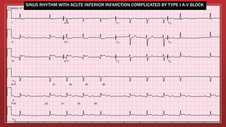
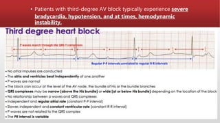
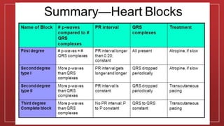
HEART
BLOCKS
NOEL CHRISTIAN
GROUP 6
▪ Heart block is an abnormal heart rhythm where the heart beats
too slowly (bradycardia).
▪ In this condition, the electrical signals that tell the heart to
contract are partially or totally blocked between the atria and
the ventricles. For this reason, it is also called atrioventricular
block (AV block).
What is Heart Block?
▪ A normal heartbeat is initiated by an electrical signal that
comes from the heart’s natural pacemaker, the sinoatrial
(SA) node, located at the top of the right atrium.
▪ The electrical signal travels through the atria and reaches the
atrioventricular (AV) node.
▪ After crossing the AV node, the electrical signal passes
through the bundle of His.
▪ This bundle then divides into thin, wire-like structures called
bundle branches that extend into the right and left
▪ The electrical signal travels down the bundle branches and
eventually reach the muscle cells of the ventricles, causing
them to contract and pump blood to the body.
▪ Heart block occurs when this passage of electricity from top
to bottom of the heart is delayed or interrupted.
Types of Heart Block: First Degree Heart Block
▪ The electrical impulses are slowed as they pass through the
conduction system, but they all successfully reach the ventricles.
▪ First-degree heart block rarely causes symptoms or problems.
▪ Well-trained athletes may have first-degree heart block. Some
medications can also cause this condition. No treatment is
generally needed for first-degree heart block.
▪ PR prolongation, is a disease of the electrical conduction
system of the heart in which the PR interval is lengthened
beyond 0.20 seconds.
Cause
▪ AV nodal disease, enhanced vagal tone (for example in
athletes), myocarditis, acute myocardial infarction (especially
acute inferior MI), electrolyte disturbances and medication.
▪ The drugs which can cause first-degree heart block are those
that increase the refractory time of the AV node, thereby
AV conduction.
▪ These include calcium channel blockers, beta-blockers.
Diagnosis
▪ In normal individuals, the AV node slows the conduction of
electrical impulse through the heart. This is manifest on a
surface electrocardiogram (ECG) as the PR interval.
▪ The normal PR interval is from 120 ms to 200 ms in length.
▪ In first-degree heart block, the diseased AV node conducts the
electrical activity more slowly. This is seen as a PR interval
greater than 200 ms in length on the surface ECG. It is usually
an incidental finding on a routine ECG not always present.
Second-degree heart block
▪ The electrical impulses are delayed further and further with each
subsequent heartbeat until a beat fails to reach to the ventricles
entirely.
▪ This type of block most often is physiologic and is seen in a
highly relaxed state and during sleep. It rarely causes symptoms.
It sometimes causes dizziness and/or other symptoms.
▪ There are two non-distinct types of second-degree AV block,
called Type 1 and Type 2.
Type 1 (Mobitz I/Wenckebach)
▪ Type 1 Second-degree AV block, also known as Mobitz I or
Wenckebach periodicity, is almost always a disease of the AV
node.
▪ Mobitz I heart block is characterized by progressive
prolongation of the PR interval on consecutive beats
by a blocked P wave (i.e., a 'dropped' QRS complex). After the
dropped QRS complex, the PR interval resets and the cycle
repeats.
▪ Mobitz I heart block is that the atrial rhythm has to be regular.
▪ This is almost always a benign condition for which no specific
treatment is needed for the rhythm itself.
▪ It can be seen in myocardial ischemia, propranolol use,
rheumatic fever, and chronically in ischemic heart disease and
other structural diseases (amyloidosis, mitral valve prolapse,
aortic valve disease, and atrial septal defect.
▪ In symptomatic cases, intravenous atropine or isoproterenol
may transiently improve conduction.
SINUS RHYTHM WITH ACUTE INFERIOR INFARCTION COMPLICATED BY TYPE I A-V BLOCK
Type 2 (Mobitz II/Hay)
▪ Type 2 Second-degree AV block, also known as "Mobitz II," is
almost always a disease of the distal conduction system (His-
Purkinje System).
▪ With this condition, some of the electrical impulses are unable to
reach the ventricles.
▪ This condition is less common than Type I, and is more serious.
Usually, your doctor will recommend a pacemaker to treat type II
second degree heart block, as it frequently progresses to third
degree heart block
▪ Mobitz II heart block is characterized on a surface ECG by
intermittently nonconducted P waves not preceded by PR
prolongation and not followed by PR shortening i.e, fixed P-R
interval.
▪ The medical significance of this type of AV block is that it may
progress rapidly to complete heart block, in which no escape
rhythm may emerge. In this case, the person may experience a
Stokes-Adams attack, cardiac arrest, or sudden cardiac
▪ The definitive treatment for this form of AV Block is an
implanted pacemaker.
Third Degree Heart Block
▪ With this condition, also called complete heart block, none of
the electrical impulses from the atria reach the ventricles.
▪ When the ventricles do not receive electrical impulses from the
atria, they may generate some impulses on their own, called
junctional or ventricular escape beats.
▪ Ventricular escape beats, the heart’s naturally occurring backups,
are usually very slow.
▪ Patients frequently feel fatigue, lightheadedness, and
decreased stamina in complete heart block. Patients are usually
treated by implanting a permanent pacemaker.
Two independent rhythms can be noted on the electrocardiogram
(ECG).
▪ The P waves with a regular P-to-P interval (in other words, a
sinus rhythm) represent the first rhythm.
▪ The QRS complexes with a regular R-to-R interval represent the
second rhythm.
▪ The PR interval will be variable, as the hallmark of complete
heart block is lack of any apparent relationship between P waves
and QRS complexes.
▪ Patients with third-degree AV block typically experience severe
bradycardia, hypotension, and at times, hemodynamic
instability.
ATRIAL TACHYCARDIA WITH COMPLETE A-V BLOCK AND RESULTING JUNCTIONAL ESCAPE
Cause
▪ Many conditions can cause third-degree heart block, but the
most common cause is coronary ischemia. Progressive
degeneration of the electrical conduction system of the heart
can lead to third-degree heart block.
▪ An inferior wall myocardial infarction may cause damage to
the AV node, causing third-degree heart block.
▪ An anterior wall myocardial infarction may damage the
distal conduction system of the heart, causing third-degree
heart block. This is typically extensive, permanent damage to the
conduction system, necessitating a permanent pacemaker to
be placed.
Symptoms of Heart Block
Some people with heart block will not experience any symptoms.
Others will have symptoms that may include the following:
•Fainting (syncope )
•Dizziness Lightheadedness
•Shortness of breath
•Decline in exercise capacity
Risk factors for Heart Block
Some medical conditions increase the risk for developing heart block.
These medical conditions include:
▪ Heart failure
▪ Prior heart attack
▪ Heart valve abnormalities
▪ Heart valve surgery
▪ Congenital heart diseases
▪ Some medications or exposure to toxic substances
▪ Lyme disease
▪ Aging
References
▪ "ECG Conduction Abnormalities". Retrieved 2009-01-07.
▪ Jump up^ "Heart Block". NHS Choices. National Health Service (UK).
Retrieved 25 August 2015.
▪ Jump up^ Sclarovsky, S; Strasberg, B; Hirshberg, A; Arditi, A; Lewin, RF;
Agmon, J (July 1984). "Advanced early and late atrioventricular block in
acute inferior wall myocardial infarction". American Heart
Journal. 108 (1): 19–24. doi:10.1016/0002-8703(84)90539-
8. PMID 6731277.
▪ Dretzke, J.; et al. "Compared to single chamber ventricular pacemakers,
dual chamber pacemakers may reduce the incidence of complications in
people with sick sinus syndrome and atrioventricular block". Cochrane
reviews. Retrieved 25 August2015.
▪ Jump up^ "Peri-arrest arrhythmias". Resuscitation guidelines.
Resuscitation Council UK. Retrieved 25 August 2015.
Heart blocks

Heart blocks

  • 1.
  • 2.
    ▪ Heart blockis an abnormal heart rhythm where the heart beats too slowly (bradycardia). ▪ In this condition, the electrical signals that tell the heart to contract are partially or totally blocked between the atria and the ventricles. For this reason, it is also called atrioventricular block (AV block).
  • 3.
    What is HeartBlock? ▪ A normal heartbeat is initiated by an electrical signal that comes from the heart’s natural pacemaker, the sinoatrial (SA) node, located at the top of the right atrium. ▪ The electrical signal travels through the atria and reaches the atrioventricular (AV) node. ▪ After crossing the AV node, the electrical signal passes through the bundle of His.
  • 4.
    ▪ This bundlethen divides into thin, wire-like structures called bundle branches that extend into the right and left ▪ The electrical signal travels down the bundle branches and eventually reach the muscle cells of the ventricles, causing them to contract and pump blood to the body. ▪ Heart block occurs when this passage of electricity from top to bottom of the heart is delayed or interrupted.
  • 6.
    Types of HeartBlock: First Degree Heart Block ▪ The electrical impulses are slowed as they pass through the conduction system, but they all successfully reach the ventricles. ▪ First-degree heart block rarely causes symptoms or problems. ▪ Well-trained athletes may have first-degree heart block. Some medications can also cause this condition. No treatment is generally needed for first-degree heart block. ▪ PR prolongation, is a disease of the electrical conduction system of the heart in which the PR interval is lengthened beyond 0.20 seconds.
  • 8.
    Cause ▪ AV nodaldisease, enhanced vagal tone (for example in athletes), myocarditis, acute myocardial infarction (especially acute inferior MI), electrolyte disturbances and medication. ▪ The drugs which can cause first-degree heart block are those that increase the refractory time of the AV node, thereby AV conduction. ▪ These include calcium channel blockers, beta-blockers.
  • 9.
    Diagnosis ▪ In normalindividuals, the AV node slows the conduction of electrical impulse through the heart. This is manifest on a surface electrocardiogram (ECG) as the PR interval. ▪ The normal PR interval is from 120 ms to 200 ms in length. ▪ In first-degree heart block, the diseased AV node conducts the electrical activity more slowly. This is seen as a PR interval greater than 200 ms in length on the surface ECG. It is usually an incidental finding on a routine ECG not always present.
  • 11.
    Second-degree heart block ▪The electrical impulses are delayed further and further with each subsequent heartbeat until a beat fails to reach to the ventricles entirely. ▪ This type of block most often is physiologic and is seen in a highly relaxed state and during sleep. It rarely causes symptoms. It sometimes causes dizziness and/or other symptoms. ▪ There are two non-distinct types of second-degree AV block, called Type 1 and Type 2.
  • 12.
    Type 1 (MobitzI/Wenckebach) ▪ Type 1 Second-degree AV block, also known as Mobitz I or Wenckebach periodicity, is almost always a disease of the AV node. ▪ Mobitz I heart block is characterized by progressive prolongation of the PR interval on consecutive beats by a blocked P wave (i.e., a 'dropped' QRS complex). After the dropped QRS complex, the PR interval resets and the cycle repeats. ▪ Mobitz I heart block is that the atrial rhythm has to be regular.
  • 14.
    ▪ This isalmost always a benign condition for which no specific treatment is needed for the rhythm itself. ▪ It can be seen in myocardial ischemia, propranolol use, rheumatic fever, and chronically in ischemic heart disease and other structural diseases (amyloidosis, mitral valve prolapse, aortic valve disease, and atrial septal defect. ▪ In symptomatic cases, intravenous atropine or isoproterenol may transiently improve conduction.
  • 15.
    SINUS RHYTHM WITHACUTE INFERIOR INFARCTION COMPLICATED BY TYPE I A-V BLOCK
  • 16.
    Type 2 (MobitzII/Hay) ▪ Type 2 Second-degree AV block, also known as "Mobitz II," is almost always a disease of the distal conduction system (His- Purkinje System). ▪ With this condition, some of the electrical impulses are unable to reach the ventricles. ▪ This condition is less common than Type I, and is more serious. Usually, your doctor will recommend a pacemaker to treat type II second degree heart block, as it frequently progresses to third degree heart block
  • 17.
    ▪ Mobitz IIheart block is characterized on a surface ECG by intermittently nonconducted P waves not preceded by PR prolongation and not followed by PR shortening i.e, fixed P-R interval. ▪ The medical significance of this type of AV block is that it may progress rapidly to complete heart block, in which no escape rhythm may emerge. In this case, the person may experience a Stokes-Adams attack, cardiac arrest, or sudden cardiac ▪ The definitive treatment for this form of AV Block is an implanted pacemaker.
  • 19.
    Third Degree HeartBlock ▪ With this condition, also called complete heart block, none of the electrical impulses from the atria reach the ventricles. ▪ When the ventricles do not receive electrical impulses from the atria, they may generate some impulses on their own, called junctional or ventricular escape beats. ▪ Ventricular escape beats, the heart’s naturally occurring backups, are usually very slow. ▪ Patients frequently feel fatigue, lightheadedness, and decreased stamina in complete heart block. Patients are usually treated by implanting a permanent pacemaker.
  • 20.
    Two independent rhythmscan be noted on the electrocardiogram (ECG). ▪ The P waves with a regular P-to-P interval (in other words, a sinus rhythm) represent the first rhythm. ▪ The QRS complexes with a regular R-to-R interval represent the second rhythm. ▪ The PR interval will be variable, as the hallmark of complete heart block is lack of any apparent relationship between P waves and QRS complexes.
  • 21.
    ▪ Patients withthird-degree AV block typically experience severe bradycardia, hypotension, and at times, hemodynamic instability.
  • 22.
    ATRIAL TACHYCARDIA WITHCOMPLETE A-V BLOCK AND RESULTING JUNCTIONAL ESCAPE
  • 23.
    Cause ▪ Many conditionscan cause third-degree heart block, but the most common cause is coronary ischemia. Progressive degeneration of the electrical conduction system of the heart can lead to third-degree heart block. ▪ An inferior wall myocardial infarction may cause damage to the AV node, causing third-degree heart block. ▪ An anterior wall myocardial infarction may damage the distal conduction system of the heart, causing third-degree heart block. This is typically extensive, permanent damage to the conduction system, necessitating a permanent pacemaker to be placed.
  • 24.
    Symptoms of HeartBlock Some people with heart block will not experience any symptoms. Others will have symptoms that may include the following: •Fainting (syncope ) •Dizziness Lightheadedness •Shortness of breath •Decline in exercise capacity
  • 25.
    Risk factors forHeart Block Some medical conditions increase the risk for developing heart block. These medical conditions include: ▪ Heart failure ▪ Prior heart attack ▪ Heart valve abnormalities ▪ Heart valve surgery ▪ Congenital heart diseases ▪ Some medications or exposure to toxic substances ▪ Lyme disease ▪ Aging
  • 28.
    References ▪ "ECG ConductionAbnormalities". Retrieved 2009-01-07. ▪ Jump up^ "Heart Block". NHS Choices. National Health Service (UK). Retrieved 25 August 2015. ▪ Jump up^ Sclarovsky, S; Strasberg, B; Hirshberg, A; Arditi, A; Lewin, RF; Agmon, J (July 1984). "Advanced early and late atrioventricular block in acute inferior wall myocardial infarction". American Heart Journal. 108 (1): 19–24. doi:10.1016/0002-8703(84)90539- 8. PMID 6731277. ▪ Dretzke, J.; et al. "Compared to single chamber ventricular pacemakers, dual chamber pacemakers may reduce the incidence of complications in people with sick sinus syndrome and atrioventricular block". Cochrane reviews. Retrieved 25 August2015. ▪ Jump up^ "Peri-arrest arrhythmias". Resuscitation guidelines. Resuscitation Council UK. Retrieved 25 August 2015.