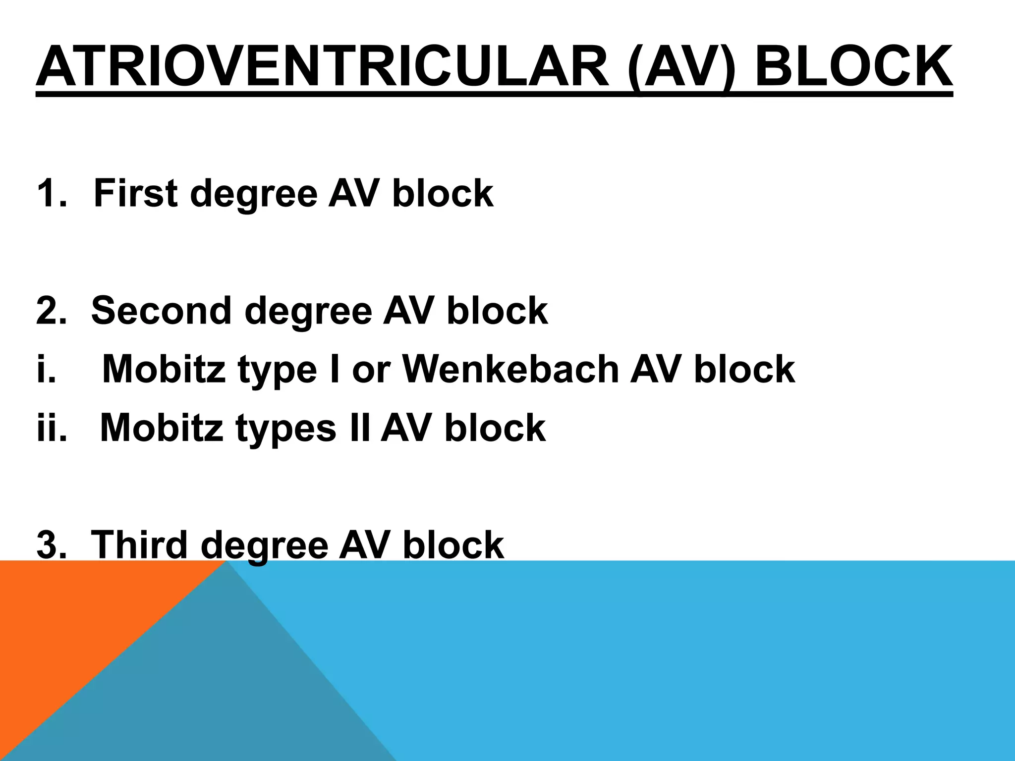
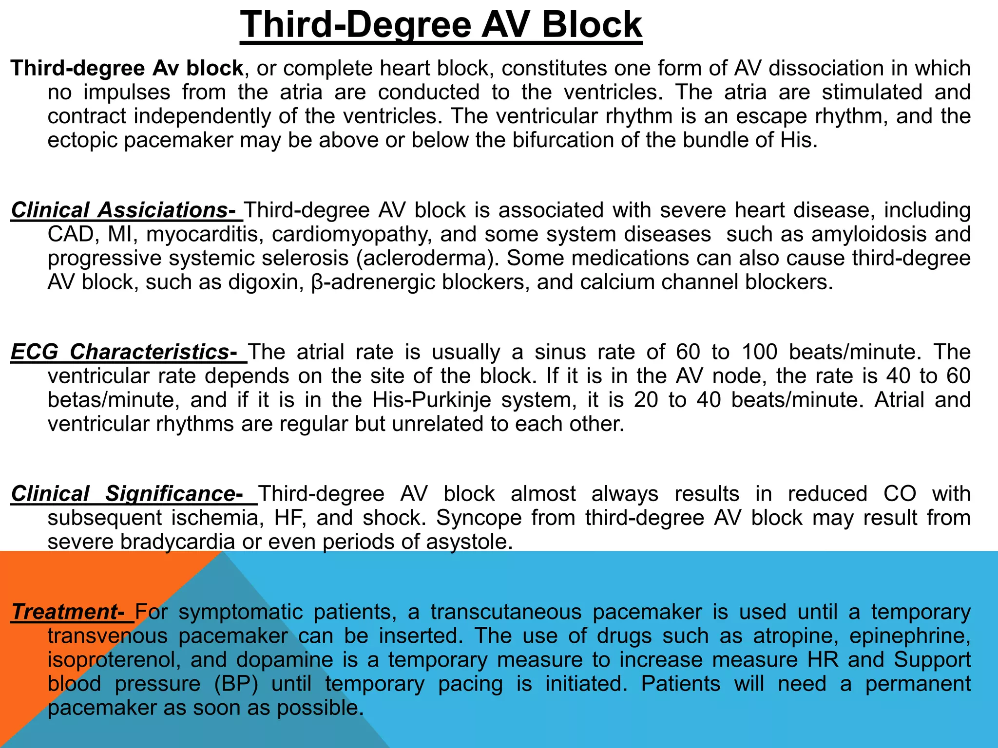
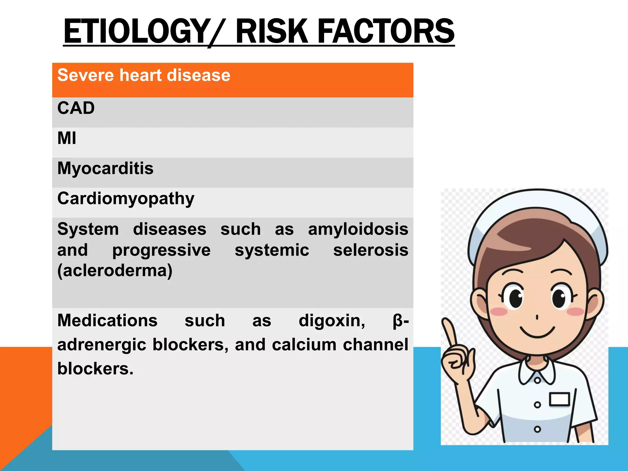
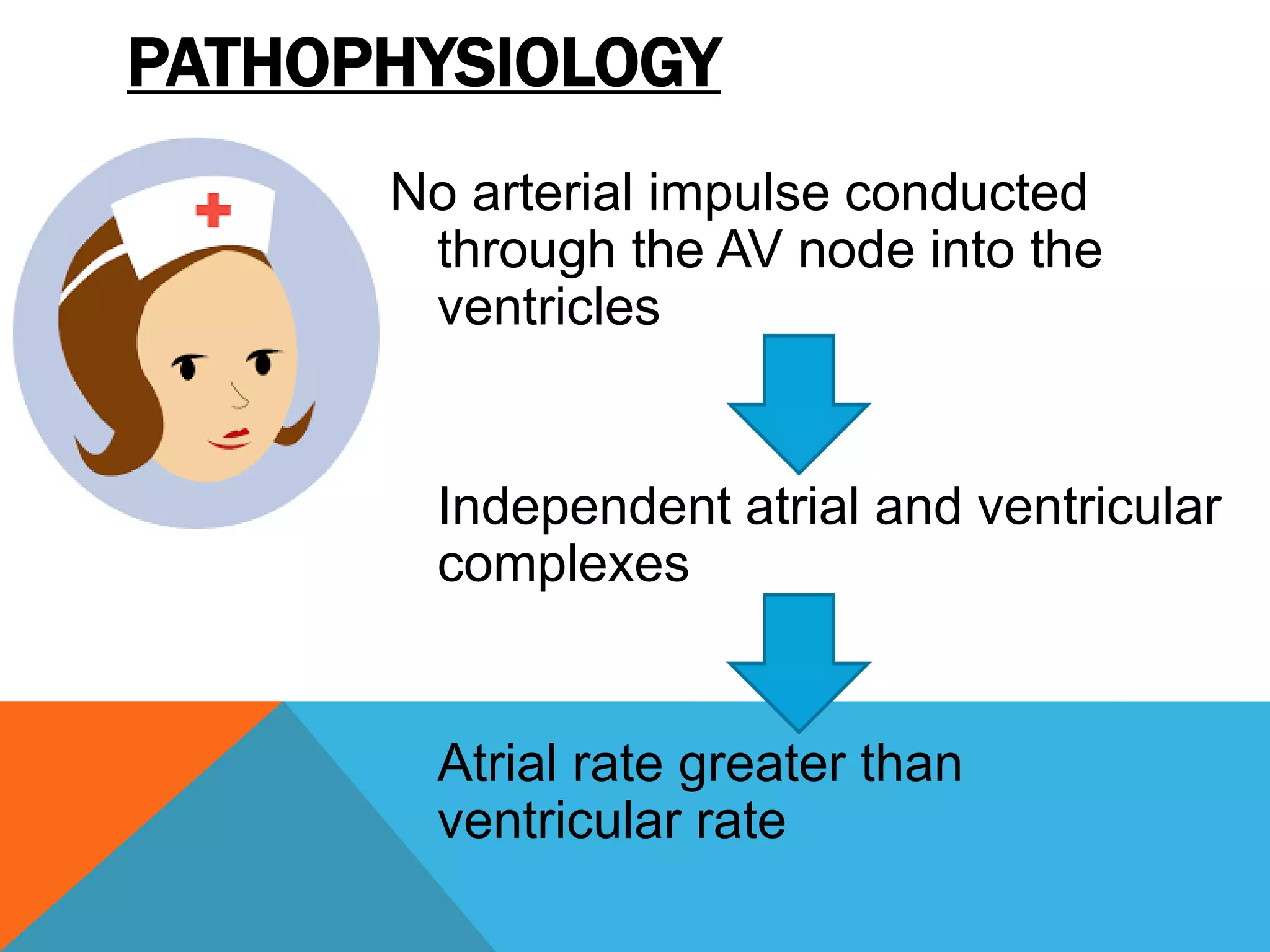
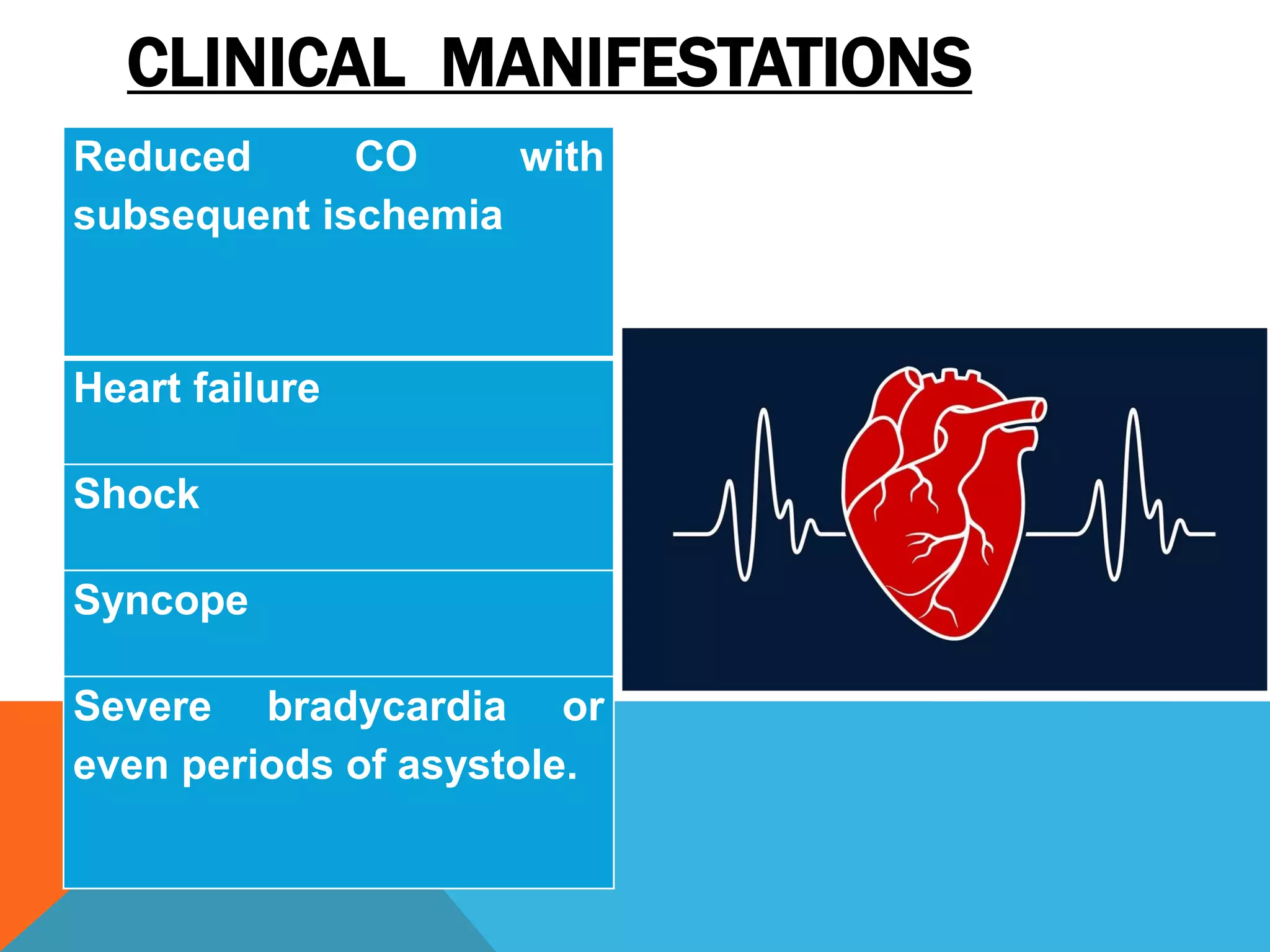
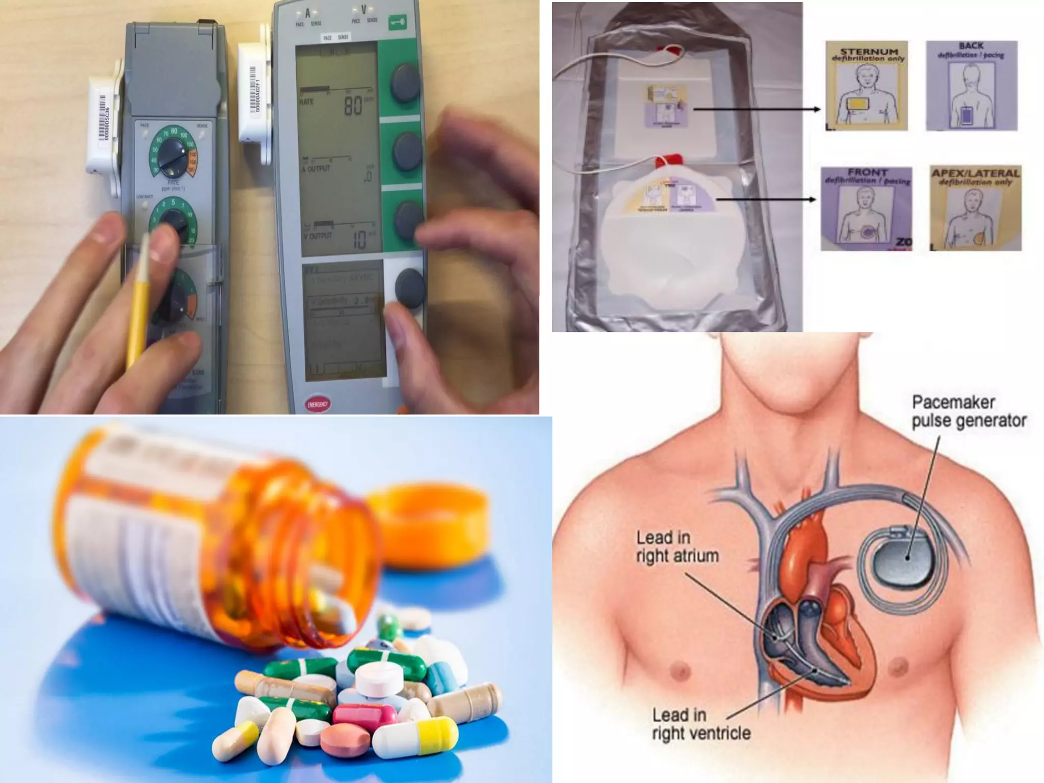
Cardiogenic shock : Medical Surgical NursingRaksha Yadav
This
presentation is designed for Nursing students and it gives a brief
about what you should know while caring for a client with Cardiogenic
shock and also its prevention.
Cardiogenic shock is a condition of diminished cardiac output that severely impairs cardiac perfusion. In this condition in which the heart suddenly can't pump enough blood to meet the body's needs.
Nursing management of patient with cardiac surgeries.PrashantSalve10
It will be helpful to overview cardiac surgeries like CABG, Valvular surgeries and heart transplant. It also enumerates the nursing diagnoses and its brief description.
Cardiomyopathy, or heart muscle disease, is a type of progressive heart disease in which the heart is abnormally enlarged, thickened, and/or stiffened. As a result, the heart muscle's ability to pump blood is less efficient, often causing heart failure and the backup of blood into the lungs or rest of the body. The disease can also cause abnormal heart rhythms.
Myocardial infarction (MI), commonly known as a heart attack, occurs when blood flow decreases or stops to a part of the heart, causing damage to the heart muscle. The most common symptom is chest pain or discomfort which may travel into the shoulder, arm, back, neck, or jaw.
Small intestine perforation- Easy ppt for student nurses
definition
causes
clinical manifestations
diagnostic tests
management of small intestine perforation
Introduction to Nursing-
Concept
Meaning
Definition of Nursing-
1. According to Florence Nightingale
2. According to American Nurses Association
Scope of Nursing
Aims of Nursing
Roles, Responsibilities and functions of nursing
Review of lungs anatomy and physiology- Easy explanation for students
Introduction
Anatomy
Air movement through Lungs
Physiology of Respiration
Assessment of Respiratory System
Types of bed in Nursing- easy explanation for Student Nurses
CLOSED BED
OPEN BED
ADMISSION BED
OCCUPIED BED
OPERATION BED/POST ANESTHESIA BED/RECOVERY BED
CARDIAC BED
FRACTURE BED
AMPUTATION BED/STUMP BED
BURN BED
Cancer of Breast - easy ppt for Nursing Students
Definition
Risk factors
Clinical manifestations
Assessment & diagnostic findings
Management
Surgery
Breast cancer rehabilitation
Pharyngitis- Easy PPT for Nursing StudentsSwatilekha Das
Pharyngitis- Easy PPT for Nursing Students
organs of the respiratory system
Definition
Acute Pharyngitis definition
Causes
Clinical features
Diagnosis
Treatment
Nutritional therapy
Chronic Pharyngitis
types
Clinical manifestations
Medical management
Cancer of Prostate- Easy PPT for Nursing StudentsSwatilekha Das
Cancer of Prostate- Easy PPT for Nursing Students
Definition & picture
Risk factors
Clinical manifestations
Assessment & diagnostic methods
Medical management
Surgical Management
-Radical prostatectomy
Radiation therapy
Hormone therapy
Other therapies
Thank you
CHAPTER 1 SEMESTER V PREVENTIVE-PEDIATRICS.pdfSachin Sharma
This content provides an overview of preventive pediatrics. It defines preventive pediatrics as preventing disease and promoting children's physical, mental, and social well-being to achieve positive health. It discusses antenatal, postnatal, and social preventive pediatrics. It also covers various child health programs like immunization, breastfeeding, ICDS, and the roles of organizations like WHO, UNICEF, and nurses in preventive pediatrics.
Antibiotic Stewardship by Anushri Srivastava.pptxAnushriSrivastav
Stewardship is the act of taking good care of something.
Antimicrobial stewardship is a coordinated program that promotes the appropriate use of antimicrobials (including antibiotics), improves patient outcomes, reduces microbial resistance, and decreases the spread of infections caused by multidrug-resistant organisms.
WHO launched the Global Antimicrobial Resistance and Use Surveillance System (GLASS) in 2015 to fill knowledge gaps and inform strategies at all levels.
ACCORDING TO apic.org,
Antimicrobial stewardship is a coordinated program that promotes the appropriate use of antimicrobials (including antibiotics), improves patient outcomes, reduces microbial resistance, and decreases the spread of infections caused by multidrug-resistant organisms.
ACCORDING TO pewtrusts.org,
Antibiotic stewardship refers to efforts in doctors’ offices, hospitals, long term care facilities, and other health care settings to ensure that antibiotics are used only when necessary and appropriate
According to WHO,
Antimicrobial stewardship is a systematic approach to educate and support health care professionals to follow evidence-based guidelines for prescribing and administering antimicrobials
In 1996, John McGowan and Dale Gerding first applied the term antimicrobial stewardship, where they suggested a causal association between antimicrobial agent use and resistance. They also focused on the urgency of large-scale controlled trials of antimicrobial-use regulation employing sophisticated epidemiologic methods, molecular typing, and precise resistance mechanism analysis.
Antimicrobial Stewardship(AMS) refers to the optimal selection, dosing, and duration of antimicrobial treatment resulting in the best clinical outcome with minimal side effects to the patients and minimal impact on subsequent resistance.
According to the 2019 report, in the US, more than 2.8 million antibiotic-resistant infections occur each year, and more than 35000 people die. In addition to this, it also mentioned that 223,900 cases of Clostridoides difficile occurred in 2017, of which 12800 people died. The report did not include viruses or parasites
VISION
Being proactive
Supporting optimal animal and human health
Exploring ways to reduce overall use of antimicrobials
Using the drugs that prevent and treat disease by killing microscopic organisms in a responsible way
GOAL
to prevent the generation and spread of antimicrobial resistance (AMR). Doing so will preserve the effectiveness of these drugs in animals and humans for years to come.
being to preserve human and animal health and the effectiveness of antimicrobial medications.
to implement a multidisciplinary approach in assembling a stewardship team to include an infectious disease physician, a clinical pharmacist with infectious diseases training, infection preventionist, and a close collaboration with the staff in the clinical microbiology laboratory
to prevent antimicrobial overuse, misuse and abuse.
to minimize the developme
QA Paediatric dentistry department, Hospital Melaka 2020Azreen Aj
QA study - To improve the 6th monthly recall rate post-comprehensive dental treatment under general anaesthesia in paediatric dentistry department, Hospital Melaka
We understand the unique challenges pickleball players face and are committed to helping you stay healthy and active. In this presentation, we’ll explore the three most common pickleball injuries and provide strategies for prevention and treatment.
How many patients does case series should have In comparison to case reports.pdfpubrica101
Pubrica’s team of researchers and writers create scientific and medical research articles, which may be important resources for authors and practitioners. Pubrica medical writers assist you in creating and revising the introduction by alerting the reader to gaps in the chosen study subject. Our professionals understand the order in which the hypothesis topic is followed by the broad subject, the issue, and the backdrop.
https://pubrica.com/academy/case-study-or-series/how-many-patients-does-case-series-should-have-in-comparison-to-case-reports/
One of the most developed cities of India, the city of Chennai is the capital of Tamilnadu and many people from different parts of India come here to earn their bread and butter. Being a metropolitan, the city is filled with towering building and beaches but the sad part as with almost every Indian city
2. INTRODUCTION
Heart block is a disturbance of impulse conduction
that can be permanent or transient owing to
anatomical or functional impairment. It must be
distinguished from interference, a normal
phenomenon that is a disturbance of impulse
conduction caused by physiological refractoriness
due to in excitability from a preceding impulse.
3. ELECTRICAL CONDUCTION SYSTEM OF
HEART
The heart is able to create its own electrical impulses and
control the route the impulses take via a specialised
conduction pathway.
This pathway is made up of 5 elements:
1.The sino-atrial (SA) node
2.The atrio-ventricular (AV) node
3. The bundle of His
4.The left and right bundle branches
5. The Purkinje fibres
5. The SA node is the natural pacemaker of the heart. The SA node releases
electrical stimuli at a regular rate, the rate is dictated by the needs of
the body. Each stimulus passes through the myocardial cells of the atria
creating a wave of contraction which spreads rapidly through both atria.
The electrical stimulus from the SA node eventually reaches the AV
node and is delayed briefly so that the contracting atria have enough
time to pump all the blood into the ventricles. Once the atria are empty
of blood the valves between the atria and ventricles close.
At this point the atria begin to refill and the electrical stimulus passes
through the AV node and Bundle of His into the Bundle branches and
Purkinje fibres. As the ventricles contract, the right ventricle pumps
blood to the lungs where carbon dioxide is released and oxygen is
absorbed, whilst the left ventricle pumps blood into the aorta from
where it passes into the coronary and arterial circulation.
At this point the ventricles are empty, the atria are full and the valves
between them are closed. The SA node is about to release another
electrical stimulus and the process is about to repeat itself.
6. The 3 stages of a single heart beat are:
Atrial depolarisation
Ventricular depolarisation
Atrial and ventricular repolarisation.
Normal sinus rhythm of heart
7. ATRIOVENTRICULAR (AV) BLOCK
1. First degree AV block
2. Second degree AV block
i. Mobitz type I or Wenkebach AV block
ii. Mobitz types II AV block
3. Third degree AV block
8. First-Degree AV Block
First-degree AV block is a type of AV block in which every impulse is conducted
to the ventricles but the duration of AV conduction is prolonged. After the
impulse moves through the AV node, it is usually conducted normally through
the ventricles.
Clinical Associations- First-degree AV block is associated with MI, CAD,
rheumatic fever, hyperthyroidism, vagal stimulation, and drugs such as
digoxin, β-adrenergic blockers, calcium channel blockers, flecainide.
ECG Characteristics- In first-degree AV block, HR is normal and rhythm is
regular.
Clinical Significance- First-degree AV block is usually not serious but can be a
precursor of higher degrees of AV block. Patients with first AV degree block
are asymptomatic.
Treatment- There is no treatment for first-degree AV block, modification to
causative medications may be considered. Patients should continue to be
monitored for any new changes in heart rhythm.
10. Second-degree AV block, Type I
Type I second-degree AV block (Mobitz I or Wenckebach heart block) includes a
gradual lengthening of the PR interval. It occurs because of a prolonged AV
conduction time until an atrial impulse is non-conducted and a QRS complex is
block (missing). Type I AV block most commonly occurs in the AV node, but it can
also occur in the His-Purkinje system.
Clinical Associations- Type I AV block may result from use of drugs such as digoxin
or β-adrenergic blockers. It may also be associated with CAD and other diseases
that can slow AV conduction.
ECG Characteristics- Atrial rate is normal, but ventricular rate may be slower as a
result of non-conducted or blocked QRS complexes.
Clinical Significances- Type I AV block is usually a result of myocardial ischemia or
infarction. It is almost transient and is usually well tolerated. However, in some
patients (e.g following MI) it may be a warning signal of a more serious AV
conduction disturbance.
Treatment- If the patient is symptomatic, atropine is used to increase HR, or a
temporary pacemaker may be needed, especially if the patient has experience an
MI. if the patient is asymptomatic, the rhythm should be closely observed with a
transcutaneous pacemaker on standby. Bradycardia is more likely to become
symptomatic when one or more of the following are present: (1) hypotension, (2)
HF, or (3) shock.
12. Second-Degree AV Block, Type II
In type II second-degree AV block (Mobitz II heart block), a P wave is non-conducted without
progressive antecedent PR lengthening. This almost always occurs when a block in one of the
bundle branches is present. On conducted beats, the PR is constant. Type II second-degree AV
block is a more serious type of block in which a certain number of impulses from the SA node
are not conducted to the ventricles. This occurs in ratios of 2:1, 3:1, and so on (i.e., two P
waves to one QRS complex, three P waves to one QRS complex). It may occur with varying
ratios. Type II AV block almost always occurs in the His-Purkinje system.
Clinical Associations- Type II AV block is associated with rheumatic heart disease, CAD, anterior
MI, and digitalis toxicity.
ECG Characteristics- Atrial rate is usually normal. Ventricular rate depends on the intrinsic rate
and the degree of AV block. Atrial rhythm is regular, but ventricular rhythm may be irregular.
Clinical Significance- Type II AV block often progresses to third-degree AV block and is associated
with a poor prognosis. The reduced HR often results in decreased CO with subsequent
hypotension and myocardial ischemia. Type II Av block is an indication for therapy with a
permanent pacemaker.
Treatment- Temporary treatment before the insertion of a permanent pacemaker may be necessary
if the patient becomes symptomatic (e.g, hypotension, angina), and involves the use of a
temporary transvenous or transcutaneous pacemaker.
14. Third-Degree AV Block
Third-degree Av block, or complete heart block, constitutes one form of AV dissociation in which
no impulses from the atria are conducted to the ventricles. The atria are stimulated and
contract independently of the ventricles. The ventricular rhythm is an escape rhythm, and the
ectopic pacemaker may be above or below the bifurcation of the bundle of His.
Clinical Assiciations- Third-degree AV block is associated with severe heart disease, including
CAD, MI, myocarditis, cardiomyopathy, and some system diseases such as amyloidosis and
progressive systemic selerosis (acleroderma). Some medications can also cause third-degree
AV block, such as digoxin, β-adrenergic blockers, and calcium channel blockers.
ECG Characteristics- The atrial rate is usually a sinus rate of 60 to 100 beats/minute. The
ventricular rate depends on the site of the block. If it is in the AV node, the rate is 40 to 60
betas/minute, and if it is in the His-Purkinje system, it is 20 to 40 beats/minute. Atrial and
ventricular rhythms are regular but unrelated to each other.
Clinical Significance- Third-degree AV block almost always results in reduced CO with
subsequent ischemia, HF, and shock. Syncope from third-degree AV block may result from
severe bradycardia or even periods of asystole.
Treatment- For symptomatic patients, a transcutaneous pacemaker is used until a temporary
transvenous pacemaker can be inserted. The use of drugs such as atropine, epinephrine,
isoproterenol, and dopamine is a temporary measure to increase measure HR and Support
blood pressure (BP) until temporary pacing is initiated. Patients will need a permanent
pacemaker as soon as possible.
16. ETIOLOGY/ RISK FACTORS
Severe heart disease
CAD
MI
Myocarditis
Cardiomyopathy
System diseases such as amyloidosis
and progressive systemic selerosis
(acleroderma)
Medications such as digoxin, β-
adrenergic blockers, and calcium channel
blockers.
17. PATHOPHYSIOLOGY
No arterial impulse conducted
through the AV node into the
ventricles
Independent atrial and ventricular
complexes
Atrial rate greater than
ventricular rate
19. DIAGNOSTIC EVALUATION
1. ECG (electrocardiogram)
2. Holter monitoring
3. Echocardiogram
4. Electrophysiology test
5. Tilt-table test
20. MANAGEMENT
1. A transcutaneous pacemaker is used until a temporary
transvenous pacemaker can be inserted.
2. The use of drugs such as atropine, epinephrine,
isoproterenol, and dopamine is a temporary measure to
increase measure HR and Support blood pressure (BP)
until temporary pacing is initiated.
3. Patients will need a permanent pacemaker as soon as
possible.
23. Nursing
Diagnosis
Goals Implementation Expected
outcome
Impaired
physical
mobility
related to
activity
restriction
To prevent
complicati
ons of
immobility
o Assess patient’s general
condition.
o Implement therapies to prevent
complications of immobility.
o Instruct on range of motion
exercise, isometric exercises of
the lower extremities, deep
breathing exercises, and
shifting weight from side to side.
o Apply antiembolism stockings
and pressure-relieving pads and
appliances to bed.
o Assist patient with repositioning
and daily care activity
Patient will
participate in
therapies.
NURSING MANAGEMENT
24. Nursing
Diagnosis
Goals Implementation Expected
outcome
Anxiety
related to
cardiac
disorder and
the impending
temporary
pacemaker
insertion as
evidenced by
verbalisation
To relieve
anxiety
o Assess patient’s anxiety level.
o Teach about the dysrhythmia and its
cause and treatment, the temporary
pacemaker insertion procedure, the
local anesthesia and analgesia
used, places the patient will go after
the procedure, ways the patient will
feel the temporary lead in place, and
restriction of the affected extremity.
o Assess patient’s ability and
willingness to participate in
relaxation sessions before and
during the pacemaker insertion.
o Use guided imagery to ‘walk’ patient
through the procedure.
o Assess patient’s level of anxiety
after session.
o Take feedback from the patient.
Patient will
verbalize an
understanding
of the disease
and
management.
Patient will
participate in
relaxation
sessions.
25. Nursing
Diagnosis
Goals Implementation Expected
outcome
High risk for
fluid volume
deficit
related to
bleeding due
to the
transvenous,
epicardial, or
transthoracic
lead
insertion
To
maintain
fluid
balance
o Monitor insertion site for signs
and symptoms of hematoma
formation or decreased arterial
perfusion.
o Apply manual pressure to
insertion site
o Assess arterial pulses distal to
insertion site
o Monitor for signs and symptoms
of intrapericardial bleeding,
including decreasing blood
pressure, rising venous
pressure, pulus paradoxus, and
distal heart sounds.
o Monitor for signs and symptoms
of hemothorax, including a fall in
blood pressure, diaphoresis, or
pallor, rise in respiratory rate and
effort, and chest discomfort.
Patient will
not develop
bleeding/hem
atoma or
hemothroax
26. Nursing
Diagnosis
Goals Implementation Expected
outcome
High risk for
decrease
cardiac
output
related to
bradydysrhy
thmias and a
delay in the
insertion of
the
temporary
pacing
system
To prevent
signs and
symptoms
of
decreased
cardiac
output,
and
maintain
hemodyna
mical
stability
o Monitor for signs and symptoms
of decreases cardiac output
such as systolic blood pressure
less than 90 mm Hg, change in
level of consciousness,
abnormal arterial blood gas
levels, and urinary output less
than 30 ml/hr
o Initiate therapy to maintain or
achieves hemodynamic stability.
o For bradydysrhythmias
administer atropine and
isoproterenol and assess for
ventricular irritability.
o For tachydysrhythmia administer
digitalis, perfrom synchronized
cardioversion, and administer
lidocaine.
Patient will
be
hemodynami
cally stable.
27. Nursing
Diagnosis
Goals Implementation Expected
outcome
High risk for
infection
related to
precutaneou
s placement
To prevent
infection
o Assess patient’s vital signs and
record.
o Initiate appropriate infection
control measures and monitor for
signs and symptoms of infection.
o Apply antibacterial ointment and
sterile dressing to insertion site.
o Change sterile dressing daily
o Assess for pain, redness,
swelling, or purulent drainage.
o Culture drainage tip of lead on
removal.
Patient will
not develop
infection
28. Nursing
Diagnosis
Goals Implementation Expected
outcome
High risk for
microshock
related to the
presence of
the
temporary
pacing lead
To prevent
atrial or
ventricular
tachydysrh
ythmia
o Prevent microshock by
maintaining electrical safety
o Insulate all exposed parts of the
lead
o Wear rubber gloves when
handling pacemaker terminals or
the lead and when changing
batteries
o Use nonelectric beds
o Avoid applying 2 different line-
powered electrical devices to the
patient at one time
o Disconnect pacemaker from the
lead during defibrillation
o Instruct patient to avoid contact
with all other electrical
equipment
Patient will
not develop
atrial or
ventricular
tachydysrhyt
hmia.
29. CONCLUSION
In left bundle branch block (LBBB), the heart's two ventricles are
being stimulated by the cardiac electrical impulse in sequence instead
of simultaneously. Specifically, the left ventricle in a person with LBBB
is stimulated to contract only after the right ventricle is stimulated. This
loss of normal coordination between the two ventricles decreases the
efficiency of the heart beat, so that the heart has to do more work to
achieve its normal pumping capacity. This can be managed by drugs
and artificial pacemakers.