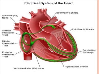
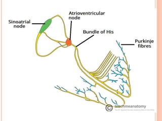
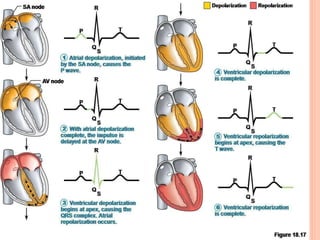
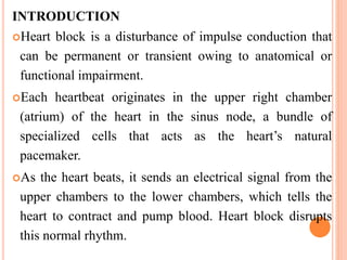
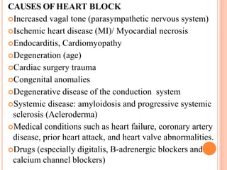
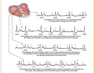
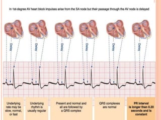
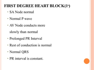
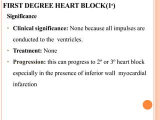
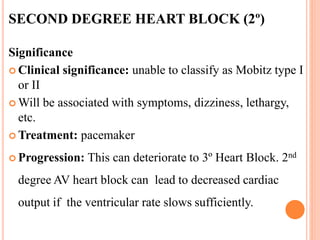
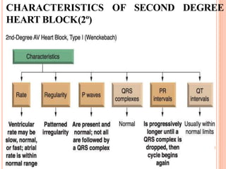
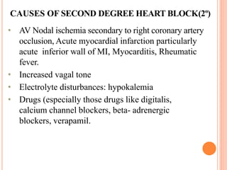
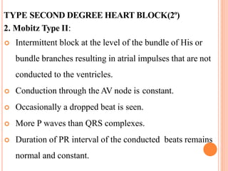
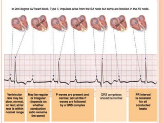
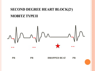
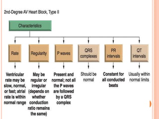
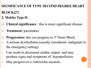
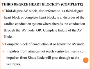
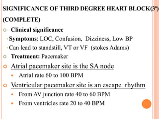
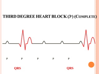
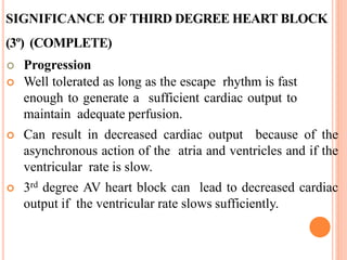
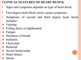
SlideShare a Scribd company logo
1 of 86
HEARTBLOCK
PREPARED BY
Ms. Netra Gautam
M. Sc. Nursing 2nd year
SNSR
OBJECTIVES
General Objectives: At the end of the class, students will be able to explain about
heart block.
Specific Objective: At the end of this class, students will be able:
 To introduce heart block.
 To define the heart block.
 To state the etiological factors of heart block.
 To describe the types of heart block.
 To explain pathophysiology of heart block.
 To enumerate the clinical features of heart block.
 To state the diagnostic evaluation of heart block.
 To describe the management of heart block.
 To enlist complication of heart block.
 To elaborate peacemaker.
 To explain nursing management of heart block.
 To enlist health education for patient with heart block.
 To summarize.
ELECTRICAL SYSTEM OF THE HEART:
The atria and ventricles work together, alternately contracting and
relaxing to pump blood through the heart. The electrical system of
the heart is the power source that makes this possible. Normally, the
electrical impulse begins at the Sino Atrial (SA) node, located in the
right atrium. The electrical activity spreads through the walls of the
atria, causing them to contract. Next, the electrical impulse travels
through the AV node, located between the atria and ventricles. The
AV node acts like a gate that slows the electrical signal before it
enters the ventricles. This delay gives the atria time to contract before
the ventricles do. From the AV node, the electrical impulse travels
through the His-Purkinje network, a pathway of specialized
electricity- conducting fibers. Then the impulse travels into the
muscular walls of the ventricles, causing them to contract. This
sequence occurs with every heartbeat (usually 60-100 times per
minute).
ELECTRICAL CONDUCTION SYSTEM OF HEART
NORMAL CONDUCTION PATHWAY:
SA NODE -> ATRIAL MUSCLE -> AV NODE -> BUNDLE
OF HIS -> LEFT AND RIGHT BUNDLE BRANCHES ->
VENTRICULAR MUSCLE
CARDIAC CONDUCTION SYSTEM
 The cardiac conduction system is a collection of nodes
and specialised conduction cells that initiate and co-
ordinate contraction of the heart muscle. It consists of:
 Sinoatrial node
 Atrioventricular node
 Atrioventricular bundle (bundle of His)
 Purkinje fibres
 In this article, we shall look at the anatomy of the cardiac
conduction system – its structure, function and clinical
correlations.
 The sequence of electrical events during one full
contraction of the heart muscle:
 An excitation signal (an action potential) is created by
the sinoatrial (SA) node.
 The wave of excitation spreads across the atria, causing
them to contract.
 Upon reaching the atrioventricular (AV) node, the signal
is delayed.
 It is then conducted into the bundle of His, down the
interventricular septum.
 The bundle of His and the Purkinje fibres spread the
wave impulses along the ventricles, causing them to
contract.
 We will now discuss the anatomy of the individual
components involved in the conducting system.
Components of the Cardiac Conduction System
 Sinoatrial Node
 The sinoatrial (SA) node is a collection of specialised cells
(pacemaker cells), and is located in the upper wall of the right
atrium, at the junction where the superior vena cava enters.
 These pacemaker cells can spontaneously generate electrical
impulses. The wave of excitation created by the SA node spreads
via gap junctions across both atria, resulting in atrial
contraction (atrial systole) – with blood moving from the atria into
the ventricles.
 The rate at which the SA node generates impulses is influenced by
the autonomic nervous system:
 Sympathetic nervous system – increases firing rate of the SA
node, and thus increases heart rate.
 Parasympathetic nervous system – decreases firing rate of the
SA node, and thus decreases heart rate.
Atrioventricular Node
 After the electrical impulses spread across the atria,
they converge at the atrioventricular node – located
within the atrioventricular septum, near the opening of
the coronary sinus.
 The AV node acts to delay the impulses by
approximately 120ms, to ensure the atria have enough
time to fully eject blood into the ventricles before
ventricular systole.
 The wave of excitation then passes from the
atrioventricular node into the atrioventricular bundle.
Atrioventricular Bundle
 The atrioventricular bundle (bundle of His) is a
continuation of the specialised tissue of the AV node,
and serves to transmit the electrical impulse from the
AV node to the Purkinje fibres of the ventricles.
 It descends down the membranous part of the
interventricular septum, before dividing into two main
bundles:
 Right bundle branch – conducts the impulse to the
Purkinje fibres of the right ventricle
 Left bundle branch – conducts the impulse to the
Purkinje fibres of the left ventricle.
Purkinje Fibres
 The Purkinje fibres (sub-endocardial plexus of
conduction cells) are a network of specialised cells.
They are abundant with glycogen and have extensive
gap junctions.
 These cells are located in the subendocardial
surface of the ventricular walls, and are able to
rapidly transmit cardiac action potentials from the
atrioventricular bundle to the myocardium of the
ventricles.
 This rapid conduction allows coordinated
ventricular contraction (ventricular systole) and
blood is moved from the right and left ventricles to
the pulmonary artery and aorta respectively.
INTRODUCTION
Heart block is a disturbance of impulse conduction that
can be permanent or transient owing to anatomical or
functional impairment.
Each heartbeat originates in the upper right chamber
(atrium) of the heart in the sinus node, a bundle of
specialized cells that acts as the heart’s natural
pacemaker.
As the heart beats, it sends an electrical signal from the
upper chambers to the lower chambers, which tells the
heart to contract and pump blood. Heart block disrupts
this normal rhythm.
INTRODUCTION
It must be distinguished from interference, a normal
phenomenon that is a disturbance of impulse conduction
caused by physiological refractoriness due to in excitability
from a preceding impulse.
AV Node : AV nodal conduction time is represented on the
ECG as the PR segment. But always measured in the PR
interval. This conduction defect can be seen on the ECG.
Disturbances of the conduction through the heart, occurring
at the AV Node. AV Node damaged/diseased delay or total
block of impulses at the AV Node.
The degree of block defines the type and classification of
heart block.
DEFINITION
Heart block is an abnormal heart rhythm where the heart
beats too slowly, which results in the electrical signals
being partially or totally blocked between the upper
chambers (atria) and lower chambers (ventricles). Heart
block is also called atrioventricular (AV) block.
Heart block is an abnormal heart rhythm where the heart
beats too slowly (bradycardia).
In this condition, the electrical signals that tell the heart
to contract are partially or totally blocked between the
upper chambers and the lower chambers.
Partial delays or complete interruptions in the cardiac
conduction pathway between the atria and ventricles.
CAUSES OFHEART BLOCK
Increased vagal tone (parasympathetic nervous system)
Ischemic heart disease (MI)/ Myocardial necrosis
Endocarditis, Cardiomyopathy
Degeneration (age)
Cardiac surgery trauma
Congenital anomalies
Degenerative disease of the conduction system
Systemic disease: amyloidosis and progressive systemic
sclerosis (Acleroderma)
Medical conditions such as heart failure, coronary artery
disease, prior heart attack, and heart valve abnormalities.
Drugs (especially digitalis, B-adrenergic blockers and
calcium channel blockers)
TYPES
They are further classified as:-
First degree heart block ( first degree AV block)
Second degree heart block (second degree AV block)
Third degree heart block (third degree AV block)
FIRST DEGREE HEART BLOCK
• First-degree atrio-ventricular block (AV block), or PR
prolongation, is a disease of the electrical conduction
system of the heart in which the PR interval is
lengthened beyond 0.20 seconds ( 5 small square).
• Heart rate and rhythm are normal and there may be
nothing wrong with the heart.
• Not a true block.
• A consistent delay of conduction at the level of the AV
node.
FIRST DEGREE HEART BLOCK(1º)
• SA Node normal
• Normal P wave
• AV Node conducts more
slowly than normal
• Prolonged PR Interval
• Rest of conduction is normal
• Normal QRS
• PR interval is constant.
CHARACTERISTICS OF FIRST DEGREE HEART
BLOCK(1º)
CAUSES OF FIRST DEGREE AV HEART BLOCK:
• Intrinsic AV Node disease
• Acute myocardial infarction (MI), particularly acute
inferior wall MI, Myocarditis
• Increased vagal tone
• Electrolyte disturbances: hypokalemia,
hypomagnesemia
• Drugs (especially those drugs like digitalis, calcium
channel blockers, beta- adrenergic blockers, quinidine,
procainamide, amiodarone that increase the refractory
time of the AV Node, thereby slowing conduction of the
impulses from the atria to the ventricles and cause 1st
degree AV block.
FIRST DEGREE HEART BLOCK(1º)
Significance
• Clinical significance: None because all impulses are
conducted to the ventricles.
• Treatment: None
• Progression: this can progress to 2º or 3º heart block
especially in the presence of inferior wall myocardial
infarction
SECOND DEGREE HEART BLOCK(2º)
Second-degree Atrio-ventricular (Av) Block, or Second-degree
Heart Block, is characterized by disturbance, delay, or
interruption of atrial impulse conduction through the AV node to
the ventricles.
 Ratio 2 P waves 1 QRS.
TYPE SECOND DEGREE HEART BLOCK(2º)
1. Mobitz Type I ( Karl Wenkebach)
2. Mobitz Type II
SECOND DEGREE HEART BLOCK (2º)
Significance
 Clinical significance: unable to classify as Mobitz type I
or II
 Will be associated with symptoms, dizziness, lethargy,
etc.
 Treatment: pacemaker
 Progression: This can deteriorate to 3º Heart Block. 2nd
degree AV heart block can lead to decreased cardiac
output if the ventricular rate slows sufficiently.
CAUSES
Drugs (beta-blockers, calcium channel blockers, amiodarone)
 Cardiomyopathy
 Rheumatic Fever
 Myocarditis
Varicella-zoster virus infection
Rheumatic diseases
Hypoxia
Hyperkalemia
Hypothyroidism
Inferior wall myocardial infraction
TYPE SECOND DEGREE HEART BLOCK(2º)
1. Mobitz Type I ( Karl Wenkebach):
 More P waves than QRS complexes and the rhythm has
patterned irregularity.
 Conduction through the AV Node is progressively delayed
until a drop beat is seen.
 Intermittent block at the level of the AV node.
 PR Interval prolongs with each beat until a QRS complex
dropped beat is seen. The PR Interval is NOT constant.
 After each dropped beat, the next PR interval is normal
and shorter.
 As each subsequent impulse generated there is a
progressively longer PR interval until again, a QRS is
dropped.
 Cycle starts again.
SECOND DEGREE HEART BLOCK(2º)
1. Mobitz typeI (wenkebach)
PR PR PR DROPPED BEAT
CHARACTERISTICS OF SECOND DEGREE
HEART BLOCK(2º)
CAUSES OF SECOND DEGREE HEART BLOCK(2º)
• AV Nodal ischemia secondary to right coronary artery
occlusion, Acute myocardial infarction particularly
acute inferior wall of MI, Myocarditis, Rheumatic
fever.
• Increased vagal tone
• Electrolyte disturbances: hypokalemia
• Drugs (especially those drugs like digitalis,
calcium channel blockers, beta- adrenergic
blockers, verapamil.
SIGNIFICANCE OFSECOND DEGREE HEART BLOCK
1. Mobitz Type I (Wenkebach):
Clinical Significance: If dropped ventricular beats occur
frequently, patient may show signs and symptoms of
decreased cardiac output, Lethargy, Confusion.
Treatment
• Pacemaker if during day &/or symptoms
• No treatment if at night
Progression:
• Usually transient and reversible, mostly resolving when the
underlying condition is corrected.
• This can progress to 3º Heart Block if it occurs early in
myocardial infarction.
TYPE SECOND DEGREE HEART BLOCK(2º)
2. Mobitz Type II:
 Intermittent block at the level of the bundle of His or
bundle branches resulting in atrial impulses that are not
conducted to the ventricles.
 Conduction through the AV node is constant.
 Occasionally a dropped beat is seen.
 More P waves than QRS complexes.
 Duration of PR interval of the conducted beats remains
normal and constant.
SECOND DEGREE HEART BLOCK(2º)
MOBITZ TYPEII
PR PR DROPPED BEAT PR
SIGNIFICANCE OFTYPE SECOND DEGREE HEART
BLOCK(2º)
2. Mobitz Type II:
 Clinical significance : this is more significant disease
 Treatment: pacemaker.
 Progression: this can progress to 3º Heart Block.
 A serious dysrhythmia (usually considered malignant in
the emergency setting).
 Can result in decreased cardiac output and may
produce signs and symptoms of hypoperfusion.
 May progress to a ventricular asystole.
THIRD DEGREE HEART BLOCK(3º) (COMPLETE)
Third-degree AV block, also referred to as third-degree
heart block or complete heart block, is a disorder of the
cardiac conduction system where there is no conduction
through the AV node. OR, Complete failure of the AV
Node.
 Complete block of conduction at or below the AV node.
 Impulses from atria cannot reach ventricles means no
impulses from Sinus Node will pass through to the
ventricles.
THIRD DEGREE HEART BLOCK(3º) (COMPLETE)
 Some part if the conducting system will take over as
pacemaker of the heart (even a myocardial cell 10-15 bpm).
 P wave rate: normal
 Ventricular rate: slow
 Ventricular complex may be broad
 Idioventricular rhythm
 Complete dissociation between P waves & QRS. OR,
Upright and round P waves seem to “march right through
the QRS complexes.
CAUSES OF THIRD DEGREE HEART BLOCK(3º)
(COMPLETE)
• Coronary artery occlusion, myocardial infarction inferior
and posterior wall of MI, Degenerative changes in the
heart, septal necrosis, Myocarditis.
• Increased vagal tone
• Electrolyte disturbances: hypokalemia
• Drugs (especially those drugs like digitalis, calcium
channel blockers, beta- adrenergic blockers,
• Surgical injury.
SIGNIFICANCE OF THIRD DEGREE HEART BLOCK(3º)
(COMPLETE)
 Clinical significance
•Symptoms: LOC, Confusion, Dizziness, Low BP
•Can lead to standstill, VT or VF (stokes Adams)
 Treatment: Pacemaker
 Atrial pacemaker site is the SA node
 Atrial rate 60 to 100 BPM
 Ventricular pacemaker site is an escape rhythm
 From AV junction rate 40 to 60 BPM
 From ventricles rate 20 to 40 BPM
THIRDDEGREEHEARTBLOCK(3º) (COMPLETE)
P P P P P
QRS QRS
SIGNIFICANCE OF THIRD DEGREE HEART BLOCK
(3º) (COMPLETE)
 Progression
 Well tolerated as long as the escape rhythm is fast
enough to generate a sufficient cardiac output to
maintain adequate perfusion.
 Can result in decreased cardiac output because of the
asynchronous action of the atria and ventricles and if the
ventricular rate is slow.
 3rd degree AV heart block can lead to decreased cardiac
output if the ventricular rate slows sufficiently.
PATHOPHYSIOLOGY OF HEART BLOCK
No arterial impulse conducted through the AV node into the
ventricles
Independent atrial and ventricular complexes
Atrial rate greater than ventricular rate
Heart Block
CLINICAL FEATURES OF HEART BLOCK
• Signs and symptoms depends on type of heart block.
• First degree heart block rarely causes symptoms.
• Symptoms of second and third degree heart block
includes:
 Fainting
 Felling dizzy or lightheaded
 Fatigue
 Shortness of breath
 Ischemia
 Chest pain
 Reduced
 Severe bradycardia
 Heart failure
 Shock
DIAGNOSTIC TESTS AND PROCEDURE
• History collection
• Physical examination
• ECG
• Echocardiogram
• Electrophysiology test
• Tilt-table test
• Holter and event monitors: Holter and event monitors are
small, portable electrocardiogram devices that record
your heart’s electrical activity for long periods of time
while you do your normal activities.
MANAGEMENT OF HEART BLOCK
• Treatment depends on type of heart block.
• First degree heart block usually needs no treatment.
• For second and third degree heart block, pacemaker is
the only choice of treatment.
1. A transcutaneous pacemaker is used until a temporary
transvenous pacemaker can be inserted.
2. The use of drugs such as atropine, epinephrine,
isoproterenol, and dopamine is a temporary measure to
increase measure HR and Support BP until temporary
pacing is initiated.
3. Patients will need a permanent pacemaker as soon as
possible.
PACEMAKER
INTRODUCTION ARTIFICIAL PACEMAKER
A artificial pacemaker is an electronic device that's placed
under the skin of chest to help control heartbeat. It's used to
help heart beat more regularly in case of irregular heartbeat
(arrhythmia), particularly a slow one. Implanting a
pacemaker in chest requires a surgical procedure.
Parts of pacemaker
Most pacemaker have 2 parts:
1. The generator contains the battery and the information
to control the heartbeat.
2. The leads are wires that connect the heart to the
generator and carry the electrical messages to the heart.
DEFINITION
A pacemaker is a small, battery-operated device. This
device senses when your heart is beating irregularly or
too slowly. It sends a signal to your heart that makes
your heart beat at the correct pace.
FUNCTION OF PACEMAKER
• The Sino Atrial (SA) node or sinus node is the heart's
natural pacemaker. It's a small mass of specialized cells
in the top of the right atrium (upper chamber of the
heart).
• It produces the electrical impulses that cause your heart
to beat.
• A chamber of the heart contracts when an electrical
impulse or signal moves across it. For the heart to beat
properly, the signal must travel down a specific path to
reach the ventricles (the heart's lower chambers).
• When the heart's natural pacemaker is defective, the
heartbeat may be too fast, too slow or irregular.
• Rhythm problems also can occur because of a blockage
of heart's electrical pathways.
FUNCTION OF PACEMAKER
https://www.youtube.com/watch?v=SMXBR_YFocs
• The pacemaker's pulse generator sends electrical impulses
to the heart to help it pump properly.
• An electrode is placed next to the heart wall and small
electrical charges travel through the wire to the heart.
• Most pacemakers have a sensing mode that inhibits the
pacemaker from sending impulses when the heartbeat is
above a certain level. It allows the pacemaker to fire when
the heartbeat is too slow.
• These are called demand pacemakers.
TYPES OF
PACEMAKER
Permanent
Pacemaker
Bivenricular
Pacemaker
https://www.youtube.com/watch?v=GIMOa0HvLE0
PERMANENT PACEMAKER
It is implanted totally in the body. Power source is implanted
subcutaneously usually over the pectoral muscle on the
patient non dominant side.
Types of permanent pacemaker
• Single-chamber pacemaker: In this type, only one pacing
lead is placed into a chamber of the heart, either the
atrium or the ventricle.
• Dual-chamber pacemaker:
 Wires are placed in two chambers of the heart.
 One lead paces the atrium and one paces the ventricle.
 Closely resembles the natural pacing of the heart.
• Rate-responsive pacemaker: It has sensors that detect
changes in the patient's physical activity and
automatically adjust the pacing rate to fulfil the body's
metabolic needs.
INDICATION OF PERMANENT PACEMAKER
• Chronic atrial fibrillation with slow ventricular response
• Hypersensitive carotid sinus syndrome
• Fibrosis or sclerotic changes of cardiac conduction
system
• Sick sinus syndrome
• Tachyarrhythmia
• Third degree AV block
TEMPORARY PACEMAKER
Temporary pacemaker is used to treat a bradysrhythmia when
the condition is temporary and when a permanent pacemaker
is either not required. It is one that has the power source
outside the body.
Types
There are 4 types of temporary pacemaker.
• Transvenous invasive pacemaker (endocardial):
It consists of lead or leads that are threaded transvenously to
the right atrium and or right ventricle and attached to
external power source.
• Epicardial Pacemaker:
Wires are attached to the endocardium of the heart, brought
out through a surgical incision onto the chest, connected to
an external pulse generator. Are commonly used when
patient is undergoing cardiac surgery.
TEMPORARY PACEMAKER
Types
• Trans cutaneous pacemaker(Non-invasive pacing):
It is used to provide adequate heart rate and rhythm to the
patient in and emergency situation.
• Transthoracic invasive pacing(Epicardial pacing ):
It is achieved by attaching an atrial and ventricle and
attached to epicardium during heart surgery . The leads
are passed through the chest wall and attached to the
external power source.
INDICATIONS OF TEMPORARY PACEMAKER
• Maintenance of adequate heart rate and rhythm during
special circumstances such as surgery and postoperative
recovery, cardiac catheterization or coronary angioplasty.
• Before implantation of a permanent pacemaker.
• As prophylaxis after open heart surgery.
• Acute anterior MI with second degree or third degree
AV block or bundle branch block.
• Acute inferior MI with symptomatic bradycardia and AV
block
BIVENRICULAR PACEMAKER
•Bivenricular pacemaker is also referred to as cardiac
resynchronization therapy (CRT) which is used to treat
the delay in heart ventricle contractions that occur in
some people with advanced heart failure.
•It is an electronic, battery-powered device that is
surgically implanted under the skin.
•The device has 2 or 3 leads (wires) that are positioned in
the heart to help the heart beat in a more balanced way.
The leads are implanted through a vein in the right atrium
and right ventricle and into the coronary sinus vein to
pace the left ventricle.
BIVENRICULAR PACEMAKER
CRT Device
• The CRT device (biventricular pacemaker) has 2 or 3 leads that
are positioned in the:
• Right atrium
• Right ventricle
• Left ventricle (via the coronary sinus vein)
COMPLICATION OF PACEMAKER
• Hematoma
• Pneumothorax
• Failure to sense or capture
• Perforation of atrial or ventricle septum
• Ventricular atrophy and tachycardia
• Movement or dislocation of lead
• Cardiac perforation
NURSING MANAGEMENT
Nursing Assessment
•Assess the high risk patients.
•Monitor ECG of the patient.
•Assess the family history of heart disease.
•Assess the history of smoking and alcoholism.
•Monitor lab values frequently especially serum cholesterol
levels.
•Assess for CAD.
•Monitor vital signs.
NURSING MANAGEMENT
Nursing Diagnosis
• Acute pain related to insertion site and prescribed post
procedure immobilization.
• Disturbed self-concept related to perceived loss of
health and dependence on pacemaker.
• Impaired physical mobility related to incisional site
pain, activity restrictions.
• Risk for infection related to operative site.
• Risk for ineffective therapeutic regimen management
related to insufficient knowledge of activity
restrictions, precautions.
NURSING MANAGEMENT
Nursing Intervention
• Preventing infection.
• Reliving pain
• Promoting effective coping
• Teaching self-care
• Care of pacemaker
• Encourage for physical activity
• Monitoring pacemaker function
• Promoting safety
• Avoiding electromagnetic interference
NURSING MANAGEMENT
Preoperative care
• Financial: Explain the type, technique, cost of the
procedure and hospital stay of pacemaker to the patient.
• Psychological: Explain the Process of the pacemaker
insertion. Reassure the patient
• Physical preparation:
 Obtain written consent from the patient and from
nearest relative
 Remove dentures, jewellery and contact lens.
 Clean and shave the area .
 Check vital signs: temperature, BP, pulse and
respiration
NURSING MANAGEMENT
Intraoperative care
•Check serology: HIV, HbsAg, HCV and others.
• Start an IV line with 5% Dextrose solution or NS solution.
• Check the battery in pulse generator.
• Prepare the emergency cart, the defibrillator and jelly ,
and the ECG monitor.
• Set up all equipment for the insertion of the pacemaker.
•The nurse should know about the pacemaker generator
including the power switch, indicator light for pacing and
sensing, stimulus output dial, sensitivity dial, and their
proper settings.
•Assist the doctor and the scrub nurse during the procedure
step by step.
•Observe vital signs and observe ECG monitor carefully for
arrhythmias and other complications.
NURSING MANAGEMENT
Post-operative care
• Receive the patient
• Keep the patient in comfort position
• Record the pacing parameters.
• Receiving time
• Monitor vitals signs and level of consciousness of
patient
• Other routine care
• Immobilize the affected part and keep in supine position
but allow the movement of finger and ankle joint.
• Monitor heart rate and rhythm.
• Prevent infection.
• Take ECG and X-ray chest.
• Watch for complication
PATIENT AND FAMILY TEACHING
• Maintain follow up care with a physician to check the
pacemaker site and begin regular pacemaker function
checks.
• Watch for signs of infection at incision site redness,
swelling dressing.
• Keep incision dry for 1 week after implantation.
• Avoid lifting operative side arm above shoulder level
until approved by care provider.
• Avoid direct blows to generators or to large magnets such
as MRI scanner. These device can reprogram a
pacemaker.
• Microwave oven are safe to use and do not threaten
pacemaker function.
• The patient should be taught how to take the pulse.
• Carry pacemaker information card at all the times.
SUMMARY
We discussed on definition, etiology, classification,
clinical features, diagnostic studies, management,
complications, nursing management of heart block.
CONCLUSION:
In left bundle branch block (LBBB), the heart's two
ventricles are being stimulated by the cardiac electrical
impulse in sequence instead of simultaneously. Specifically,
the left ventricle in a person with LBBB is stimulated to
contract only after the right ventricle is stimulated. This loss
of normal coordination between the two ventricles decreases
the efficiency of the heart beat, so that the heart has to do
more work to achieve its normal pumping capacity. This can
be managed by drugs and artificial pacemakers.
REFERENCES
• Brunner and suddharth’s; textbook of medical –surgical nursing; wolters kluwer
publication; 2014; 13th edition; volume 2; page no. 1959- 1963.
• Mandel. G.N; Textbook of Medical Surgical Nursing; Makalu Publication;
Kathmandu, Nepal; 5th edidition; page no.333-339.
• Monahan Sands, “ Medical-Surgical Nursing,” 8th edition, Elsevier Pvt Ltd. Page
no. 790-793.
• Smeltzer Suzanne C, Barebrenda G, Hinkle Janice L, Cheever Kerry H. “Textbook
of Medical-Surgical Nursing,” 12th edition, New Delhi: Lippincot wolter’s kluwer;
p.113-114.
• Lewis Dirksen and Heitkemper Bucher, “Medical-Surgical Nursing,” 9th edition,
Page no. 803-805.
• https://www.slideshare.net/Ratheeshkrishnakripa/heart-block-66802439
• https://www.okheart.com/about-us/ohh-news/heart-block-symptoms-
diagnosis-and-treatment
• https://www.nhlbi.nih.gov/health-topics/holter-and-event-monitors
• https://teachmeanatomy.info/thorax/organs/heart/conducting-
system/#:~:text=The%20cardiac%20conduction%20system%20is,Atriovent
ricular%20bundle%20(bundle%20of%20His)
• https://www.slideshare.net/sunnymumu/heart-block-with-nursing-
management

More Related Content

What's hot (20)

Myocarditis
MyocarditisMyocarditis
Myocarditis
 
Mitral valve stenosis powerpoint
Mitral valve stenosis powerpointMitral valve stenosis powerpoint
Mitral valve stenosis powerpoint
 
Pericarditis
PericarditisPericarditis
Pericarditis
 
Aneurysm
Aneurysm Aneurysm
Aneurysm
 
Endocarditis
EndocarditisEndocarditis
Endocarditis
 
Valvular heart disease
Valvular heart diseaseValvular heart disease
Valvular heart disease
 
Pericardial effusion
Pericardial effusionPericardial effusion
Pericardial effusion
 
coronary artery disease
coronary artery diseasecoronary artery disease
coronary artery disease
 
Raynaud’s
Raynaud’sRaynaud’s
Raynaud’s
 
Cardiac arrest bsc
Cardiac arrest bscCardiac arrest bsc
Cardiac arrest bsc
 
Heart block
Heart blockHeart block
Heart block
 
Pacemaker
PacemakerPacemaker
Pacemaker
 
Cardiomyopathy
CardiomyopathyCardiomyopathy
Cardiomyopathy
 
Cardioversion
Cardioversion Cardioversion
Cardioversion
 
Holter monitoring
Holter monitoringHolter monitoring
Holter monitoring
 
Anuerysm
AnuerysmAnuerysm
Anuerysm
 
Cardiomyopathies
CardiomyopathiesCardiomyopathies
Cardiomyopathies
 
Pericarditis
PericarditisPericarditis
Pericarditis
 
Pulmonary edema
Pulmonary edemaPulmonary edema
Pulmonary edema
 
Pulmonary embolism1
Pulmonary embolism1Pulmonary embolism1
Pulmonary embolism1
 

Similar to Heart block

Cardiovascular System.ppt
Cardiovascular System.pptCardiovascular System.ppt
Cardiovascular System.ppt
Shama
 
05 Cardiovascular System.ppt
05 Cardiovascular System.ppt05 Cardiovascular System.ppt
05 Cardiovascular System.ppt
Shama
 
Antiarrhythmic drugs class Bidya.pdf
Antiarrhythmic drugs class Bidya.pdfAntiarrhythmic drugs class Bidya.pdf
Antiarrhythmic drugs class Bidya.pdf
SaishDalvi
 
ORIGIN OF THE HEARTBEAT & THE ELECTRICAL ACTIVITY OF THE HEART.pptx
ORIGIN OF THE HEARTBEAT & THE ELECTRICAL ACTIVITY OF THE HEART.pptxORIGIN OF THE HEARTBEAT & THE ELECTRICAL ACTIVITY OF THE HEART.pptx
ORIGIN OF THE HEARTBEAT & THE ELECTRICAL ACTIVITY OF THE HEART.pptx
shreya730959
 

Similar to Heart block (20)

Conduction system of heart
Conduction system of heartConduction system of heart
Conduction system of heart
 
Conducting system of the heart
Conducting system of the heartConducting system of the heart
Conducting system of the heart
 
Cardiovascular assessment and diagnostic procedures
Cardiovascular assessment and diagnostic proceduresCardiovascular assessment and diagnostic procedures
Cardiovascular assessment and diagnostic procedures
 
Term paper on ecg and cardiac arrhythmias
Term paper on ecg  and cardiac arrhythmiasTerm paper on ecg  and cardiac arrhythmias
Term paper on ecg and cardiac arrhythmias
 
Term paper on ECG and cardiac arrhythmias
Term paper on ECG  and cardiac arrhythmiasTerm paper on ECG  and cardiac arrhythmias
Term paper on ECG and cardiac arrhythmias
 
Generation and conduction of Cardiac impulse
Generation and conduction of Cardiac impulseGeneration and conduction of Cardiac impulse
Generation and conduction of Cardiac impulse
 
Cardiac function testing
Cardiac function testingCardiac function testing
Cardiac function testing
 
Arrthymias management
Arrthymias managementArrthymias management
Arrthymias management
 
Arrthymias management
Arrthymias managementArrthymias management
Arrthymias management
 
Cardiovascular System.ppt
Cardiovascular System.pptCardiovascular System.ppt
Cardiovascular System.ppt
 
05 Cardiovascular System.ppt
05 Cardiovascular System.ppt05 Cardiovascular System.ppt
05 Cardiovascular System.ppt
 
NORMAL Electrocardiogram, ECK/EKG PPT....
NORMAL Electrocardiogram, ECK/EKG PPT....NORMAL Electrocardiogram, ECK/EKG PPT....
NORMAL Electrocardiogram, ECK/EKG PPT....
 
The conducting system of heart
The conducting system of heartThe conducting system of heart
The conducting system of heart
 
heart-180521025843.pdf
heart-180521025843.pdfheart-180521025843.pdf
heart-180521025843.pdf
 
Heart anatomy & physiology slide share, circulatory system pdf
Heart anatomy & physiology slide share, circulatory system pdfHeart anatomy & physiology slide share, circulatory system pdf
Heart anatomy & physiology slide share, circulatory system pdf
 
ANATOMY AND PHYSIOLOGY OF HEART
ANATOMY AND PHYSIOLOGY OF HEARTANATOMY AND PHYSIOLOGY OF HEART
ANATOMY AND PHYSIOLOGY OF HEART
 
Heart assessment
Heart assessmentHeart assessment
Heart assessment
 
Heart physiology, blood physiology
Heart physiology, blood physiologyHeart physiology, blood physiology
Heart physiology, blood physiology
 
Antiarrhythmic drugs class Bidya.pdf
Antiarrhythmic drugs class Bidya.pdfAntiarrhythmic drugs class Bidya.pdf
Antiarrhythmic drugs class Bidya.pdf
 
ORIGIN OF THE HEARTBEAT & THE ELECTRICAL ACTIVITY OF THE HEART.pptx
ORIGIN OF THE HEARTBEAT & THE ELECTRICAL ACTIVITY OF THE HEART.pptxORIGIN OF THE HEARTBEAT & THE ELECTRICAL ACTIVITY OF THE HEART.pptx
ORIGIN OF THE HEARTBEAT & THE ELECTRICAL ACTIVITY OF THE HEART.pptx
 

Recently uploaded

👉 Solapur Call Girls Service 👉📞 7014168258 👉📞 Just📲 Call Ruhi Call Girl Near ...
👉 Solapur Call Girls Service 👉📞 7014168258 👉📞 Just📲 Call Ruhi Call Girl Near ...👉 Solapur Call Girls Service 👉📞 7014168258 👉📞 Just📲 Call Ruhi Call Girl Near ...
👉 Solapur Call Girls Service 👉📞 7014168258 👉📞 Just📲 Call Ruhi Call Girl Near ...
Call Girls
 
TIME FOR ACTION: MAY 2024 Securing A Strong Nursing Workforce for North Carolina
TIME FOR ACTION: MAY 2024 Securing A Strong Nursing Workforce for North CarolinaTIME FOR ACTION: MAY 2024 Securing A Strong Nursing Workforce for North Carolina
TIME FOR ACTION: MAY 2024 Securing A Strong Nursing Workforce for North Carolina
Mebane Rash
 
Goa Call Girls Service +9316020077 Call GirlsGoa By Russian Call Girlsin Goa
Goa Call Girls Service  +9316020077 Call GirlsGoa By Russian Call Girlsin GoaGoa Call Girls Service  +9316020077 Call GirlsGoa By Russian Call Girlsin Goa
Goa Call Girls Service +9316020077 Call GirlsGoa By Russian Call Girlsin Goa
Real Sex Provide In Goa
 
Call Girls In Kharar 💯Call Us 🔝 9915851334🔝 💃 Top Class ☎️ Call Girl Service ...
Call Girls In Kharar 💯Call Us 🔝 9915851334🔝 💃 Top Class ☎️ Call Girl Service ...Call Girls In Kharar 💯Call Us 🔝 9915851334🔝 💃 Top Class ☎️ Call Girl Service ...
Call Girls In Kharar 💯Call Us 🔝 9915851334🔝 💃 Top Class ☎️ Call Girl Service ...
daljeetkaur2026
 
Cara Menggugurkan Kandungan Secara Alami 3 Jam Tuntas
Cara Menggugurkan Kandungan Secara Alami 3 Jam TuntasCara Menggugurkan Kandungan Secara Alami 3 Jam Tuntas
Cara Menggugurkan Kandungan Secara Alami 3 Jam Tuntas
Cara Menggugurkan Kandungan 087776558899
 
Abortion pills Buy Farwaniya (+918133066128) Cytotec 200mg tablets Al AHMEDI
Abortion pills Buy Farwaniya (+918133066128) Cytotec 200mg tablets Al AHMEDIAbortion pills Buy Farwaniya (+918133066128) Cytotec 200mg tablets Al AHMEDI
Abortion pills Buy Farwaniya (+918133066128) Cytotec 200mg tablets Al AHMEDI
Abortion pills in Kuwait Cytotec pills in Kuwait
 
Call Girls Service In Jalandhar💯Call Us 🔝 8146719683🔝 💃 Top Class ☎️ Call Gir...
Call Girls Service In Jalandhar💯Call Us 🔝 8146719683🔝 💃 Top Class ☎️ Call Gir...Call Girls Service In Jalandhar💯Call Us 🔝 8146719683🔝 💃 Top Class ☎️ Call Gir...
Call Girls Service In Jalandhar💯Call Us 🔝 8146719683🔝 💃 Top Class ☎️ Call Gir...
daljeetkaur2026
 
@Safe Abortion pills IN Jeddah(+918133066128) Un_wanted kit Buy Jeddah
@Safe Abortion pills IN Jeddah(+918133066128) Un_wanted kit Buy Jeddah@Safe Abortion pills IN Jeddah(+918133066128) Un_wanted kit Buy Jeddah
@Safe Abortion pills IN Jeddah(+918133066128) Un_wanted kit Buy Jeddah
Abortion pills in Kuwait Cytotec pills in Kuwait
 

Recently uploaded (20)

Test bank for community public health nursing evidence for practice 4TH editi...
Test bank for community public health nursing evidence for practice 4TH editi...Test bank for community public health nursing evidence for practice 4TH editi...
Test bank for community public health nursing evidence for practice 4TH editi...
 
Coach Dan Quinn Commanders Feather T Shirts
Coach Dan Quinn Commanders Feather T ShirtsCoach Dan Quinn Commanders Feather T Shirts
Coach Dan Quinn Commanders Feather T Shirts
 
VIP Just Call 9548273370 Lucknow Top Class Call Girls Number | 8630512678 Esc...
VIP Just Call 9548273370 Lucknow Top Class Call Girls Number | 8630512678 Esc...VIP Just Call 9548273370 Lucknow Top Class Call Girls Number | 8630512678 Esc...
VIP Just Call 9548273370 Lucknow Top Class Call Girls Number | 8630512678 Esc...
 
👉 Solapur Call Girls Service 👉📞 7014168258 👉📞 Just📲 Call Ruhi Call Girl Near ...
👉 Solapur Call Girls Service 👉📞 7014168258 👉📞 Just📲 Call Ruhi Call Girl Near ...👉 Solapur Call Girls Service 👉📞 7014168258 👉📞 Just📲 Call Ruhi Call Girl Near ...
👉 Solapur Call Girls Service 👉📞 7014168258 👉📞 Just📲 Call Ruhi Call Girl Near ...
 
Agra Call Girl 📲 ( 9084454195 ) ⏎ Independent Call Girls In Agra By Meera
Agra Call Girl 📲 ( 9084454195 ) ⏎ Independent Call Girls In Agra By MeeraAgra Call Girl 📲 ( 9084454195 ) ⏎ Independent Call Girls In Agra By Meera
Agra Call Girl 📲 ( 9084454195 ) ⏎ Independent Call Girls In Agra By Meera
 
TIME FOR ACTION: MAY 2024 Securing A Strong Nursing Workforce for North Carolina
TIME FOR ACTION: MAY 2024 Securing A Strong Nursing Workforce for North CarolinaTIME FOR ACTION: MAY 2024 Securing A Strong Nursing Workforce for North Carolina
TIME FOR ACTION: MAY 2024 Securing A Strong Nursing Workforce for North Carolina
 
Goa Call Girls Service +9316020077 Call GirlsGoa By Russian Call Girlsin Goa
Goa Call Girls Service  +9316020077 Call GirlsGoa By Russian Call Girlsin GoaGoa Call Girls Service  +9316020077 Call GirlsGoa By Russian Call Girlsin Goa
Goa Call Girls Service +9316020077 Call GirlsGoa By Russian Call Girlsin Goa
 
zencortex suppliment-health and benefit (1).pdf
zencortex suppliment-health and benefit (1).pdfzencortex suppliment-health and benefit (1).pdf
zencortex suppliment-health and benefit (1).pdf
 
Call Girls In Kharar 💯Call Us 🔝 9915851334🔝 💃 Top Class ☎️ Call Girl Service ...
Call Girls In Kharar 💯Call Us 🔝 9915851334🔝 💃 Top Class ☎️ Call Girl Service ...Call Girls In Kharar 💯Call Us 🔝 9915851334🔝 💃 Top Class ☎️ Call Girl Service ...
Call Girls In Kharar 💯Call Us 🔝 9915851334🔝 💃 Top Class ☎️ Call Girl Service ...
 
The Events of Cardiac Cycle - Wigger's Diagram
The Events of Cardiac Cycle - Wigger's DiagramThe Events of Cardiac Cycle - Wigger's Diagram
The Events of Cardiac Cycle - Wigger's Diagram
 
Cara Menggugurkan Kandungan Secara Alami 3 Jam Tuntas
Cara Menggugurkan Kandungan Secara Alami 3 Jam TuntasCara Menggugurkan Kandungan Secara Alami 3 Jam Tuntas
Cara Menggugurkan Kandungan Secara Alami 3 Jam Tuntas
 
TEST BANK For Little and Falace's Dental Management of the Medically Compromi...
TEST BANK For Little and Falace's Dental Management of the Medically Compromi...TEST BANK For Little and Falace's Dental Management of the Medically Compromi...
TEST BANK For Little and Falace's Dental Management of the Medically Compromi...
 
Bobath Technique (Samrth Pareta) .ppt.pptx
Bobath Technique (Samrth Pareta) .ppt.pptxBobath Technique (Samrth Pareta) .ppt.pptx
Bobath Technique (Samrth Pareta) .ppt.pptx
 
Abortion pills Buy Farwaniya (+918133066128) Cytotec 200mg tablets Al AHMEDI
Abortion pills Buy Farwaniya (+918133066128) Cytotec 200mg tablets Al AHMEDIAbortion pills Buy Farwaniya (+918133066128) Cytotec 200mg tablets Al AHMEDI
Abortion pills Buy Farwaniya (+918133066128) Cytotec 200mg tablets Al AHMEDI
 
TEST BANK For Robbins & Kumar Basic Pathology, 11th Edition by Vinay Kumar, A...
TEST BANK For Robbins & Kumar Basic Pathology, 11th Edition by Vinay Kumar, A...TEST BANK For Robbins & Kumar Basic Pathology, 11th Edition by Vinay Kumar, A...
TEST BANK For Robbins & Kumar Basic Pathology, 11th Edition by Vinay Kumar, A...
 
Call Girls Service In Jalandhar💯Call Us 🔝 8146719683🔝 💃 Top Class ☎️ Call Gir...
Call Girls Service In Jalandhar💯Call Us 🔝 8146719683🔝 💃 Top Class ☎️ Call Gir...Call Girls Service In Jalandhar💯Call Us 🔝 8146719683🔝 💃 Top Class ☎️ Call Gir...
Call Girls Service In Jalandhar💯Call Us 🔝 8146719683🔝 💃 Top Class ☎️ Call Gir...
 
ACNE VULGARIS , ALLERGIES, ECZEMA, PEMPHIGUS.pdf
ACNE VULGARIS , ALLERGIES, ECZEMA, PEMPHIGUS.pdfACNE VULGARIS , ALLERGIES, ECZEMA, PEMPHIGUS.pdf
ACNE VULGARIS , ALLERGIES, ECZEMA, PEMPHIGUS.pdf
 
CALCIUM - ELECTROLYTE IMBALANCE (HYPERCALCEMIA & HYPOCALCEMIA).pdf
CALCIUM - ELECTROLYTE IMBALANCE (HYPERCALCEMIA & HYPOCALCEMIA).pdfCALCIUM - ELECTROLYTE IMBALANCE (HYPERCALCEMIA & HYPOCALCEMIA).pdf
CALCIUM - ELECTROLYTE IMBALANCE (HYPERCALCEMIA & HYPOCALCEMIA).pdf
 
@Safe Abortion pills IN Jeddah(+918133066128) Un_wanted kit Buy Jeddah
@Safe Abortion pills IN Jeddah(+918133066128) Un_wanted kit Buy Jeddah@Safe Abortion pills IN Jeddah(+918133066128) Un_wanted kit Buy Jeddah
@Safe Abortion pills IN Jeddah(+918133066128) Un_wanted kit Buy Jeddah
 
Test Bank -Medical-Surgical Nursing Concepts for Interprofessional Collaborat...
Test Bank -Medical-Surgical Nursing Concepts for Interprofessional Collaborat...Test Bank -Medical-Surgical Nursing Concepts for Interprofessional Collaborat...
Test Bank -Medical-Surgical Nursing Concepts for Interprofessional Collaborat...
 

Heart block

  • 1. HEARTBLOCK PREPARED BY Ms. Netra Gautam M. Sc. Nursing 2nd year SNSR
  • 2. OBJECTIVES General Objectives: At the end of the class, students will be able to explain about heart block. Specific Objective: At the end of this class, students will be able:  To introduce heart block.  To define the heart block.  To state the etiological factors of heart block.  To describe the types of heart block.  To explain pathophysiology of heart block.  To enumerate the clinical features of heart block.  To state the diagnostic evaluation of heart block.  To describe the management of heart block.  To enlist complication of heart block.  To elaborate peacemaker.  To explain nursing management of heart block.  To enlist health education for patient with heart block.  To summarize.
  • 3.
  • 4.
  • 5.
  • 6. ELECTRICAL SYSTEM OF THE HEART: The atria and ventricles work together, alternately contracting and relaxing to pump blood through the heart. The electrical system of the heart is the power source that makes this possible. Normally, the electrical impulse begins at the Sino Atrial (SA) node, located in the right atrium. The electrical activity spreads through the walls of the atria, causing them to contract. Next, the electrical impulse travels through the AV node, located between the atria and ventricles. The AV node acts like a gate that slows the electrical signal before it enters the ventricles. This delay gives the atria time to contract before the ventricles do. From the AV node, the electrical impulse travels through the His-Purkinje network, a pathway of specialized electricity- conducting fibers. Then the impulse travels into the muscular walls of the ventricles, causing them to contract. This sequence occurs with every heartbeat (usually 60-100 times per minute).
  • 8. NORMAL CONDUCTION PATHWAY: SA NODE -> ATRIAL MUSCLE -> AV NODE -> BUNDLE OF HIS -> LEFT AND RIGHT BUNDLE BRANCHES -> VENTRICULAR MUSCLE
  • 9.
  • 10. CARDIAC CONDUCTION SYSTEM  The cardiac conduction system is a collection of nodes and specialised conduction cells that initiate and co- ordinate contraction of the heart muscle. It consists of:  Sinoatrial node  Atrioventricular node  Atrioventricular bundle (bundle of His)  Purkinje fibres  In this article, we shall look at the anatomy of the cardiac conduction system – its structure, function and clinical correlations.
  • 11.  The sequence of electrical events during one full contraction of the heart muscle:  An excitation signal (an action potential) is created by the sinoatrial (SA) node.  The wave of excitation spreads across the atria, causing them to contract.  Upon reaching the atrioventricular (AV) node, the signal is delayed.  It is then conducted into the bundle of His, down the interventricular septum.  The bundle of His and the Purkinje fibres spread the wave impulses along the ventricles, causing them to contract.  We will now discuss the anatomy of the individual components involved in the conducting system.
  • 12. Components of the Cardiac Conduction System  Sinoatrial Node  The sinoatrial (SA) node is a collection of specialised cells (pacemaker cells), and is located in the upper wall of the right atrium, at the junction where the superior vena cava enters.  These pacemaker cells can spontaneously generate electrical impulses. The wave of excitation created by the SA node spreads via gap junctions across both atria, resulting in atrial contraction (atrial systole) – with blood moving from the atria into the ventricles.  The rate at which the SA node generates impulses is influenced by the autonomic nervous system:  Sympathetic nervous system – increases firing rate of the SA node, and thus increases heart rate.  Parasympathetic nervous system – decreases firing rate of the SA node, and thus decreases heart rate.
  • 13. Atrioventricular Node  After the electrical impulses spread across the atria, they converge at the atrioventricular node – located within the atrioventricular septum, near the opening of the coronary sinus.  The AV node acts to delay the impulses by approximately 120ms, to ensure the atria have enough time to fully eject blood into the ventricles before ventricular systole.  The wave of excitation then passes from the atrioventricular node into the atrioventricular bundle.
  • 14. Atrioventricular Bundle  The atrioventricular bundle (bundle of His) is a continuation of the specialised tissue of the AV node, and serves to transmit the electrical impulse from the AV node to the Purkinje fibres of the ventricles.  It descends down the membranous part of the interventricular septum, before dividing into two main bundles:  Right bundle branch – conducts the impulse to the Purkinje fibres of the right ventricle  Left bundle branch – conducts the impulse to the Purkinje fibres of the left ventricle.
  • 15.
  • 16. Purkinje Fibres  The Purkinje fibres (sub-endocardial plexus of conduction cells) are a network of specialised cells. They are abundant with glycogen and have extensive gap junctions.  These cells are located in the subendocardial surface of the ventricular walls, and are able to rapidly transmit cardiac action potentials from the atrioventricular bundle to the myocardium of the ventricles.  This rapid conduction allows coordinated ventricular contraction (ventricular systole) and blood is moved from the right and left ventricles to the pulmonary artery and aorta respectively.
  • 17.
  • 18.
  • 19.
  • 20.
  • 21. INTRODUCTION Heart block is a disturbance of impulse conduction that can be permanent or transient owing to anatomical or functional impairment. Each heartbeat originates in the upper right chamber (atrium) of the heart in the sinus node, a bundle of specialized cells that acts as the heart’s natural pacemaker. As the heart beats, it sends an electrical signal from the upper chambers to the lower chambers, which tells the heart to contract and pump blood. Heart block disrupts this normal rhythm.
  • 22. INTRODUCTION It must be distinguished from interference, a normal phenomenon that is a disturbance of impulse conduction caused by physiological refractoriness due to in excitability from a preceding impulse. AV Node : AV nodal conduction time is represented on the ECG as the PR segment. But always measured in the PR interval. This conduction defect can be seen on the ECG. Disturbances of the conduction through the heart, occurring at the AV Node. AV Node damaged/diseased delay or total block of impulses at the AV Node. The degree of block defines the type and classification of heart block.
  • 23. DEFINITION Heart block is an abnormal heart rhythm where the heart beats too slowly, which results in the electrical signals being partially or totally blocked between the upper chambers (atria) and lower chambers (ventricles). Heart block is also called atrioventricular (AV) block. Heart block is an abnormal heart rhythm where the heart beats too slowly (bradycardia). In this condition, the electrical signals that tell the heart to contract are partially or totally blocked between the upper chambers and the lower chambers. Partial delays or complete interruptions in the cardiac conduction pathway between the atria and ventricles.
  • 24. CAUSES OFHEART BLOCK Increased vagal tone (parasympathetic nervous system) Ischemic heart disease (MI)/ Myocardial necrosis Endocarditis, Cardiomyopathy Degeneration (age) Cardiac surgery trauma Congenital anomalies Degenerative disease of the conduction system Systemic disease: amyloidosis and progressive systemic sclerosis (Acleroderma) Medical conditions such as heart failure, coronary artery disease, prior heart attack, and heart valve abnormalities. Drugs (especially digitalis, B-adrenergic blockers and calcium channel blockers)
  • 25.
  • 26. TYPES They are further classified as:- First degree heart block ( first degree AV block) Second degree heart block (second degree AV block) Third degree heart block (third degree AV block)
  • 27.
  • 28.
  • 29. FIRST DEGREE HEART BLOCK • First-degree atrio-ventricular block (AV block), or PR prolongation, is a disease of the electrical conduction system of the heart in which the PR interval is lengthened beyond 0.20 seconds ( 5 small square). • Heart rate and rhythm are normal and there may be nothing wrong with the heart. • Not a true block. • A consistent delay of conduction at the level of the AV node.
  • 30.
  • 31. FIRST DEGREE HEART BLOCK(1º) • SA Node normal • Normal P wave • AV Node conducts more slowly than normal • Prolonged PR Interval • Rest of conduction is normal • Normal QRS • PR interval is constant.
  • 32. CHARACTERISTICS OF FIRST DEGREE HEART BLOCK(1º)
  • 33. CAUSES OF FIRST DEGREE AV HEART BLOCK: • Intrinsic AV Node disease • Acute myocardial infarction (MI), particularly acute inferior wall MI, Myocarditis • Increased vagal tone • Electrolyte disturbances: hypokalemia, hypomagnesemia • Drugs (especially those drugs like digitalis, calcium channel blockers, beta- adrenergic blockers, quinidine, procainamide, amiodarone that increase the refractory time of the AV Node, thereby slowing conduction of the impulses from the atria to the ventricles and cause 1st degree AV block.
  • 34. FIRST DEGREE HEART BLOCK(1º) Significance • Clinical significance: None because all impulses are conducted to the ventricles. • Treatment: None • Progression: this can progress to 2º or 3º heart block especially in the presence of inferior wall myocardial infarction
  • 35. SECOND DEGREE HEART BLOCK(2º) Second-degree Atrio-ventricular (Av) Block, or Second-degree Heart Block, is characterized by disturbance, delay, or interruption of atrial impulse conduction through the AV node to the ventricles.  Ratio 2 P waves 1 QRS. TYPE SECOND DEGREE HEART BLOCK(2º) 1. Mobitz Type I ( Karl Wenkebach) 2. Mobitz Type II
  • 36. SECOND DEGREE HEART BLOCK (2º) Significance  Clinical significance: unable to classify as Mobitz type I or II  Will be associated with symptoms, dizziness, lethargy, etc.  Treatment: pacemaker  Progression: This can deteriorate to 3º Heart Block. 2nd degree AV heart block can lead to decreased cardiac output if the ventricular rate slows sufficiently.
  • 37. CAUSES Drugs (beta-blockers, calcium channel blockers, amiodarone)  Cardiomyopathy  Rheumatic Fever  Myocarditis Varicella-zoster virus infection Rheumatic diseases Hypoxia Hyperkalemia Hypothyroidism Inferior wall myocardial infraction
  • 38. TYPE SECOND DEGREE HEART BLOCK(2º) 1. Mobitz Type I ( Karl Wenkebach):  More P waves than QRS complexes and the rhythm has patterned irregularity.  Conduction through the AV Node is progressively delayed until a drop beat is seen.  Intermittent block at the level of the AV node.  PR Interval prolongs with each beat until a QRS complex dropped beat is seen. The PR Interval is NOT constant.  After each dropped beat, the next PR interval is normal and shorter.  As each subsequent impulse generated there is a progressively longer PR interval until again, a QRS is dropped.  Cycle starts again.
  • 39.
  • 40. SECOND DEGREE HEART BLOCK(2º) 1. Mobitz typeI (wenkebach) PR PR PR DROPPED BEAT
  • 41. CHARACTERISTICS OF SECOND DEGREE HEART BLOCK(2º)
  • 42. CAUSES OF SECOND DEGREE HEART BLOCK(2º) • AV Nodal ischemia secondary to right coronary artery occlusion, Acute myocardial infarction particularly acute inferior wall of MI, Myocarditis, Rheumatic fever. • Increased vagal tone • Electrolyte disturbances: hypokalemia • Drugs (especially those drugs like digitalis, calcium channel blockers, beta- adrenergic blockers, verapamil.
  • 43. SIGNIFICANCE OFSECOND DEGREE HEART BLOCK 1. Mobitz Type I (Wenkebach): Clinical Significance: If dropped ventricular beats occur frequently, patient may show signs and symptoms of decreased cardiac output, Lethargy, Confusion. Treatment • Pacemaker if during day &/or symptoms • No treatment if at night Progression: • Usually transient and reversible, mostly resolving when the underlying condition is corrected. • This can progress to 3º Heart Block if it occurs early in myocardial infarction.
  • 44. TYPE SECOND DEGREE HEART BLOCK(2º) 2. Mobitz Type II:  Intermittent block at the level of the bundle of His or bundle branches resulting in atrial impulses that are not conducted to the ventricles.  Conduction through the AV node is constant.  Occasionally a dropped beat is seen.  More P waves than QRS complexes.  Duration of PR interval of the conducted beats remains normal and constant.
  • 45.
  • 46. SECOND DEGREE HEART BLOCK(2º) MOBITZ TYPEII PR PR DROPPED BEAT PR
  • 47.
  • 48. SIGNIFICANCE OFTYPE SECOND DEGREE HEART BLOCK(2º) 2. Mobitz Type II:  Clinical significance : this is more significant disease  Treatment: pacemaker.  Progression: this can progress to 3º Heart Block.  A serious dysrhythmia (usually considered malignant in the emergency setting).  Can result in decreased cardiac output and may produce signs and symptoms of hypoperfusion.  May progress to a ventricular asystole.
  • 49. THIRD DEGREE HEART BLOCK(3º) (COMPLETE) Third-degree AV block, also referred to as third-degree heart block or complete heart block, is a disorder of the cardiac conduction system where there is no conduction through the AV node. OR, Complete failure of the AV Node.  Complete block of conduction at or below the AV node.  Impulses from atria cannot reach ventricles means no impulses from Sinus Node will pass through to the ventricles.
  • 50. THIRD DEGREE HEART BLOCK(3º) (COMPLETE)  Some part if the conducting system will take over as pacemaker of the heart (even a myocardial cell 10-15 bpm).  P wave rate: normal  Ventricular rate: slow  Ventricular complex may be broad  Idioventricular rhythm  Complete dissociation between P waves & QRS. OR, Upright and round P waves seem to “march right through the QRS complexes.
  • 51.
  • 52. CAUSES OF THIRD DEGREE HEART BLOCK(3º) (COMPLETE) • Coronary artery occlusion, myocardial infarction inferior and posterior wall of MI, Degenerative changes in the heart, septal necrosis, Myocarditis. • Increased vagal tone • Electrolyte disturbances: hypokalemia • Drugs (especially those drugs like digitalis, calcium channel blockers, beta- adrenergic blockers, • Surgical injury.
  • 53. SIGNIFICANCE OF THIRD DEGREE HEART BLOCK(3º) (COMPLETE)  Clinical significance •Symptoms: LOC, Confusion, Dizziness, Low BP •Can lead to standstill, VT or VF (stokes Adams)  Treatment: Pacemaker  Atrial pacemaker site is the SA node  Atrial rate 60 to 100 BPM  Ventricular pacemaker site is an escape rhythm  From AV junction rate 40 to 60 BPM  From ventricles rate 20 to 40 BPM
  • 55. SIGNIFICANCE OF THIRD DEGREE HEART BLOCK (3º) (COMPLETE)  Progression  Well tolerated as long as the escape rhythm is fast enough to generate a sufficient cardiac output to maintain adequate perfusion.  Can result in decreased cardiac output because of the asynchronous action of the atria and ventricles and if the ventricular rate is slow.  3rd degree AV heart block can lead to decreased cardiac output if the ventricular rate slows sufficiently.
  • 56.
  • 57. PATHOPHYSIOLOGY OF HEART BLOCK No arterial impulse conducted through the AV node into the ventricles Independent atrial and ventricular complexes Atrial rate greater than ventricular rate Heart Block
  • 58. CLINICAL FEATURES OF HEART BLOCK • Signs and symptoms depends on type of heart block. • First degree heart block rarely causes symptoms. • Symptoms of second and third degree heart block includes:  Fainting  Felling dizzy or lightheaded  Fatigue  Shortness of breath  Ischemia  Chest pain  Reduced  Severe bradycardia  Heart failure  Shock
  • 59. DIAGNOSTIC TESTS AND PROCEDURE • History collection • Physical examination • ECG • Echocardiogram • Electrophysiology test • Tilt-table test • Holter and event monitors: Holter and event monitors are small, portable electrocardiogram devices that record your heart’s electrical activity for long periods of time while you do your normal activities.
  • 60. MANAGEMENT OF HEART BLOCK • Treatment depends on type of heart block. • First degree heart block usually needs no treatment. • For second and third degree heart block, pacemaker is the only choice of treatment. 1. A transcutaneous pacemaker is used until a temporary transvenous pacemaker can be inserted. 2. The use of drugs such as atropine, epinephrine, isoproterenol, and dopamine is a temporary measure to increase measure HR and Support BP until temporary pacing is initiated. 3. Patients will need a permanent pacemaker as soon as possible.
  • 61.
  • 63. INTRODUCTION ARTIFICIAL PACEMAKER A artificial pacemaker is an electronic device that's placed under the skin of chest to help control heartbeat. It's used to help heart beat more regularly in case of irregular heartbeat (arrhythmia), particularly a slow one. Implanting a pacemaker in chest requires a surgical procedure. Parts of pacemaker Most pacemaker have 2 parts: 1. The generator contains the battery and the information to control the heartbeat. 2. The leads are wires that connect the heart to the generator and carry the electrical messages to the heart.
  • 64.
  • 65. DEFINITION A pacemaker is a small, battery-operated device. This device senses when your heart is beating irregularly or too slowly. It sends a signal to your heart that makes your heart beat at the correct pace.
  • 66. FUNCTION OF PACEMAKER • The Sino Atrial (SA) node or sinus node is the heart's natural pacemaker. It's a small mass of specialized cells in the top of the right atrium (upper chamber of the heart). • It produces the electrical impulses that cause your heart to beat. • A chamber of the heart contracts when an electrical impulse or signal moves across it. For the heart to beat properly, the signal must travel down a specific path to reach the ventricles (the heart's lower chambers). • When the heart's natural pacemaker is defective, the heartbeat may be too fast, too slow or irregular. • Rhythm problems also can occur because of a blockage of heart's electrical pathways.
  • 67. FUNCTION OF PACEMAKER https://www.youtube.com/watch?v=SMXBR_YFocs • The pacemaker's pulse generator sends electrical impulses to the heart to help it pump properly. • An electrode is placed next to the heart wall and small electrical charges travel through the wire to the heart. • Most pacemakers have a sensing mode that inhibits the pacemaker from sending impulses when the heartbeat is above a certain level. It allows the pacemaker to fire when the heartbeat is too slow. • These are called demand pacemakers.
  • 69. PERMANENT PACEMAKER It is implanted totally in the body. Power source is implanted subcutaneously usually over the pectoral muscle on the patient non dominant side. Types of permanent pacemaker • Single-chamber pacemaker: In this type, only one pacing lead is placed into a chamber of the heart, either the atrium or the ventricle. • Dual-chamber pacemaker:  Wires are placed in two chambers of the heart.  One lead paces the atrium and one paces the ventricle.  Closely resembles the natural pacing of the heart. • Rate-responsive pacemaker: It has sensors that detect changes in the patient's physical activity and automatically adjust the pacing rate to fulfil the body's metabolic needs.
  • 70. INDICATION OF PERMANENT PACEMAKER • Chronic atrial fibrillation with slow ventricular response • Hypersensitive carotid sinus syndrome • Fibrosis or sclerotic changes of cardiac conduction system • Sick sinus syndrome • Tachyarrhythmia • Third degree AV block
  • 71. TEMPORARY PACEMAKER Temporary pacemaker is used to treat a bradysrhythmia when the condition is temporary and when a permanent pacemaker is either not required. It is one that has the power source outside the body. Types There are 4 types of temporary pacemaker. • Transvenous invasive pacemaker (endocardial): It consists of lead or leads that are threaded transvenously to the right atrium and or right ventricle and attached to external power source. • Epicardial Pacemaker: Wires are attached to the endocardium of the heart, brought out through a surgical incision onto the chest, connected to an external pulse generator. Are commonly used when patient is undergoing cardiac surgery.
  • 72. TEMPORARY PACEMAKER Types • Trans cutaneous pacemaker(Non-invasive pacing): It is used to provide adequate heart rate and rhythm to the patient in and emergency situation. • Transthoracic invasive pacing(Epicardial pacing ): It is achieved by attaching an atrial and ventricle and attached to epicardium during heart surgery . The leads are passed through the chest wall and attached to the external power source.
  • 73. INDICATIONS OF TEMPORARY PACEMAKER • Maintenance of adequate heart rate and rhythm during special circumstances such as surgery and postoperative recovery, cardiac catheterization or coronary angioplasty. • Before implantation of a permanent pacemaker. • As prophylaxis after open heart surgery. • Acute anterior MI with second degree or third degree AV block or bundle branch block. • Acute inferior MI with symptomatic bradycardia and AV block
  • 74. BIVENRICULAR PACEMAKER •Bivenricular pacemaker is also referred to as cardiac resynchronization therapy (CRT) which is used to treat the delay in heart ventricle contractions that occur in some people with advanced heart failure. •It is an electronic, battery-powered device that is surgically implanted under the skin. •The device has 2 or 3 leads (wires) that are positioned in the heart to help the heart beat in a more balanced way. The leads are implanted through a vein in the right atrium and right ventricle and into the coronary sinus vein to pace the left ventricle.
  • 75. BIVENRICULAR PACEMAKER CRT Device • The CRT device (biventricular pacemaker) has 2 or 3 leads that are positioned in the: • Right atrium • Right ventricle • Left ventricle (via the coronary sinus vein)
  • 76. COMPLICATION OF PACEMAKER • Hematoma • Pneumothorax • Failure to sense or capture • Perforation of atrial or ventricle septum • Ventricular atrophy and tachycardia • Movement or dislocation of lead • Cardiac perforation
  • 77. NURSING MANAGEMENT Nursing Assessment •Assess the high risk patients. •Monitor ECG of the patient. •Assess the family history of heart disease. •Assess the history of smoking and alcoholism. •Monitor lab values frequently especially serum cholesterol levels. •Assess for CAD. •Monitor vital signs.
  • 78. NURSING MANAGEMENT Nursing Diagnosis • Acute pain related to insertion site and prescribed post procedure immobilization. • Disturbed self-concept related to perceived loss of health and dependence on pacemaker. • Impaired physical mobility related to incisional site pain, activity restrictions. • Risk for infection related to operative site. • Risk for ineffective therapeutic regimen management related to insufficient knowledge of activity restrictions, precautions.
  • 79. NURSING MANAGEMENT Nursing Intervention • Preventing infection. • Reliving pain • Promoting effective coping • Teaching self-care • Care of pacemaker • Encourage for physical activity • Monitoring pacemaker function • Promoting safety • Avoiding electromagnetic interference
  • 80. NURSING MANAGEMENT Preoperative care • Financial: Explain the type, technique, cost of the procedure and hospital stay of pacemaker to the patient. • Psychological: Explain the Process of the pacemaker insertion. Reassure the patient • Physical preparation:  Obtain written consent from the patient and from nearest relative  Remove dentures, jewellery and contact lens.  Clean and shave the area .  Check vital signs: temperature, BP, pulse and respiration
  • 81. NURSING MANAGEMENT Intraoperative care •Check serology: HIV, HbsAg, HCV and others. • Start an IV line with 5% Dextrose solution or NS solution. • Check the battery in pulse generator. • Prepare the emergency cart, the defibrillator and jelly , and the ECG monitor. • Set up all equipment for the insertion of the pacemaker. •The nurse should know about the pacemaker generator including the power switch, indicator light for pacing and sensing, stimulus output dial, sensitivity dial, and their proper settings. •Assist the doctor and the scrub nurse during the procedure step by step. •Observe vital signs and observe ECG monitor carefully for arrhythmias and other complications.
  • 82. NURSING MANAGEMENT Post-operative care • Receive the patient • Keep the patient in comfort position • Record the pacing parameters. • Receiving time • Monitor vitals signs and level of consciousness of patient • Other routine care • Immobilize the affected part and keep in supine position but allow the movement of finger and ankle joint. • Monitor heart rate and rhythm. • Prevent infection. • Take ECG and X-ray chest. • Watch for complication
  • 83. PATIENT AND FAMILY TEACHING • Maintain follow up care with a physician to check the pacemaker site and begin regular pacemaker function checks. • Watch for signs of infection at incision site redness, swelling dressing. • Keep incision dry for 1 week after implantation. • Avoid lifting operative side arm above shoulder level until approved by care provider. • Avoid direct blows to generators or to large magnets such as MRI scanner. These device can reprogram a pacemaker. • Microwave oven are safe to use and do not threaten pacemaker function. • The patient should be taught how to take the pulse. • Carry pacemaker information card at all the times.
  • 84. SUMMARY We discussed on definition, etiology, classification, clinical features, diagnostic studies, management, complications, nursing management of heart block.
  • 85. CONCLUSION: In left bundle branch block (LBBB), the heart's two ventricles are being stimulated by the cardiac electrical impulse in sequence instead of simultaneously. Specifically, the left ventricle in a person with LBBB is stimulated to contract only after the right ventricle is stimulated. This loss of normal coordination between the two ventricles decreases the efficiency of the heart beat, so that the heart has to do more work to achieve its normal pumping capacity. This can be managed by drugs and artificial pacemakers.
  • 86. REFERENCES • Brunner and suddharth’s; textbook of medical –surgical nursing; wolters kluwer publication; 2014; 13th edition; volume 2; page no. 1959- 1963. • Mandel. G.N; Textbook of Medical Surgical Nursing; Makalu Publication; Kathmandu, Nepal; 5th edidition; page no.333-339. • Monahan Sands, “ Medical-Surgical Nursing,” 8th edition, Elsevier Pvt Ltd. Page no. 790-793. • Smeltzer Suzanne C, Barebrenda G, Hinkle Janice L, Cheever Kerry H. “Textbook of Medical-Surgical Nursing,” 12th edition, New Delhi: Lippincot wolter’s kluwer; p.113-114. • Lewis Dirksen and Heitkemper Bucher, “Medical-Surgical Nursing,” 9th edition, Page no. 803-805. • https://www.slideshare.net/Ratheeshkrishnakripa/heart-block-66802439 • https://www.okheart.com/about-us/ohh-news/heart-block-symptoms- diagnosis-and-treatment • https://www.nhlbi.nih.gov/health-topics/holter-and-event-monitors • https://teachmeanatomy.info/thorax/organs/heart/conducting- system/#:~:text=The%20cardiac%20conduction%20system%20is,Atriovent ricular%20bundle%20(bundle%20of%20His) • https://www.slideshare.net/sunnymumu/heart-block-with-nursing- management