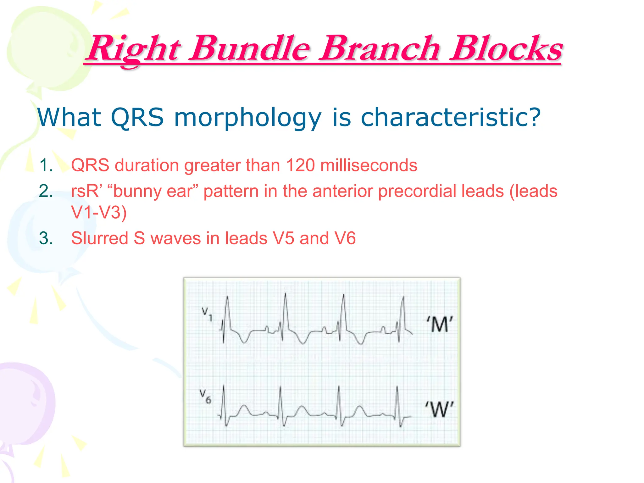
Bradycardias & heart
block
Jane Nader, MD
Lecturer of cardiology, BUC
Definitions
Bradycardia is decrease HR < 60/min
Classification of Brdaycardia
Sinus bradycardia
Junctional rhythm
Sick sinus syndrome
Sinoatrial exit block and sinus arrest
Slow AF
AV block
Bundle branch block
Sinus Bradycardia
 Definition:
A heart rate of 60/min. or less provided that the heart is
activated by a sinus mechanism.
 AE:
1. Drug induced:
1.Digitalis.
2.Quinidine.
3.B-blockers.
4.Hyperkalemia.
2. Vagal stimulation and
carotid sinus syncope.
3. Metabolic disorders:
• Hypothyroidism.
• Obstructive jaundice.
• Hypothermia.
4. Normal individuals:
• Athletes.
• Elderly subjects.
5. Diseases of the atrium
& SAN:
• Coronary artery disease.
• Sick sinus syndrome.
Junctional Rhythm
Junctional Escape Rhythm produces a heart rate between 40-60
beats per minute and has a relatively narrow QRS. The P waves may
be hidden, inverted, retrograde, or short/upright.
Sick Sinus Syndrome
• Definition:
Abnormal cardiac impulse formation and by abnormal
propagation from the SA node which prevents it from
performing its pace-making function.
It is characterized by sinus node dysfunction with an atrial rate
inappropriate for physiologic requirements.
Causes:
ischemia
fibrosis (often age-related)
drug-induced (beta blockers, calcium channel blockers, digoxin,
sympatholytic medications, antiarrhythmics, lithium)
infiltrative disorders (haemochromatosis, sarcoidosis,
connective tissue diseases)
autonomic dysfunction.
Clinical Features
Usually no symptoms.
Rarely: symptoms of low cardiac output.
Syncope may occur due to excessive bradycardia.
A slow heart rate in the presence of fever, left ventricular failure or
pulmonary edema may be suggestive of sick-sinus syndrome.
Treatment
Treatment of recurrent symptomatic bradycardia or prolonged
pauses requires implantation of a permanent pacemaker.
Sinoatrial exit block and sinus arrest
 Failure of impulse formation (arrest) or conduction from
(exit block) the SA node.
 Absent P waves on ECG.
 Escape rhythm may take over: junctional — narrow QRS
45–60bpm — or ventricular — wide QRS 30–45 bpm.
 Its causes overlap with those of sinus bradycardia,
including sick sinus, ↑K+, inferior MI, and CV drugs.
Heart block is defined as block of impulse
transmission any where allover the
conducting system of the heart
Sino-atrial block
A-V Block
Intra ventricular /bundle branch block (BBB)
Heart Block
Heart Block
Aetiology:
1. Acute:
 Cardiac:
• AMI.
• Myocarditis.
• Bacterial endocarditis.
 Drug intoxication or over-dosage:
• Digitalis.
• Anti-arrhythmic drugs.
• Tricyclic antidpressant.
• Phenothiazines.
 Electrolyte derangements:
• Hyperkalemia.
Heart Block
2. Chronic:
 Cardiac:
1. CAD.
2. Cardiomyopathy.
3. Aortic valve disease.
4. Congenital.
5. Idiopathic fibrosis of the conductive system.
 Connective tissue disorders:
1. Sarcoidosis.
2. Scleroderma.
3. Amyloidosis.
4. Systemic lupus.
 Endocrinal:
• Myxoedema.
Atrioventricular Block (A-V Block)
Classification:
 1-First A-V:
Prolonged A-V conduction, manifested by prolonged P-R interval
in the ECG (> 0.22 msec).
1st degree HB
Atrioventricular Block (A-V Block)
Classification:
 2- Second degree:
This results in dropped beats i.e. non conduction of all atrial activities.
Such dropping of beats may be regular or irregular.
Accordingly it has been subdivided into:
• Wenckbach or Mobitz type I:
It is manifested as progressive prolongation of the P-R
interval until there is a dropped QRS complex.
• Mobitz type II:
It is manifested by dropping of some beats but the
conducted one has a fixed P-R interval.
Second Degree Heart Block
Mobitz T
ype I
(Wenkebach)
PR PR PR DROPPED BEAT
2nd degree Mobitz type 1
Second Degree Heart Block
Mobitz T
ype II
PR PR DROPPED BEAT PR
2nd degree Mobitz type 2
Atrioventricular Block (A-V Block)
Classification:
3. Complete or third degree A-V block:
 There is complete dissociation between atrial rate and
ventricular rate.
 The atrium is controlled by the SAN rate and the ventricle by
the idioventricular rate.
3rd degree CHB
3rd degree CHB
Clinical Features of A-V Block
Symptoms:
1.No symptoms, even in CHB, as in most
cases. the bradycardia is compensated for by
the increased stroke volume due to prolonged
filling time.
2.Palpitation: either due to:
Dropped beats in partial heart block. Or
Slow forceful regular heart beats in complete
heart block.
3.Giddiness and faintness: may occur due
to transient asystole.
Clinical Features of A-V Block
4.If asystole is prolonged up to 15 seconds
Stockes-Adam attacks occur due to cerebral
anoxia; which is characterized by:
 Loss of consciousness.
 Incontinence.
 Extensor planter response.
 During the attack the patient is pale, flaccid,
without heart sound, and the pupils are dilated.
 Immediate on recovery the patient becomes
bright flushed due to reactive hyperaemia.
 Recovery is complete and rapid.
Extensor planter response.
Signs of HB
 1-First degree:
1. The heart rate is normal.
2. B.P. is maintained.
3. Neck veins reveal prolonged A-C interval.
4. Weak S 1.
 2- Second degree:- may be
 Pulse: irregular due to dropped beats (type 1 Mobitz) and
regular in Mobitz type 2.
 B.P. is maintained.
 "A" waves may appear which may be followed by C-V waves.
 S 1 variable intensity due to variable conduction (in Mobitz
type 1).
 3-Third degree:
 Pulse: rate between 30-40 b/min.
 BP wide pulse pressure in chronic cases
 Neck veins: Canon "A" wave due to atrial contraction against closed
A-V valves.
 S1:
• Variable intensity due to variability of position of A-V valves at the
onset of ventricular contraction.
• Canon sounds.
Treatment
-Treatment of the underlying cause if possible.
-The first degree of A-V block needs no further
treatment.
-Second degree A-V block:
1. Acute management when indicated
2. Permanent ventricular pacing is usually indicated
in Mobitz type II
-Third degree A-V block:
Acute management when indicated
Transient or permanent pacemaker must be
considered.
Acute management of bradycardia
* Atropine 500 mcg IV
• In inadequate response or risk of asystole:
• further atropine (up to 3 mg)
• transcutaenous pacing,
• adrenaline infusion, or isoprenaline infusion (β1 agonist).
Risk of asystole is defined as severe AV block (3rd degree or 2nd
degree type 2), recent asystole, or ventricular pauses (> 3 s).
* Definitive management with transient and/or permanent pacemaker.
Cardiac Pacemakers
Components
Pacemaker components combine with the
body tissue to form a complete circuit
• Pulse generator: power
source / battery
• Leads or wires
• Cathode (negative
electrode)
• Anode (positive electrode)
• Body tissue
Indications
2nd degree AV block, Mobitz type 2
Complications
Bundle Branch Blocks
Why Important?
Normal Impulse Conduction
Sinoatrial node
AV node
Bundle of His
Bundle Branches
Purkinje fibers
Bundle Branch Blocks
With Bundle Branch Blocks you will see two
changes on the ECG.
1. QRS complex widens (>
2. QRS morphology changes (v
s. left bundle branch block
Why does the QRS complex widen?
When the conduction pathway is blocked it will take longer
for the electrical signal to pass throughout the ventricles.
Depolarization of the Bundle Branches and Purkinje fibers is
seen as the QRS complex on the ECG.
Therefore, a conduction block of the Bundle Branches would
be reflected as a change in the QRS complex.
Right Bundle Branch Blocks
What QRS morphology is characteristic?
1. QRS duration greater than 120 milliseconds
2. rsR’ “bunny ear” pattern in the anterior precordial leads (leads
V1-V3)
3. Slurred S waves in leads V5 and V6
Left Bundle Branch Blocks
What QRS morphology is characteristic?
1. QRS duration > 120ms
2. Deep slurred S wave in V1
3. Broad monophasic R wave in lateral leads (I, aVL, V5-6)
CARDIAC ARREST
DEFINITIONS
• CARDIAC ARREST: Abrupt cessation of
cardiac pump function which may be
reversible by a rapid intervention but will
lead to death in its absence.
• DEATH: Irreversible cessation of all
biologic functions
MECHANISMS OF CARDIAC
ARREST
• 50-80%: VF and PULSLESS VT
• 20-30%: Asystole, severe bradycardia
and pulseless electrical activity
48
Decision to start CPR:
 Decision to start CPR is made if a victim is
unresponsive and not breathing normally.
 Pulse check is no longer required, and is NOT
recommended for lay persons.
 Pulse check has been shown to be unreliable,
with unacceptably high rates of false positives
and negatives.
Cardio-pulmonary resuscitation (CPR)
is a means to provide temporary
support to the coronary and cerebral
circulation, till normal cardiac output
is restored
Bradycardias & heart block Jane Nader.pdf

Bradycardias & heart block Jane Nader.pdf

  • 1.
    Bradycardias & heart block JaneNader, MD Lecturer of cardiology, BUC
  • 2.
  • 3.
    Classification of Brdaycardia Sinusbradycardia Junctional rhythm Sick sinus syndrome Sinoatrial exit block and sinus arrest Slow AF AV block Bundle branch block
  • 4.
    Sinus Bradycardia  Definition: Aheart rate of 60/min. or less provided that the heart is activated by a sinus mechanism.  AE: 1. Drug induced: 1.Digitalis. 2.Quinidine. 3.B-blockers. 4.Hyperkalemia. 2. Vagal stimulation and carotid sinus syncope. 3. Metabolic disorders: • Hypothyroidism. • Obstructive jaundice. • Hypothermia. 4. Normal individuals: • Athletes. • Elderly subjects. 5. Diseases of the atrium & SAN: • Coronary artery disease. • Sick sinus syndrome.
  • 5.
    Junctional Rhythm Junctional EscapeRhythm produces a heart rate between 40-60 beats per minute and has a relatively narrow QRS. The P waves may be hidden, inverted, retrograde, or short/upright.
  • 7.
    Sick Sinus Syndrome •Definition: Abnormal cardiac impulse formation and by abnormal propagation from the SA node which prevents it from performing its pace-making function. It is characterized by sinus node dysfunction with an atrial rate inappropriate for physiologic requirements. Causes: ischemia fibrosis (often age-related) drug-induced (beta blockers, calcium channel blockers, digoxin, sympatholytic medications, antiarrhythmics, lithium) infiltrative disorders (haemochromatosis, sarcoidosis, connective tissue diseases) autonomic dysfunction.
  • 8.
    Clinical Features Usually nosymptoms. Rarely: symptoms of low cardiac output. Syncope may occur due to excessive bradycardia. A slow heart rate in the presence of fever, left ventricular failure or pulmonary edema may be suggestive of sick-sinus syndrome. Treatment Treatment of recurrent symptomatic bradycardia or prolonged pauses requires implantation of a permanent pacemaker.
  • 11.
    Sinoatrial exit blockand sinus arrest  Failure of impulse formation (arrest) or conduction from (exit block) the SA node.  Absent P waves on ECG.  Escape rhythm may take over: junctional — narrow QRS 45–60bpm — or ventricular — wide QRS 30–45 bpm.  Its causes overlap with those of sinus bradycardia, including sick sinus, ↑K+, inferior MI, and CV drugs.
  • 12.
    Heart block isdefined as block of impulse transmission any where allover the conducting system of the heart Sino-atrial block A-V Block Intra ventricular /bundle branch block (BBB) Heart Block
  • 13.
    Heart Block Aetiology: 1. Acute: Cardiac: • AMI. • Myocarditis. • Bacterial endocarditis.  Drug intoxication or over-dosage: • Digitalis. • Anti-arrhythmic drugs. • Tricyclic antidpressant. • Phenothiazines.  Electrolyte derangements: • Hyperkalemia.
  • 14.
    Heart Block 2. Chronic: Cardiac: 1. CAD. 2. Cardiomyopathy. 3. Aortic valve disease. 4. Congenital. 5. Idiopathic fibrosis of the conductive system.  Connective tissue disorders: 1. Sarcoidosis. 2. Scleroderma. 3. Amyloidosis. 4. Systemic lupus.  Endocrinal: • Myxoedema.
  • 15.
    Atrioventricular Block (A-VBlock) Classification:  1-First A-V: Prolonged A-V conduction, manifested by prolonged P-R interval in the ECG (> 0.22 msec).
  • 16.
  • 17.
    Atrioventricular Block (A-VBlock) Classification:  2- Second degree: This results in dropped beats i.e. non conduction of all atrial activities. Such dropping of beats may be regular or irregular. Accordingly it has been subdivided into: • Wenckbach or Mobitz type I: It is manifested as progressive prolongation of the P-R interval until there is a dropped QRS complex. • Mobitz type II: It is manifested by dropping of some beats but the conducted one has a fixed P-R interval.
  • 18.
    Second Degree HeartBlock Mobitz T ype I (Wenkebach) PR PR PR DROPPED BEAT
  • 19.
  • 20.
    Second Degree HeartBlock Mobitz T ype II PR PR DROPPED BEAT PR
  • 21.
  • 22.
    Atrioventricular Block (A-VBlock) Classification: 3. Complete or third degree A-V block:  There is complete dissociation between atrial rate and ventricular rate.  The atrium is controlled by the SAN rate and the ventricle by the idioventricular rate.
  • 23.
  • 24.
  • 26.
    Clinical Features ofA-V Block Symptoms: 1.No symptoms, even in CHB, as in most cases. the bradycardia is compensated for by the increased stroke volume due to prolonged filling time. 2.Palpitation: either due to: Dropped beats in partial heart block. Or Slow forceful regular heart beats in complete heart block. 3.Giddiness and faintness: may occur due to transient asystole.
  • 27.
    Clinical Features ofA-V Block 4.If asystole is prolonged up to 15 seconds Stockes-Adam attacks occur due to cerebral anoxia; which is characterized by:  Loss of consciousness.  Incontinence.  Extensor planter response.  During the attack the patient is pale, flaccid, without heart sound, and the pupils are dilated.  Immediate on recovery the patient becomes bright flushed due to reactive hyperaemia.  Recovery is complete and rapid.
  • 28.
  • 29.
    Signs of HB 1-First degree: 1. The heart rate is normal. 2. B.P. is maintained. 3. Neck veins reveal prolonged A-C interval. 4. Weak S 1.  2- Second degree:- may be  Pulse: irregular due to dropped beats (type 1 Mobitz) and regular in Mobitz type 2.  B.P. is maintained.  "A" waves may appear which may be followed by C-V waves.  S 1 variable intensity due to variable conduction (in Mobitz type 1).  3-Third degree:  Pulse: rate between 30-40 b/min.  BP wide pulse pressure in chronic cases  Neck veins: Canon "A" wave due to atrial contraction against closed A-V valves.  S1: • Variable intensity due to variability of position of A-V valves at the onset of ventricular contraction. • Canon sounds.
  • 31.
    Treatment -Treatment of theunderlying cause if possible. -The first degree of A-V block needs no further treatment. -Second degree A-V block: 1. Acute management when indicated 2. Permanent ventricular pacing is usually indicated in Mobitz type II -Third degree A-V block: Acute management when indicated Transient or permanent pacemaker must be considered.
  • 32.
    Acute management ofbradycardia * Atropine 500 mcg IV • In inadequate response or risk of asystole: • further atropine (up to 3 mg) • transcutaenous pacing, • adrenaline infusion, or isoprenaline infusion (β1 agonist). Risk of asystole is defined as severe AV block (3rd degree or 2nd degree type 2), recent asystole, or ventricular pauses (> 3 s). * Definitive management with transient and/or permanent pacemaker.
  • 33.
  • 34.
  • 35.
    Pacemaker components combinewith the body tissue to form a complete circuit • Pulse generator: power source / battery • Leads or wires • Cathode (negative electrode) • Anode (positive electrode) • Body tissue
  • 36.
    Indications 2nd degree AVblock, Mobitz type 2
  • 37.
  • 38.
  • 40.
    Normal Impulse Conduction Sinoatrialnode AV node Bundle of His Bundle Branches Purkinje fibers
  • 41.
    Bundle Branch Blocks WithBundle Branch Blocks you will see two changes on the ECG. 1. QRS complex widens (> 2. QRS morphology changes (v s. left bundle branch block Why does the QRS complex widen? When the conduction pathway is blocked it will take longer for the electrical signal to pass throughout the ventricles. Depolarization of the Bundle Branches and Purkinje fibers is seen as the QRS complex on the ECG. Therefore, a conduction block of the Bundle Branches would be reflected as a change in the QRS complex.
  • 42.
    Right Bundle BranchBlocks What QRS morphology is characteristic? 1. QRS duration greater than 120 milliseconds 2. rsR’ “bunny ear” pattern in the anterior precordial leads (leads V1-V3) 3. Slurred S waves in leads V5 and V6
  • 43.
    Left Bundle BranchBlocks What QRS morphology is characteristic? 1. QRS duration > 120ms 2. Deep slurred S wave in V1 3. Broad monophasic R wave in lateral leads (I, aVL, V5-6)
  • 45.
  • 46.
    DEFINITIONS • CARDIAC ARREST:Abrupt cessation of cardiac pump function which may be reversible by a rapid intervention but will lead to death in its absence. • DEATH: Irreversible cessation of all biologic functions
  • 47.
    MECHANISMS OF CARDIAC ARREST •50-80%: VF and PULSLESS VT • 20-30%: Asystole, severe bradycardia and pulseless electrical activity
  • 48.
    48 Decision to startCPR:  Decision to start CPR is made if a victim is unresponsive and not breathing normally.  Pulse check is no longer required, and is NOT recommended for lay persons.  Pulse check has been shown to be unreliable, with unacceptably high rates of false positives and negatives.
  • 49.
    Cardio-pulmonary resuscitation (CPR) isa means to provide temporary support to the coronary and cerebral circulation, till normal cardiac output is restored