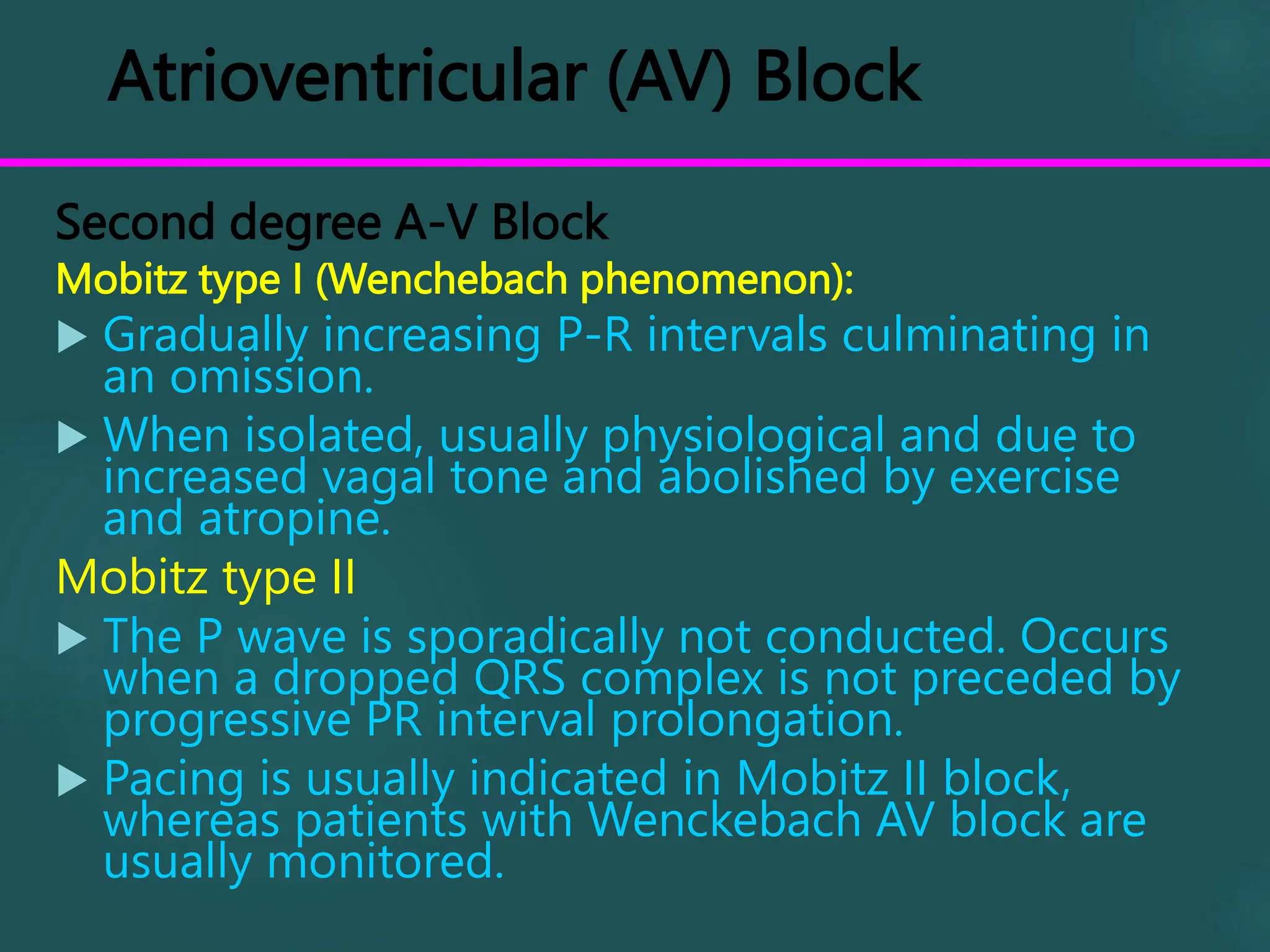
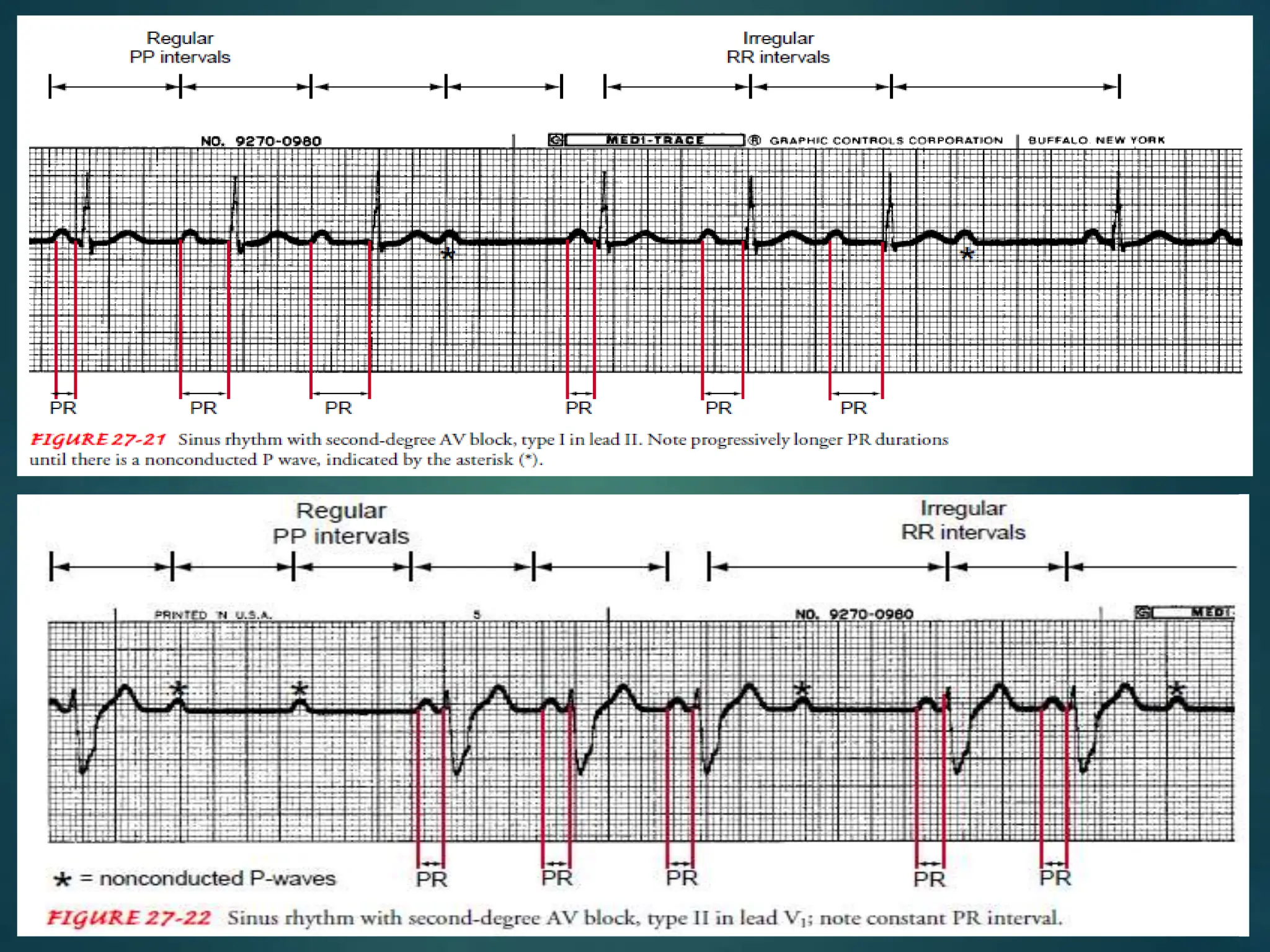
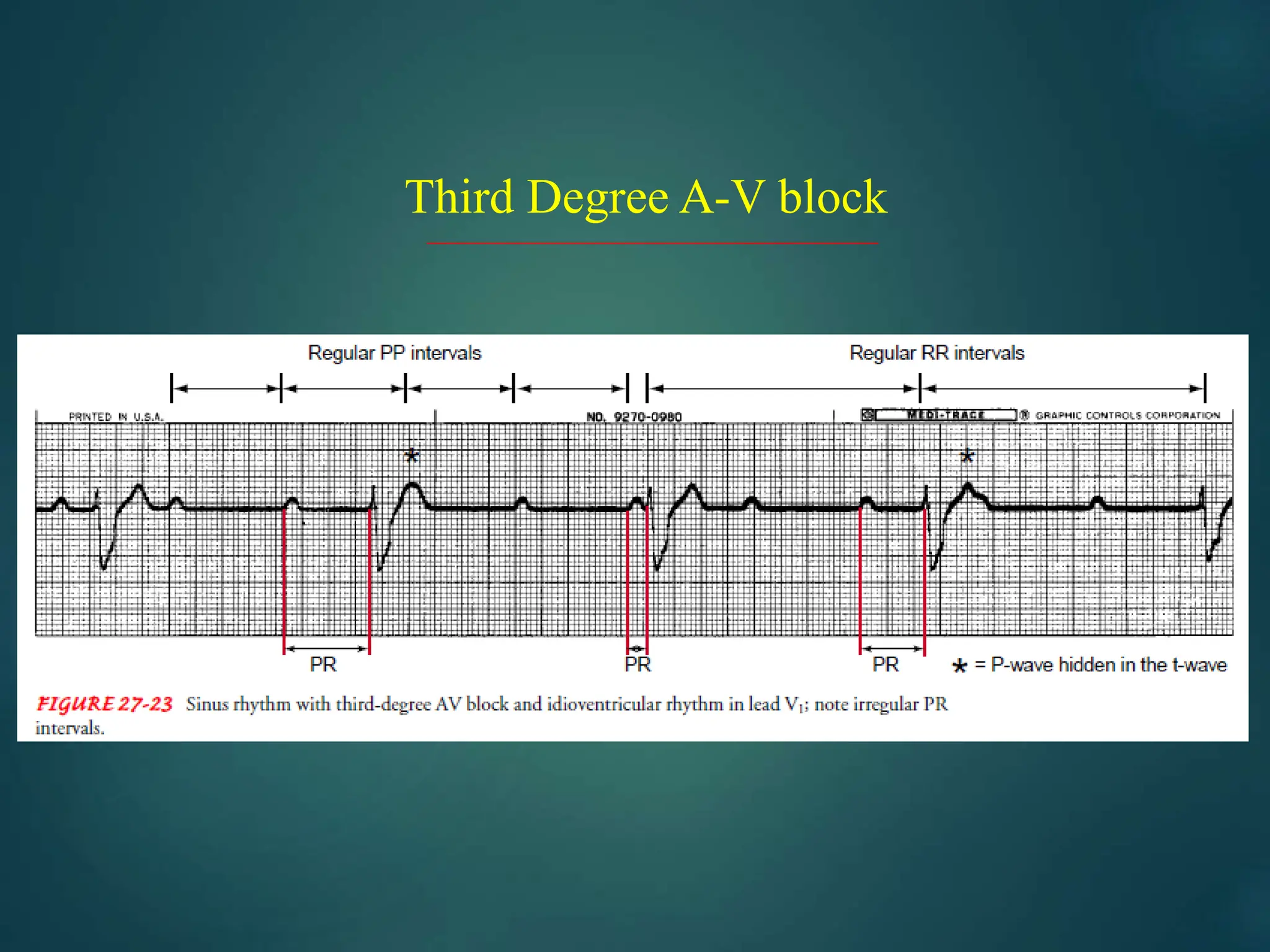
ATRIOVENTRICULAR NODE
BY
SARATHKUMAR
BSC CARDIAC TECHNOLOGY
Atrioventricular (AV) Block
First degree A-V Block
 Seldom of clinical significance, and unlikely to
progress.
 ECG shows prolonged PR interval.
 May be associated with acute rheumatic fever,
diphtheria, myocardial infarction or drugs as digoxin
Atrioventricular (AV) Block
Second degree A-V Block
Mobitz type I (Wenchebach phenomenon):
 Gradually increasing P-R intervals culminating in
an omission.
 When isolated, usually physiological and due to
increased vagal tone and abolished by exercise
and atropine.
Mobitz type II
 The P wave is sporadically not conducted. Occurs
when a dropped QRS complex is not preceded by
progressive PR interval prolongation.
 Pacing is usually indicated in Mobitz II block,
whereas patients with Wenckebach AV block are
usually monitored.
Second Degree AV Block
Acute myocardial infarction may produce second-degree
heart block. In inferior myocardial infarction, close
monitoring and transcutaneous temporary back-up pacing
are all that is required. In anterior myocardial infarction,
second-degree heart block is associated with a high risk of
progression to complete heart block, and temporary pacing
followed by permanent pacemaker implantation is usually
indicated.
Atrioventricular (AV) Block
Third degree A-V Block
 Common in elderly age groups due to idiopathic bundle branch
fibrosis.
 Other causes include coronary heart disease, calcification from aortic
valve, sarcoidosis or congenital.
 ECG shows bradycardia, P wave continue, unrelated to regular slow
idioventricular rhythm.
 Treatment is permanent pacing.
Third Degree A-V block
Atrioventricular (AV) Block
Bundle Branch Block (BBB):
 Interruption of the right or left branch of the
bundle of Hiss delays activation of the
corresponding ventricle leading to broadening of
the QRS complex
 Unlike right BBB, left BBB is always associated with
an underlying heart disease.
 Both RT and LT BBB show wide deformed QRS
complex. In RBBB there is rSR pattern in lead V1,
while in LBBB there is a broad monophasic (or
notched) R wave in leads V5 and V6.
Atrioventricular (AV) Block
Bundle Branch Block (BBB):
Hemiblock
Delay or block in the divisions of the left bundle branch
produces a swing in the direction of depolarization
(electrical axis) of the heart. When the anterior division is
blocked (left anterior hemiblock), there is left axis
deviation. Delay or block in the postero-inferior division
causes(right axis deviation).
Bifascicular block
This is a combination of a block of any two of the
following: the right bundle branch, the left antero-superior
division and the left postero-inferior division. Block of the
remaining fascicle will result in complete AV block.
Atrioventricular node by sarathkumar bsx

Atrioventricular node by sarathkumar bsx

  • 1.
  • 2.
    Atrioventricular (AV) Block Firstdegree A-V Block  Seldom of clinical significance, and unlikely to progress.  ECG shows prolonged PR interval.  May be associated with acute rheumatic fever, diphtheria, myocardial infarction or drugs as digoxin
  • 3.
    Atrioventricular (AV) Block Seconddegree A-V Block Mobitz type I (Wenchebach phenomenon):  Gradually increasing P-R intervals culminating in an omission.  When isolated, usually physiological and due to increased vagal tone and abolished by exercise and atropine. Mobitz type II  The P wave is sporadically not conducted. Occurs when a dropped QRS complex is not preceded by progressive PR interval prolongation.  Pacing is usually indicated in Mobitz II block, whereas patients with Wenckebach AV block are usually monitored.
  • 4.
    Second Degree AVBlock Acute myocardial infarction may produce second-degree heart block. In inferior myocardial infarction, close monitoring and transcutaneous temporary back-up pacing are all that is required. In anterior myocardial infarction, second-degree heart block is associated with a high risk of progression to complete heart block, and temporary pacing followed by permanent pacemaker implantation is usually indicated.
  • 6.
    Atrioventricular (AV) Block Thirddegree A-V Block  Common in elderly age groups due to idiopathic bundle branch fibrosis.  Other causes include coronary heart disease, calcification from aortic valve, sarcoidosis or congenital.  ECG shows bradycardia, P wave continue, unrelated to regular slow idioventricular rhythm.  Treatment is permanent pacing.
  • 7.
  • 8.
    Atrioventricular (AV) Block BundleBranch Block (BBB):  Interruption of the right or left branch of the bundle of Hiss delays activation of the corresponding ventricle leading to broadening of the QRS complex  Unlike right BBB, left BBB is always associated with an underlying heart disease.  Both RT and LT BBB show wide deformed QRS complex. In RBBB there is rSR pattern in lead V1, while in LBBB there is a broad monophasic (or notched) R wave in leads V5 and V6.
  • 9.
    Atrioventricular (AV) Block BundleBranch Block (BBB): Hemiblock Delay or block in the divisions of the left bundle branch produces a swing in the direction of depolarization (electrical axis) of the heart. When the anterior division is blocked (left anterior hemiblock), there is left axis deviation. Delay or block in the postero-inferior division causes(right axis deviation). Bifascicular block This is a combination of a block of any two of the following: the right bundle branch, the left antero-superior division and the left postero-inferior division. Block of the remaining fascicle will result in complete AV block.