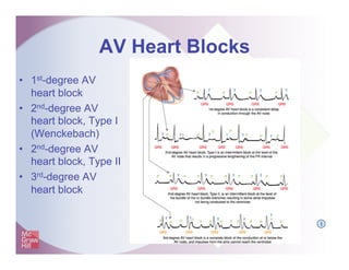
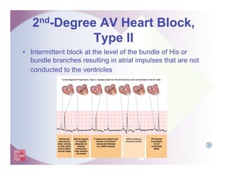
Q
I
A
12
Fast & Easy ECGs – A Self-Paced
Learning Program
Origin and Clinical Aspects of
AV Heart Blocks
Heart Blocks
•  Partial delays or complete interruptions in
the cardiac conduction pathway between
the atria and ventricles
•  The degree of block defines the type and
classification of heart block
Q
I
Heart Blocks
•  Common causes:
– Ischemia
– Myocardial necrosis
– Degenerative disease of the conduction
system
– Congenital anomalies
– Drugs (especially digitalis preparations)
I
AV Heart Blocks
•  1st-degree AV
heart block
•  2nd-degree AV
heart block, Type I
(Wenckebach)
•  2nd-degree AV
heart block, Type II
•  3rd-degree AV
heart block
I
1st-Degree AV Heart Block
•  Not a true block
•  A consistent delay of conduction at the level of the AV
node
I
1st-Degree AV Heart Block
•  Often of little or no clinical significance
because all impulses are conducted to the
ventricles
•  Can progress to higher degree block,
especially in the presence of inferior wall
myocardial infarction
I
1st-Degree AV Heart Block
Q
1st-Degree AV Heart Block
2nd-Degree AV Heart Block,
Type I
•  Intermittent block at the level of the AV node
•  Also referred to as Wenckebach
2nd-Degree AV Heart Block,
Type I
•  More P waves than QRS complexes and the
rhythm has patterned irregularity
•  PR interval increases until a QRS complex is
dropped
•  After dropped beat the next PR interval is shorter
•  As each subsequent impulse generated there is
a progressively longer PR interval until again, a
QRS is dropped
•  Cycle repeats
I
2nd-Degree AV Heart Block,
Type I
2nd-Degree AV Heart Block,
Type I
2nd-Degree AV Heart Block,
Type I
•  May occur in otherwise healthy persons
•  Usually transient and reversible, mostly
resolving when the underlying condition is
corrected
•  May progress to more serious blocks
(particularly if it occurs early in myocardial
infarction)
2nd-Degree AV Heart Block,
Type I
•  If dropped ventricular beats occur
frequently, patient may show signs and
symptoms of decreased cardiac output
I
2nd-Degree AV Heart Block,
Type II
•  Intermittent block at the level of the bundle of His or
bundle branches resulting in atrial impulses that are not
conducted to the ventricles
I
2nd-Degree AV Heart Block,
Type II
•  More P waves than QRS complexes
•  Duration of PR interval of the conducted
beats remains constant
2nd-Degree AV Heart Block,
Type II
2nd-Degree AV Heart Block,
Type II
•  A serious dysrhythmia (usually considered
malignant in the emergency setting)
•  Can result in decreased cardiac output
and may produce signs and symptoms of
hypoperfusion
•  May progress to a more severe heart
block and ventricular asystole
I
3rd-Degree AV Heart Block
•  Complete block of conduction at or below the AV node
•  Impulses from atria cannot reach ventricles
3rd-Degree AV Heart Block
•  Atrial pacemaker site is the SA node
– Atrial rate 60 to 100 BPM
•  Ventricular pacemaker site is an escape
rhythm
– From AV junction rate 40 to 60 BPM
– From ventricles rate 20 to 40 BPM
Q
3rd-Degree AV Heart Block
•  Upright and round P waves seem to
“march right through the QRS complexes”
I
3rd-Degree AV Heart Block
•  Well tolerated as long as the escape
rhythm is fast enough to generate a
sufficient cardiac output to maintain
adequate perfusion
•  Can result in decreased cardiac output
because of the asynchronous action of the
atria and ventricles and if the ventricular
rate is slow
I
Remember!
2nd- and 3rd-degree AV heart block can
lead to decreased cardiac output if
the ventricular rate slows sufficiently
Practice Makes Perfect
•  Determine the type of dysrhythmia
I
Practice Makes Perfect
•  Determine the type of dysrhythmia
I
Practice Makes Perfect
•  Determine the type of dysrhythmia
I
Practice Makes Perfect
•  Determine the type of dysrhythmia
I
Practice Makes Perfect
•  Determine the type of dysrhythmia
I
Practice Makes Perfect
•  Determine the type of dysrhythmia
I
Practice Makes Perfect
•  Determine the type of dysrhythmia
I
Practice Makes Perfect
•  Determine the type of dysrhythmia
I
Practice Makes Perfect
•  Determine the type of dysrhythmia
I
Practice Makes Perfect
•  Determine the type of dysrhythmia
I
Summary
•  Heart blocks are partial delays or complete interruptions
in the cardiac conduction pathway between the atria and
ventricles.
•  1st-degree AV heart block is not a true block. Instead it is
a consistent delay of conduction at the level of the AV
node which results in a PR interval that is greater than
0.20 seconds in duration.
•  2nd-degree AV heart block, Type I is an intermittent block
at the level of the AV node.
Summary
•  With 2nd-degree AV heart block, Type I, the PR interval
increases until a QRS complex is dropped. After the
dropped beat the next PR interval is shorter. Then as
each subsequent impulse is generated and transmitted
through the AV junction there is a progressively longer
PR interval until again, a QRS is dropped. This cycle
can repeat itself.
•  With 2nd-degree AV heart block, Type I, there are more
P waves than QRS complexes and the rhythm is
regularly irregular.
Summary
•  2nd-degree AV heart block, Type II is an intermittent
block at the level of the bundle of His or bundle branches
resulting in atrial impulses that are not conducted to the
ventricles.
•  With 2nd-degree AV heart block, Type II, there are more
P waves than QRS complexes and the duration of PR
interval of the conducted beats remains the same (are
constant).
•  3rd-degree AV heart block is a complete block of the
conduction at or below the AV node and impulses from
the atria cannot reach the ventricles.
Summary
•  In 3rd-degree AV heart block the pacemaker for the atria
arises from the SA node while the pacemaker for the
ventricles arises as an escape rhythm from the AV
junction or from the ventricles.
•  With 3rd-degree AV heart block the upright and round P
waves seem to “march right through the QRS
complexes." This reveals that there is no relationship
between the P waves and QRS complexes.
•  2nd- and 3rd-degree AV heart block can lead to
decreased cardiac output.

Shadechapter12.ppt [read only]

  • 1.
    Q I A 12 Fast & EasyECGs – A Self-Paced Learning Program Origin and Clinical Aspects of AV Heart Blocks
  • 2.
    Heart Blocks •  Partialdelays or complete interruptions in the cardiac conduction pathway between the atria and ventricles •  The degree of block defines the type and classification of heart block Q I
  • 3.
    Heart Blocks •  Commoncauses: – Ischemia – Myocardial necrosis – Degenerative disease of the conduction system – Congenital anomalies – Drugs (especially digitalis preparations) I
  • 4.
    AV Heart Blocks • 1st-degree AV heart block •  2nd-degree AV heart block, Type I (Wenckebach) •  2nd-degree AV heart block, Type II •  3rd-degree AV heart block I
  • 5.
    1st-Degree AV HeartBlock •  Not a true block •  A consistent delay of conduction at the level of the AV node I
  • 6.
    1st-Degree AV HeartBlock •  Often of little or no clinical significance because all impulses are conducted to the ventricles •  Can progress to higher degree block, especially in the presence of inferior wall myocardial infarction I
  • 7.
  • 8.
  • 9.
    2nd-Degree AV HeartBlock, Type I •  Intermittent block at the level of the AV node •  Also referred to as Wenckebach
  • 10.
    2nd-Degree AV HeartBlock, Type I •  More P waves than QRS complexes and the rhythm has patterned irregularity •  PR interval increases until a QRS complex is dropped •  After dropped beat the next PR interval is shorter •  As each subsequent impulse generated there is a progressively longer PR interval until again, a QRS is dropped •  Cycle repeats I
  • 11.
    2nd-Degree AV HeartBlock, Type I
  • 12.
    2nd-Degree AV HeartBlock, Type I
  • 13.
    2nd-Degree AV HeartBlock, Type I •  May occur in otherwise healthy persons •  Usually transient and reversible, mostly resolving when the underlying condition is corrected •  May progress to more serious blocks (particularly if it occurs early in myocardial infarction)
  • 14.
    2nd-Degree AV HeartBlock, Type I •  If dropped ventricular beats occur frequently, patient may show signs and symptoms of decreased cardiac output I
  • 15.
    2nd-Degree AV HeartBlock, Type II •  Intermittent block at the level of the bundle of His or bundle branches resulting in atrial impulses that are not conducted to the ventricles I
  • 16.
    2nd-Degree AV HeartBlock, Type II •  More P waves than QRS complexes •  Duration of PR interval of the conducted beats remains constant
  • 17.
    2nd-Degree AV HeartBlock, Type II
  • 18.
    2nd-Degree AV HeartBlock, Type II •  A serious dysrhythmia (usually considered malignant in the emergency setting) •  Can result in decreased cardiac output and may produce signs and symptoms of hypoperfusion •  May progress to a more severe heart block and ventricular asystole I
  • 19.
    3rd-Degree AV HeartBlock •  Complete block of conduction at or below the AV node •  Impulses from atria cannot reach ventricles
  • 20.
    3rd-Degree AV HeartBlock •  Atrial pacemaker site is the SA node – Atrial rate 60 to 100 BPM •  Ventricular pacemaker site is an escape rhythm – From AV junction rate 40 to 60 BPM – From ventricles rate 20 to 40 BPM Q
  • 21.
    3rd-Degree AV HeartBlock •  Upright and round P waves seem to “march right through the QRS complexes” I
  • 22.
  • 23.
  • 24.
    3rd-Degree AV HeartBlock •  Well tolerated as long as the escape rhythm is fast enough to generate a sufficient cardiac output to maintain adequate perfusion •  Can result in decreased cardiac output because of the asynchronous action of the atria and ventricles and if the ventricular rate is slow I
  • 25.
    Remember! 2nd- and 3rd-degreeAV heart block can lead to decreased cardiac output if the ventricular rate slows sufficiently
  • 27.
    Practice Makes Perfect • Determine the type of dysrhythmia I
  • 28.
    Practice Makes Perfect • Determine the type of dysrhythmia I
  • 29.
    Practice Makes Perfect • Determine the type of dysrhythmia I
  • 30.
    Practice Makes Perfect • Determine the type of dysrhythmia I
  • 31.
    Practice Makes Perfect • Determine the type of dysrhythmia I
  • 32.
    Practice Makes Perfect • Determine the type of dysrhythmia I
  • 33.
    Practice Makes Perfect • Determine the type of dysrhythmia I
  • 34.
    Practice Makes Perfect • Determine the type of dysrhythmia I
  • 35.
    Practice Makes Perfect • Determine the type of dysrhythmia I
  • 36.
    Practice Makes Perfect • Determine the type of dysrhythmia I
  • 37.
    Summary •  Heart blocksare partial delays or complete interruptions in the cardiac conduction pathway between the atria and ventricles. •  1st-degree AV heart block is not a true block. Instead it is a consistent delay of conduction at the level of the AV node which results in a PR interval that is greater than 0.20 seconds in duration. •  2nd-degree AV heart block, Type I is an intermittent block at the level of the AV node.
  • 38.
    Summary •  With 2nd-degreeAV heart block, Type I, the PR interval increases until a QRS complex is dropped. After the dropped beat the next PR interval is shorter. Then as each subsequent impulse is generated and transmitted through the AV junction there is a progressively longer PR interval until again, a QRS is dropped. This cycle can repeat itself. •  With 2nd-degree AV heart block, Type I, there are more P waves than QRS complexes and the rhythm is regularly irregular.
  • 39.
    Summary •  2nd-degree AVheart block, Type II is an intermittent block at the level of the bundle of His or bundle branches resulting in atrial impulses that are not conducted to the ventricles. •  With 2nd-degree AV heart block, Type II, there are more P waves than QRS complexes and the duration of PR interval of the conducted beats remains the same (are constant). •  3rd-degree AV heart block is a complete block of the conduction at or below the AV node and impulses from the atria cannot reach the ventricles.
  • 40.
    Summary •  In 3rd-degreeAV heart block the pacemaker for the atria arises from the SA node while the pacemaker for the ventricles arises as an escape rhythm from the AV junction or from the ventricles. •  With 3rd-degree AV heart block the upright and round P waves seem to “march right through the QRS complexes." This reveals that there is no relationship between the P waves and QRS complexes. •  2nd- and 3rd-degree AV heart block can lead to decreased cardiac output.